


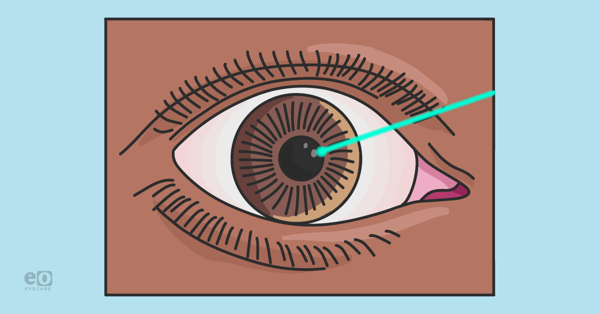
When thinking of refractive surgery, our minds gravitate towards laser-assisted in situ keratomileusis (LASIK)—and for a good reason. LASIK is the most commonly performed refractive surgery and has been for over 25 years. It is the single most studied procedure in the history of medicine, supporting its stellar reputation for safety and visual outcomes. However, not everyone is a candidate for LASIK. Today, there are several options for those who want clear vision without glasses and contact lenses.1
Alternate choices for refractive surgery
With the advent of an aging population, the popularity of correcting presbyopia through refractive surgery is becoming more commonplace in today’s practice. Patients typically over 40, who might be experiencing presbyopic shifts in vision, become ideal candidates to explore options beyond glasses and contacts to manage their visual demand requirements.
The question becomes: should the refractive error be corrected at the cornea or the lens? At this stage in life, refractive lens exchange (RLE) can improve the quality of vision at potentially all ranges of sight by removing the natural lens prior to cataract formation with an intraocular lens (IOL). Laser vision correction could be considered more advantageous since this type of surgical procedure is limited to distance management.2
“As the human lens matures, this tissue becomes dysfunctional in multiple ways, including visual obscurations and decreased vision.”
With advancements in multifocal, trifocal, and extended depth of focus (EDOF) IOLs, patients can experience the full range of distance to near vision while maintaining stereopsis. RLE provides the satisfaction of depth perception, summed uncorrected visual acuity, and enhanced contrast sensitivity. The patient can better understand this by explaining options such as LASIK, small incision lenticule extraction (SMILE), and photorefractive keratectomy (PRK).
Co-managing refractive surgery
Let’s take a moment to discuss the importance of collaborative relationships between ophthalmologists and optometrists. As we know, each profession plays a crucial role in co-managing these surgical patients to ensure they receive premium care at each step of their cataract and/or refractive journey.
“The development of mutual trust and respect are pivotal when communicating ahead of a potential referral from an OD to MD, or internally in an OD/MD clinical setting.”
Invariably, an essential piece of a successful partnership is the “all ships rise” mentality when sharing knowledge and keeping up on the latest advancements through continued educational enrichment. Now that you have a lay of the land and appreciation for collaborative care read on to learn more about these individual surgical procedures and techniques coupled with a glimpse into the future of refractive surgery.
Download the cheat sheet here!

Comparative Guide of Refractive Surgery
Use this cheat sheet to compare a variety of procedures within refractive surgery.

LASIK
Overview
LASIK is a surgical procedure designed to correct refractive error by reshaping corneal tissue. The ideal patient profile includes a stable refractive error for 1 year, 18 years or older, corneal thickness greater than 450 to 550 microns, a refractive error that ranges between 0.50 to 12.00 diopters of myopia, less than 6.00 diopters of hyperopia, and astigmatism less than 5.00 diopters.3,4
Procedure
The technique creates a corneal flap using an excimer or femtosecond laser, which is then lifted and folded back on the hinge side. Subsequently, the laser is used to reshape tissue by ablating the underlying exposed stromal bed, followed by carefully placing the corneal flap back into position while smoothing the edges.4 Procedure time is brief, usually clocking in around 8 to 10 minutes. The patient typically has functional vision on the same day.
Contraindications
Contraindications for LASIK include:
- Unstable refractive error (>0.50D) within 1 year of surgery
- Meibomian Gland Dysfunction (MGD)
- Unstable glaucoma
- Fuchs’ Endothelial Dystrophy
- Epithelial basement membrane dystrophy (EBMD)
- Anterior blepharitis
- Significant cataract
- Dry eye
Absolute contraindications include:
- Abnormalities of the cornea, such as keratoconus
- Pellucid marginal degeneration
- Corneal ectasias
- Edema
A few relative contraindications include a history of herpes simplex keratitis (HSK), patients with systemic autoimmune disease, and pregnant women or nursing mothers, as hormonal influences may result in changes in refractive error. Patients taking medications with a high risk of ocular side effects (amiodarone, isotretinoin, sumatriptan, etc.) are generally advised to avoid refractive surgeries.1
Complications
Immediately following the procedure, patients may experience tearing, photophobia, blurred vision, and discomfort, but this usually resolves the following day. With these symptoms in mind, the most common postoperative challenge is dry eye disease (DED).
Other complications that could potentially occur at the interface between the flap and stromal bed are diffuse lamellar keratitis (DLK), infection, and epithelial ingrowth, along with longer-term visual consequences, such as regression (clinically observed in mid to high myopes). In rare instances, complications could occur, leading to significant permanent visual loss, such as progressive ectasia, but these are not common.1
Advanced surface ablation and PRK
Overview
Advanced surface ablation (ASA), a form of PRK, is a surgical procedure to correct refractive errors when there are contraindications for LASIK, such as large refractive errors or abnormally thin corneas.
The ideal patient profile includes a stable refractive error for 1 year and a range between 0.50 to 12.00 diopters of myopia, less than 6.00 diopters of hyperopia, and astigmatism less than 5.00 diopters.1,5 Additionally, patients who participate in contact sports or other impact activities are recommended ASA over LASIK.
Procedure
The procedure begins by applying a diluted alcohol solution that loosens and removes the corneal epithelium with a brushing action. Then, an excimer laser treatment is applied to the exposed stromal bed to reshape the cornea. The customary postoperative regimen is topical antibiotics, steroids, NSAIDs, and a disposable soft bandage contact lens placed over the cornea for 5 to 7 days.6
Contraindications
Contraindications for ASA and PRK include:
- Unstable refractive error (>0.50D) within 1 year of surgery
- Significant cataract
- Unstable glaucoma
- Anterior blepharitis
- MGD
- Dry eye
Absolute contraindications include:
- Abnormalities of the cornea, such as keratoconus, corneal ectasias, and edema
A few relative contraindications include a history of herpes simplex keratitis (HSK) and pregnant women or nursing mothers, as hormonal influences may result in changes in refractive error. Patients taking medications with a high risk of ocular side effects (amiodarone, isotretinoin, sumatriptan, etc.) are generally advised to avoid refractive surgeries.1,7
Complications
Postoperatively, it is common to experience eye irritation and/or watering and light sensitivity immediately following the procedure.8 Similar to LASIK, the most common potential postoperative issue is DED. Other adverse events include under- or overcorrection, regression, corneal haze, corneal ectasia, infection, or glare/halos, which could be most noticeable at night.
SMILE
Overview
SMILE is a surgical procedure that uses a femtosecond laser to create a corneal lenticule (thin layer of stroma) which is then removed through a small incision without using an excimer laser.1 SMILE gained US FDA approval in 2016 and 2018 for spherical myopic correction and compound myopic astigmatism, respectively. This procedure achieves results similar to LASIK but with additional benefits, including shorter recovery of postoperative dry eye and potential re-innervation of corneal nerves.
Procedure
As mentioned, this surgical technique uses a laser to create a thin contact lens-shaped “lenticule” layer within the cornea.1 This lenticule is removed through a tiny 2 to 3mm opening, allowing the surrounding tissues to heal together.
SMILE theoretically combines the advantages of ASA and LASIK. It requires only one small incision, does not require a flap, and has the added benefit of minimal to no postoperative restrictions. An important point to note, enhancement procedures for residual refractive error following SMILE are done by performing ASA.9
Contraindications
Contraindications for SMILE include:
- Those with unstable refraction over a 12-month period
- Under the age of 18 years
- Insufficient corneal thickness
- Visually significant cataract
- Uncontrolled glaucoma
- Unrealistic expectations of surgical outcome
- Hyperopic refractive errors7
Complications
Potential postoperative issues include under- or overcorrection of refractive error, DED, visual distortion, and/or halo/glare. Corneal interface complications seen in SMILE could be diffuse lamellar keratitis, corneal ectasia, epithelial ingrowth, transient light sensitivity syndrome (TLSS), pressure-induced stromal keratitis, interface fluid syndrome, interface debris, or infectious keratitis.9
Laser epithelial keratomileusis
Overview
Laser epithelial keratomileusis (LASEK) theoretically has all the advantages of LASIK and PRK but is structured to prevent some of the more commonly-found complications.
Both LASEK and PRK procedures interact with the epithelial layer of the cornea—PRK removes the targeted epithelial layer, while LASEK creates a thin epithelial flap which is peeled aside and replaced following the stroma reshaping. The flap created during LASEK is only epithelial, while the flap created for LASIK includes epithelium and stroma.1
Procedure
Laser epithelial keratomileusis (LASEK) is a variant form of PRK that utilizes a diluted alcohol solution to loosen the corneal epithelium and peel a thin layer to the side.3 An excimer laser is used to reshape the underlying exposed stromal bed, and once complete, the corneal epithelium is repositioned back over the stroma. Following the procedure, a soft bandage contact lens is placed over the cornea and removed after re-epithelialization is complete.10
Contraindications
Contraindications for LASEK include:
- Fuchs’ Endothelial Dystrophy
- Keratoconus
- Pellucid marginal degeneration
- Epithelial basement membrane dystrophy (EBMD)
- Systemic autoimmune disease
- Pregnancy
- Lactation
- Anterior blepharitis
- MGD
- Significant dry eye7
Separately, other adverse events might include under- or overcorrection, regression, corneal haze, corneal ectasia, infection, and glare/halos, most noticeable at night.
Complications
Postoperatively, it is common to experience eye irritation and/or watering and light sensitivity immediately following the procedure.3 Thematically, the most common potential postoperative issue is dry eye.
Check out the Comparative Guide of Refractive Surgery Cheat Sheet
Conductive and laser thermal keratoplasty
Overview
Conductive keratoplasty (CK) is a refractive procedure treating the mid-peripheral cornea 360 degrees that use small amounts of radiofrequency energy in a circular pattern. On the other hand, laser thermal keratoplasty (LTK) utilizes holmium-YAG laser energy pulsed in a short radial line pattern.
Procedure
This causes the treated region to “tighten or shrink,” leading to an increase in central coverage and overall more power. This surgical technique aims to correct presbyopia and small amounts of hyperopia with or without astigmatism. The typical patient population includes those around the age of 40 who are prematurely impacted by the effects of presbyopia.11
Contraindications
Contraindications for CK and LTK include:
- Pregnancy
- Breastfeeding
- Corneal dystrophies
- Corneal scarring within the central 6 to 7mm optical zone
- History of herpetic keratitis
- Autoimmune or collagen vascular disease
- Significant atopic disease
- Insulin-dependent diabetes
- Immunocompromised states
Complications
Potential postoperative issues that might occur are foreign body sensation, light sensitivity during the first few days after surgery, and surgically induced astigmatism.7,11
Intracorneal ring segments or Intacs
Overview
Intracorneal ring segments (ICRS), or Intacs, is a refractive surgery specific for correcting patients with myopic refractive errors and keratoconus.
Procedure
Two crescent-shaped plastic rings are placed in the stromal tissue through a small incision made in the cornea.12 The rings are placed in the periphery of the cornea. This is not an ideal option for patients who can achieve functional vision on a daily basis using contact lenses.
Contraindications
Contraindications for Intacs include:
- Under the age of 21
- Abnormally thin cornea
- Collagen vascular disease
- Recurrent corneal erosion
- Patients taking isotretinoin or amiodarone
- Patients with autoimmune or immunodeficiency diseases
Complications
Potential postoperative issues include infection, lamellar channel deposits, incisional haze, visual symptoms, non-infectious lamellar keratitis, and neovascularization.12
Presbyopic lens exchange
Overview
Presbyopic lens exchange (RLE or PRELEX) is very similar to traditional cataract surgery, with the only exception being that the natural lens is not clouded with a cataract. The surgical and patient goals are to reduce dependence on glasses or contacts. This procedure targets patients around 40 to 50 who are beginning to or are currently experiencing the effects of presbyopia.2
Procedure
Presbyopic lens exchange (RLE or PRELEX) is a procedure in which the natural lens of the eye is removed and replaced with an artificial intraocular lens. RLE provides patients with a relatively predictable refractive procedure with a quick recovery that addresses all types of refractive errors, including presbyopia, with the added benefit of not experiencing cataract maturation.
A brief overview of commonly used lens types:
- Monofocal IOLs - Lens selection for patients who want to have their distance vision corrected with a need for glasses for computers and reading. These lenses provide excellent quality of vision at distance.
- Multifocal IOLs (i.e., AcrySof IQ PanOptix, TECNIS Synergy) - These lenses are best for patients who prioritize a full range of vision. Multifocal IOLs provide an excellent array of vision, including distance, intermediate, and reading, with a reduced need for glasses. These lenses focus light over a range of vision rather than only in one place by splitting light. Drawbacks include: patients may start to notice glare or halo effects after surgery (days to weeks) around car headlights or street lamps, facilitating an overall decrease in image quality, which can lead to patient dissatisfaction with the surgical outcome.
- Extended Depth of Focus IOLs (i.e., AcrySof IQ Vivity, TECNIS Symfony OptiBlue) - These lenses also focus light over a range of vision, rather than only in one place, with the noted exception that the proprietary designs do not split light. The extended depth of focus lens achieves a smoother range of vision by elongating the focal point of the lens. EDOF IOLs generally cause less glare and halo effect around light sources but do not have quality of vision for small print reading compared to multifocal lens implants.
Contraindications
Systemic contraindications for RLE include:
- Pregnancy
- Breastfeeding
- Systemic conditions, such as osteoarthritis
- Autoimmune diseases, such as systemic lupus erythematosus
- Immunodeficiency diseases
Ophthalmic manifestations might be corneal disease such as keratoconus, Pellucid marginal degeneration that cause ectasia, age-related macular degeneration, diabetic retinopathy, severe retinal thinning possibly secondary to high myopia, pigmentary dispersion syndrome (especially in the presence of glaucoma), or elevated IOP.
Complications
Potential postoperative issues are dysphotopsia, the necessity for reading glasses (likely case dependent on IOL selection), posterior capsular opacification, residual refractive error requiring a refractive enhancement, lens dislocation, endophthalmitis, cystoid macular edema, or retinal tear or detachment.2
Limbal or corneal relaxing incisions
Overview
Often, astigmatic patients with spherical monofocal IOLs implanted are left with one common source of postoperative refractive astigmatism at the anterior corneal surface plane. Limbal or corneal relaxing incisions (LRI/CRI) are partial-thickness incisions made on the periphery of the cornea to treat corneal astigmatism. Another term you might see in the surgical record is “AK” or astigmatic keratotomies, a synonymous term for the procedures mentioned above.
Procedure
Today, LRIs/CRIs can be performed intraoperatively by flattening the steep corneal meridian using certain femtosecond laser suites when incorporated into the preoperative surgical plan.13 If the ambulatory surgery center (ASC) or hospital does not have this technology available, it can still be performed manually at the time of surgery or at the slit lamp postoperatively if not initially planned by the surgeon.
Contraindications
Contraindications for LRI/CRI include:
- Keratoconus
- Autoimmune disease
- Peripheral corneal disease
- Terrien's marginal or furrow degeneration
- Prior corneal surgery
Complications
Potential postoperative issues that may occur are infections, under- or overcorrection, corneal perforation, induced astigmatism, ocular discomfort, and decreased corneal sensation.13,8
Phakic intraocular lens implants or implantable collamer lens
Overview
Phakic intraocular lens implants (pIOL), either anterior or posterior chamber versions, are used for appropriate candidates to correct visual dysfunctions when the patient exhibits contraindications for excimer laser, such as reduced corneal endothelium thickness.2
Conversely, patients with a refractive error of 11 to 21 diopters of myopia are likely to receive surgical counseling for phakic implantable Collamer lenses (ICL) lenses to correct their vision. An ideal candidate is over 21 years old and has pupils less than 5 to 6mm in diameter with stable refraction for at least 1 year who might be unhappy with their contact lenses and glasses. An important point to note is the larger the refractive error, the greater the amount of corneal tissue required for ablation, which could increase the risk of postoperative complications in this population.14
Procedure
The phakic IOL procedure includes placing an IOL inside the eye without removing the natural lens. The IOL is placed either in front or behind the iris, referred to as anterior chamber IOL (ACIOL) or posterior chamber IOL (PCIOL), respectively.1
The most up-to-date ICL on the market is the EVO or the EVO+ Visian ICL by STAAR Surgical. This lens gained FDA approval in March 2022 for those aged 21 to 45 with refractive correction ranging from 3.00 to 15.00 diopters of myopia and 1.00 to 4.00 diopters of astigmatism. Refractive surgery utilizing an ICL has seen recent public awareness and surgical volume growth mainly because of myopia's increased incidence.1,15
Contraindications
Contraindications for pIOL or ICL include:
- Reduced endothelial cell density
- Shallow anterior chamber depth
- Recurrent or chronic uveitis
- Cataracts
- Glaucoma
- Elevated intraocular pressure (IOP)
- Macular pathology
- Systemic diseases associated with poor healing1,15
Complications
Potential postoperative issues include corneal edema, endophthalmitis, iritis, retinal detachment, cystoid macular edema, and accelerated cataract development. Special attention must be given to patients who might experience elevated IOP since it could indicate a potential for pupillary block and iridocorneal angle scarring from the position of the lens.
Endothelial cell damage can occur due to the proximity between the ICL and posterior cornea when placed in the anterior chamber. Posteriorly placed ICLs increase the risk of induced cataract formation due to interaction between the new pIOL/ICL and the natural lens.16
Refractive surgical procedures on the horizon
Laser-induced refractive index change (LIRIC) is a new non-incisional, non-ablative laser technology used to correct optical aberrations. LIRIC utilizes a femtosecond laser at low energy levels, below the ablation threshold taking place at a wavelength of 405nm. This surgical technique applies a 2-photon absorptive step, which results in highly localized refractive index change by altering collagen fibril density within the corneal stroma.
“Essentially, instead of removing tissue this procedure is “sculpting” the tissue with a fine ink pen-like action.”
Another exciting advancement is the Gemini Refractive Capsule by Omega Ophthalmics from Gary Wörtz’s team, which uses a 3D intraocular implant that keeps the capsular bag open. The open design allows for exchangeable optics, biometric sensors, drug storage/delivery, and other lens technology. For all intents and purposes beyond light adjustable lenses, this novel implant opens the door to a wave of possibilities for customization to add or subtract when medically necessary.
Make sure to download the Comparative Guide of Refractive Surgery Cheat Sheet
Final thoughts
Surgical refractive correction spans nearly all stages of life. From laser correction in young adults to presbyopia treatments in middle-aged patients and cataract extraction in our senior population, the world of refractive technology is ever-evolving in front of our very eyes.
What does this mean for all of us? It creates exciting opportunities to continue to expand the development of new procedures and provide long-term vision solutions for patients in our chairs for consultation presently and in the future.
