Published in Ocular Surface

Neurogenic Dry Eye: The Third Category of Dry Eye Disease

This is editorially independent content
28 min read

Review the link between corneal nerve dysfunction and dry eye, and how optometrists can identify and manage neuropathic ocular pain.

Neurogenic Dry Eye: The Third Category of Dry Eye Disease
When we think of the causes of dry eye, we often think about meibomian gland dysfunction (MGD) or Sjögren’s syndrome—conditions that affect the meibomian glands and lacrimal glands, respectively. We may also consider bacterial overgrowth, Demodex mite infestation, or incomplete eyelid closure leading to chronic exposure.
However, a vital component of the ocular surface that is often overlooked is the corneal nerves, which can potentially cause an even greater disruption to the ocular surface if damaged or dysfunctional.

The link between dry eye and corneal nerve function

The cornea has over 7,000 nerve endings per square millimeter, making it the most densely innervated structure in the body.1-3 Accordingly, proper corneal nerve function is vital to maintaining ocular surface homeostasis, as the nerves are responsible for providing sensation (including mechano-nociceptors and thermoreceptors),1 routine epithelial cell turnover and maintenance,4 corneal wound healing,1 and blinking coupled with lacrimation.5,6,7
Additionally, the lacrimal gland,8 meibomian glands,9 and goblet cells10-12 receive innervation from branches of the ophthalmic nerve and have all been shown to have parasympathetic innervation. This innervation is responsible for tear production from the lacrimal gland and mucin secretion from the goblet cells,8,10-12 however, the function of this innervation in the meibomian glands is unknown.9

Therefore, corneal and ocular surface nerve dysfunction can have a profound impact on ocular surface health, as nerve damage can disrupt normal epithelial cell turnover, impair wound healing, and alter blinking and lacrimation.

Neurotrophic keratitis

This is exactly what happens in neurotrophic keratitis (NK), which is the result of damage to the corneal nerves, or any point along the trigeminal pathway from which the corneal nerves arise, that results in loss of nerve function.5,13 This loss of function is characterized by reduced or absent corneal sensation, which is pathognomonic for NK and can lead to the spontaneous breakdown of the corneal surface.13,14

NK is traditionally categorized into three stages of severity:15

  • Stage 1: Mild epithelial disruption (epithelial irregularity or superficial punctate keratitis), potentially corneal edema, and symptoms similar to those observed in dry eye.
  • Stage 2: Recurrent and/or persistent epithelial defects (PED).
  • Stage 3: Corneal ulceration with stromal involvement with eventual complications such as stromal melting and ultimately corneal perforation.
Stages 2 and 3 are relatively easy to diagnose, primarily because these stages display the classic “stain without pain” pattern indicative of NK. “Stain without pain” refers to corneal surface disruptions, such as epithelial defects or ulcers, that pick up sodium fluorescein dye, but have relatively little to no associated pain or discomfort as patients cannot feel this damage due to impaired corneal sensation.
However, Stage 1 NK is much more challenging to diagnose because the signs and symptoms are often subtle and nonspecific, might be more sectoral compared to a geographic presentation found in later stages, and mimic “textbook” findings of dry eye.

So, it is incredibly easy to overlook NK at this stage or attribute it to a more common condition, like dry eye, especially as NK is considered relatively rare.

How to screen patients for NK

It is becoming more apparent that NK is not nearly as rare as it was once thought. A major reason for this is likely due to the fact that prior to the FDA approval of Oxervate (cenegermin-bkbj ophthalmic solution 0.002% (20 mcg/mL), Dompé) in 2018, there were no treatments specifically indicated for NK.16 Therefore, fewer doctors may have been looking for NK, as it lacked a dedicated therapy.

So, if Stage 1 NK can be so subtle, how do we make sure we do not miss it?

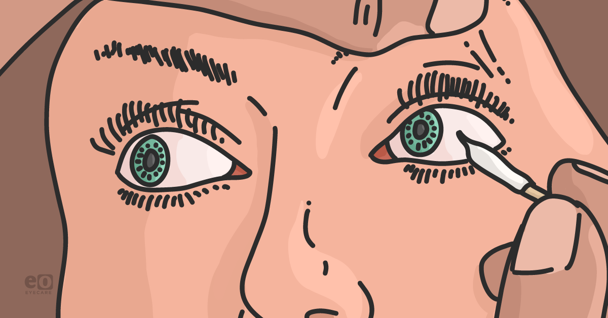
Fortunately, screening for NK is incredibly simple; the only tool required is a cotton-tipped applicator. As I previously mentioned, the diagnostic sign of NK is reduced or absent corneal sensitivity, which is true for all stages of the disease.13,14
So, an easy way to test corneal sensation is by teasing the end of a cotton-tipped applicator to form a wisp and then touching this wisp against the patient’s cornea (in midperipheral to peripheral quadrants and centrally), which allows you to assess their corneal sensitivity, and by extension, their corneal nerve function. If you find the patient has reduced sensitivity or no sensation at all when touching the wisp against their corneal surface, it is a sign you are dealing with NK.
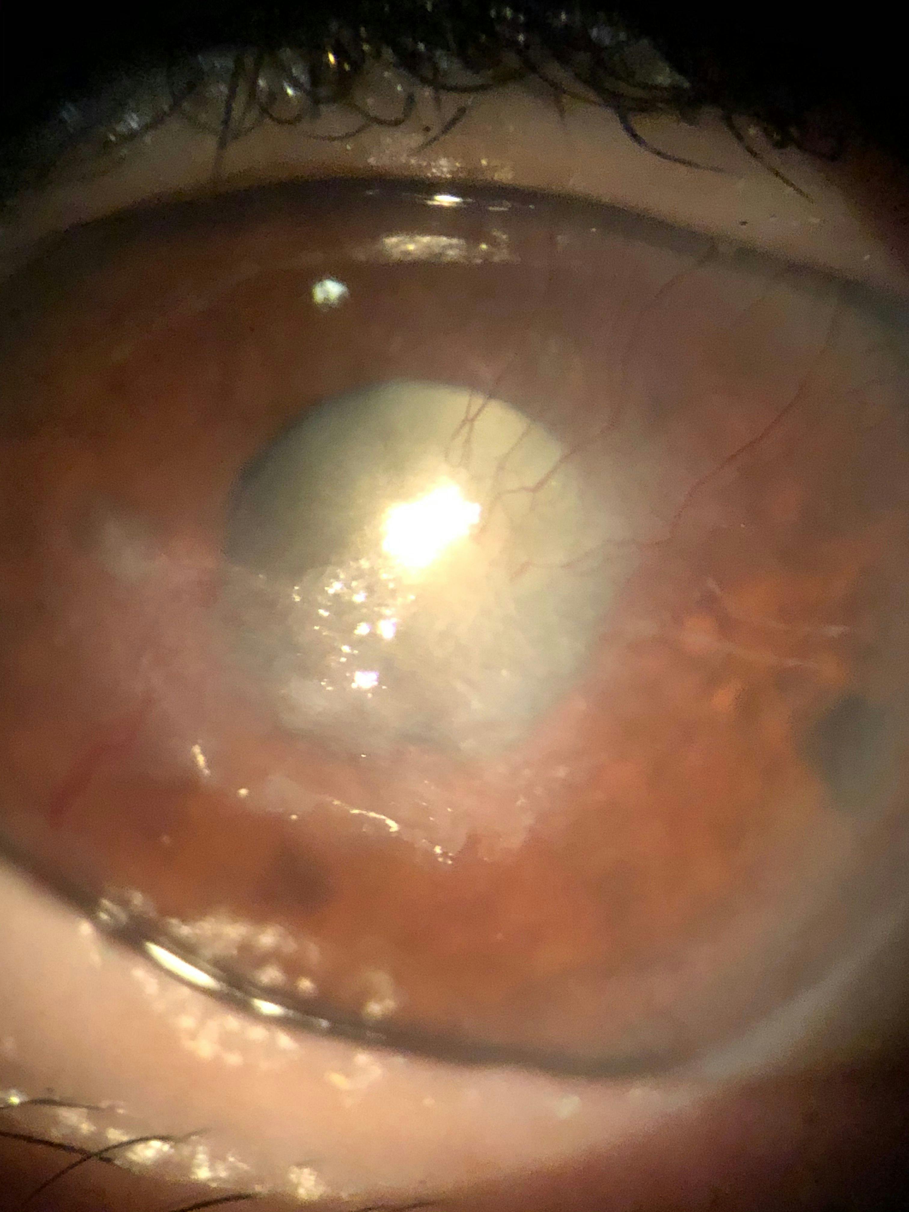
Ideally, we want to catch NK at the earliest stage possible, so we can intervene before it causes significant ocular surface damage, such as the vision-threatening ulcers present in later stages, as shown in Figure 1. This is the reason I recommend checking corneal sensitivity on all dry eye patients, and even non-dry eye patients, if they have a history of herpetic infection or refractive surgery, like LASIK, as these are two routinely encountered causes of NK.17,18
Figure 1 is a clinical image of vision-threatening ulcers in a patient with neurotrophic keratitis that has progressed to later stages.
Neurotrophic Keratitis Ulcers
Figure 1: Courtesy of Cory Lappin, OD, MS, FAAO

Pearls for measuring corneal nerve function to identify NK

Another key point to note when screening for NK in its early stages is that it is still possible for these patients to experience ocular discomfort. Even though reduced or absent corneal sensitivity is the hallmark sign of NK, that does not necessarily mean a patient will be entirely asymptomatic in regard to ocular irritation.
These patients often still retain some degree of nerve function, albeit diminished or isolated to one region, and can experience nerve damage that is localized to one area of the cornea rather than the entire surface.19 This is a crucial concept, as a common mistake made by many eyecare providers is the belief that if a patient has any irritation or discomfort associated with a corneal disruption, then it cannot be neurotrophic keratitis due to its classic description of “stain without pain,” which yet again leads to an underdiagnosis of Stage 1 NK.
Therefore, when testing corneal sensitivity, it is recommended that sensation be assessed in four quadrants rather than just centrally, in case the nerve damage is limited to only one portion of the cornea.19 Once you start routinely testing corneal sensitivity, you may be surprised how many dry eye patients actually display some degree of desensitization and nerve dysfunction, even in those you would not typically expect.

When I started implementing corneal sensitivity testing as part of my standard dry eye workup, I was astounded by how often patients displayed reduced sensation. To me, this suggests that nerve dysfunction likely plays a much larger role in dry eye and ocular surface disease than previously thought.

Understanding the impact of corneal nerves on dry eye

This implication goes beyond NK. The corneal nerves are capable of sensing more than just mechanical sensation, as they can sense other stimuli, such as temperature and inflammation as well.1,20,21 Homeostasis, the underlying mechanism that allows our ocular surface to function, is a feedback-dependent and self-regulated system that occurs subconsciously, which means it relies on sensory feedback to operate.22

Therefore, the corneal nerves’ function as the primary sensors of the ocular surface is vital to ocular surface homeostasis.

Thus, it is likely that the corneal nerves are sensing and providing feedback on stimuli we are not even aware of yet. This would be fitting given the cornea’s extremely high degree of innervation that far exceeds what would be necessary for merely providing sensation. Consequently, nerve dysfunction of any degree causes a significant disruption to the ocular surface, even beyond just the loss of sensation that is associated with NK.

Overview of neurogenic dry eye

Therefore, NK is just one form of nerve dysfunction on a spectrum of corneal and ocular surface neuropathies, and nerve dysfunction is likely a contributing factor to dry eye more broadly. This concept of nerve dysfunction-related dry eye is recognized in clinical research as cenegermin, the recombinant form of human nerve growth factor (NGF) used to treat NK, currently being investigated as a treatment for dry eye in general.23
Due to this wide-ranging impact of the corneal nerves on ocular surface health, I have begun referring to cases of dry eye that display nerve dysfunction as neurogenic dry eye (NDE). If we use the classic dry eye divisions as either evaporative or aqueous deficient, neurogenic dry eye would be the third category.
NDE refers to dry eye emanating from nervous system impairment leading to eventual nerve dysfunction or deficit. The role of nerve dysfunction in dry eye has been well established, as neurosensory abnormalities are listed as one of the factors contributing to dry eye in the TFOS DEWS II definition of dry eye.24

However, these “neurosensory abnormalities” have often been treated as separate, isolated entities, whereas the concept of NDE unifies these conditions under a single category due to their shared nature as ocular surface neuropathies. For instance, I have already discussed NK, which would fall into the category of NDE, but there is also the opposite end of the spectrum in the form of neuropathic corneal pain.

What are some examples of nde?

While NK is a loss of sensation of the corneal nerves, neuropathic corneal pain, also referred to as neuropathic ocular pain (NOP), is a hypersensitization of the corneal nerves.25 And just as NK is referred to as “stain without pain,” NOP is referred to as “pain without stain,” as patients often report significant discomfort even though the cornea is otherwise undisturbed.26
Traumatic brain injury (TBI) patients, including those with post-concussion syndrome, also often report dry eye-like symptoms,27-29 as do those afflicted by a myriad of other potentially nerve-damaging conditions listed below.
Therefore, dry eye-like symptoms associated with these conditions would too be classified as NDE. The causes of NDE are essentially any pathology that can lead to damage of the corneal nerves or the trigeminal pathway from which ocular surface innervation originates.5,25 Thus, there are numerous infectious, traumatic, systemic, genetic, and medication-related causes.

Underlying causes of neurogenic dry eye include:

  1. Infectious: Herpes simplex,30-32 herpes zoster,33-35 leprosy,36 acanthamoeba keratitis,37,38 and fungal keratitis.37
  2. Ocular Surface Disease: Chronic dry eye/ocular surface disease,39-44 chronic exposure (lid malposition, lagophthalmos, Bell’s palsy, or Parkinson’s disease),45-47 corneal dystrophies (lattice, granular),14,13 recurrent corneal erosion,48 and chronic corneal pain with blepharospasm.48
  3. Ocular Surgery: Refractive surgery (LASIK,18,49-51 PRK44,52-54), retinal surgery (PRP/laser,55-57 retinal detachment repair/scleral buckle58-60), glaucoma surgery (micropulse transscleral cyclophotocoagulation [MP-TCP]61-63), corneal incisions (cataract surgery42,48,64), corneal transplantation (keratoplasty: penetrating keratoplasty [PK] and deep anterior lamellar keratoplasty [DALK]),48,64-66 corneal collagen crosslinking (CXL),67 and blepharoplasty.48
  4. Neurosurgery: Ablative procedures for trigeminal neuralgia68-71 and tumor resection.5,72
  5. Compressive Lesions/Neoplasms: Tumors (acoustic neuroma,73,74 schwannoma,75 meningioma,75 orbital neoplasm,64 metastatic tumors,76,77 aneurysm,78 and ocular surface neoplasm).48
  6. Systemic Disease: Diabetes,41,55,79-81 stroke,13,15,64 multiple sclerosis,64,75 vitamin A deficiency,78 amyloidosis,19 degenerative disorders of the central nervous system (Alzheimer’s disease, Parkinson’s disease19), fibromyalgia,41,43 small-fiber polyneuropathy,41,43 trigeminal neuralgia,42 oculofacial pain,42 autoimmune disease (Sjögren’s syndrome, systemic lupus erythematosus [SLE], psoriatic arthritis, inflammatory bowel disease inclusive of ulcerative colitis variants and Crohn's disease, sarcoidosis, and celiac disease).41
  7. Iatrogenic: Topical timolol and betaxolol,82 topical NSAIDs,83,84 chronic preservative (benzalkonium chloride [BAK]) exposure,85,86 toxic keratopathy,48 abuse of topical anesthetics,87,88 sulfacetamide 30%,89 trifluridine,75 long-term use of oral systemic antihistamines,4,64 long-term use of systemic antipsychotics and neuroleptics,4,64 chemotherapy,25,90 and radiation therapy.48,91
  8. Traumatic: Corneal chemical burn/injury,5,13,14,48 corneal thermal burn,13 physical corneal trauma,5,13,41,64 facial trauma,92 and traumatic brain injury/concussion.27-29
  9. Genetic: Congenital corneal hypoesthesia,93,94 Riley-Day Syndrome,95 Goldenhar-Gorlin Syndrome,96 Mobius Syndrome,94 Familial Corneal Hypoesthesia,97 and congenital insensitivity to pain with anhidrosis.98
  10. Other: Contact lens wear,99 aging,78 dark eye color,78 Adie’s syndrome,100 and dihydroxypyrimidine dehydrogenase (DHPD) deficiency.101
While this list is extensive, the key takeaway is to carefully review a patient’s health history, and if any potentially nerve-damaging condition is present, then NDE should be on your radar.

Neuropathic ocular pain

Besides reduced corneal sensitivity demonstrated with a cotton wisp or Cochet-Bonnet aesthesiometer, what are some other indicators you could be dealing with NDE, specifically NOP?
While the use of in vivo confocal microscopy (IVCM) is particularly useful in identifying nerve abnormalities indicative of NDE, most clinicians do not have access to this technology.25,19 Fortunately, there are a few key signs and simple tests that suggest a patient may have NOP-related neurogenic dry eye.

Symptoms of neuropathic ocular pain

Hallmark symptoms of NOP are allodynia,102 photoallodynia,43 and hyperalgesia,44 which are a hypersensitivity to normal stimuli, hypersensitivity to light, and an elevated sensitivity and extreme response to pain, respectively.

These patients will also often report an inability to tolerate the use of topical eye drops, especially those that contain preservatives, but in some cases, they are even unable to use preservative-free artificial tears due to the hypersensitized nature of their ocular surface.

Additionally, these patients will often report significant discomfort with even the gentlest breeze or very dim lighting. So, if a patient mentions avoidance of certain environments, like a drafty office or grocery stores with bright fluorescent lighting, or they ask to keep the lights off during their exam, these are signs you may be dealing with NOP.

Testing for neuropathic ocular pain

As far as testing goes for NOP, the most straightforward is the anesthetization test. Simply instill a drop of topical anesthetic, such as proparacaine hydrochloride 0.5%, onto the eye and ask the patient if they feel better. If they report an improvement in their symptoms, then you know their irritation stems from a disruption of the ocular surface directly.
If they report no improvement in their symptoms and still feel the surface irritation even with the anesthetic, this indicates the pain is coming from a higher central level other than the ocular surface directly, as peripheral sensation is blocked.48
However, NOP can be classified as peripheral, central, or mixed/combined, depending on the source of the pain.25 While central NOP can be identified by pain that persists even after the instillation of topical anesthetic, peripheral NOP can be harder to differentiate from traditional dry eye as both will improve with anesthetic use.

Therefore, a key sign that a patient has peripheral neuropathic pain rather than traditional dry eye is a nonresponse to conventional dry eye therapies, especially symptomatically.

What to look for in patients with neuropathic ocular pain

What I typically observe in patients with NOP who use a standard dry eye treatment regimen is an improvement in their objective testing results, such as increased tear breakup time (TBUT) or improved lipid layer thickness, yet they still report no subjective improvement in their symptoms.
This suggests that the driving force behind their discomfort may be the nerves themselves, as the other elements of the ocular surface have improved but have not provided any significant relief. So, a nonresponse to traditional dry eye therapies or improvement that is limited to objective testing measures only with no subjective symptomatic relief, especially in the context of nerve damage, such as a history of LASIK or herpetic infection, is highly suggestive of NOP.
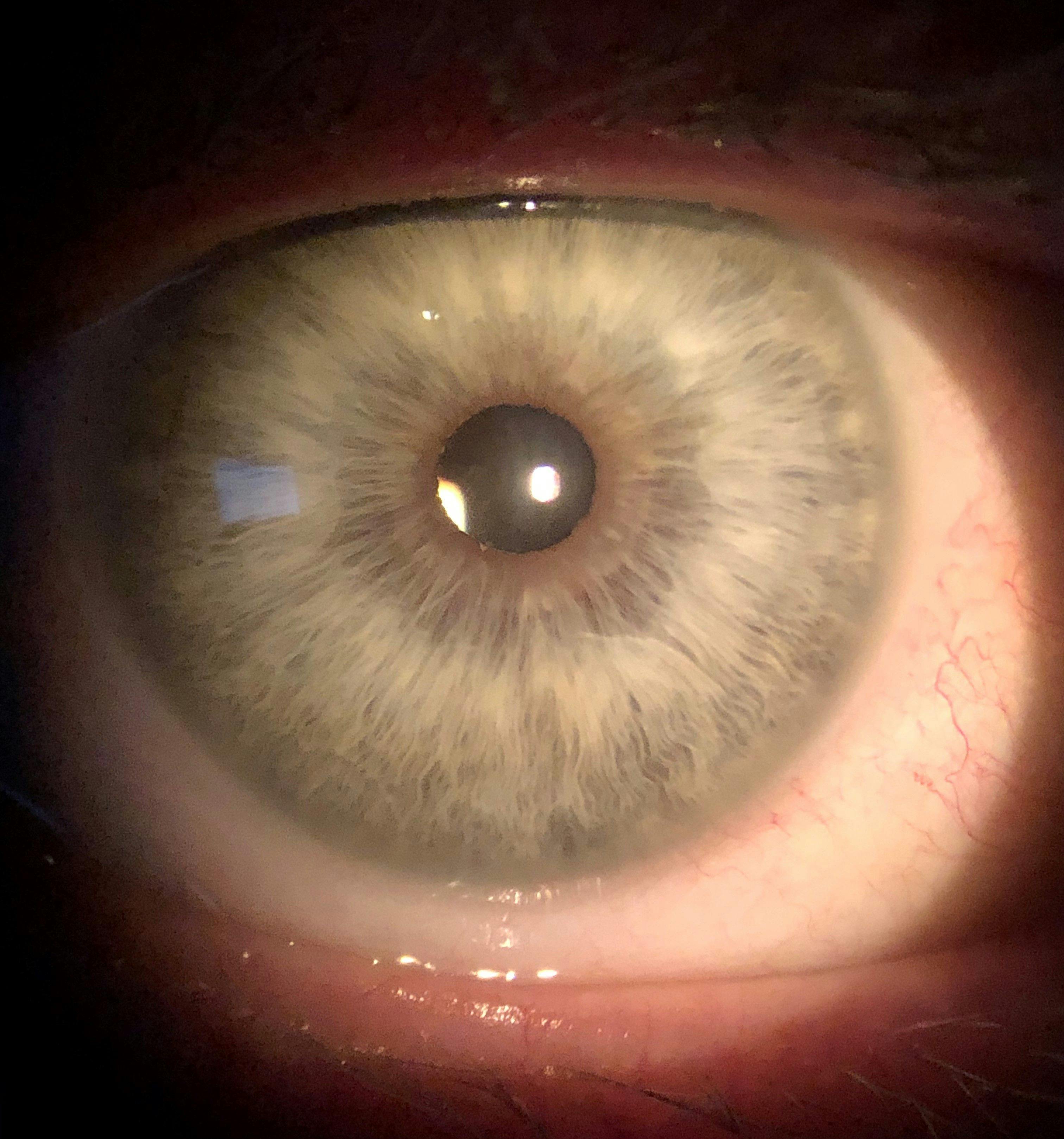
It is worth noting again that it is not unusual for these patients to have a relatively healthy appearance to their ocular surface, like in Figure 2, at the same time they are experiencing significant pain, as this pain can be entirely nervous in nature.25
Figure 2 is a clinical image of a patient with a healthy-looking ocular surface who experienced significant neuropathic ocular pain.
Healthy Ocular Surface Neuropathic Ocular Pain
Figure 2: Courtesy of Cory Lappin, OD, MS, FAAO

Managing neurogenic dry eye

Once NDE has been diagnosed, how is it managed? By its very nature, NDE is particularly challenging to treat. For instance, with NK we are attempting to heal a cornea that has essentially lost the ability to do so.
As the nerves are responsible for wound healing and epithelial cell maintenance, any treatment that does not restore near function will likely ultimately fail or regress once treatment is discontinued, as sustained healing is often not possible without proper nerve function. Additionally, because blinking and lacrimation are disrupted in NK, this can lead to an unstable tear film creating an environment that is not conducive to healing.

Available treatments for neurogenic dry eye

Although there are many supportive therapies like the use of aggressive lubrication with artificial tears, or a bandage contact lens to stabilize the corneal surface, or even surgical procedures, such as tarsorrhaphy or the use of a conjunctival flap,5,75 the best options for treating NK are those that address the root cause by repairing the damaged corneal nerves directly.

Amniotic membranes

This is perhaps best achieved using human nerve growth factor (NGF), which is capable of corneal nerve restoration.4,103 Amniotic membranes, specifically cryopreserved forms, such as PROKERA (BioTissue Ocular), contain NGF and have been shown to improve NK, especially at early stages.104
Whereas Oxervate is a recombinant form of human NGF, and is the only FDA-approved therapy specifically indicated for the treatment of NK.16,105 Further, it may be advantageous to apply a ‘one-two punch’ with an amniotic membrane followed by Oxervate thereafter for these patients.

Similar to the recurrent nature of other corneal diseases (i.e., recurrent corneal erosion, Stage 2 NK, herpetic keratitis, etc.), patients might need repeated dosing of Oxervate or combinations thereof to maintain some level of stability to possibly reduce or halt NK progression.

Adeno-associated virus vectors

The potential to use adeno-associated virus vectors (OC-101 AAV-NGF, Viatris) to introduce the genes responsible for NGF production via an intralacrimal gland injection in an attempt to increase the NGF levels produced in tears is in preclinical research.106 An alternative to NGF that is also being researched is a deleted form of hepatocyte growth factor (CSB-001, Claris Biotherapeutics), which has neurotrophic, antifibrotic, and anti-inflammatory properties, and also enhances corneal wound healing.107-110
RGN-259 (RegeneRx), a topical form of thymosin beta-4 is also being evaluated as a treatment for NK, as it promotes tissue healing and regeneration through enhanced cell migration and anti-inflammatory properties.111,112 In cases where other therapies fail, corneal neurotization surgery, where a donor nerve is transplanted to the cornea to restore sensation to the tissue, can be performed.113-118

Optimizing the ocular surface

In addition to treatments specifically targeting the nerves, the ocular surface should also be optimized when managing NK by treating any concomitant meibomian gland dysfunction or blepharitis to help create an ocular surface environment conducive to healing.5,25

Managing neuropathic ocular pain

Managing NOP can be equally, if not more, challenging than NK. The hypersensitive nature of the nerves in these patients makes their cases some of the most difficult to treat, as they often do not respond to traditional therapies.25

Determining if the patient has peripheral, central, or mixed NOP is critical to determining the appropriate treatment, as peripheral NOP is due to hypersensitization of the corneal nerves.

Whereas central NOP occurs when nerves of the central nervous system, such as the brainstem and brain, become hypersensitized and can cause the sensation of pain even when no stimulus is present.25,41,48 Consequently, peripheral NOP may improve with topical treatments directed at the ocular surface, whereas central NOP will likely require the use of systemic therapies, and mixed/combined NOP may require both.25

Available treatments for neuropathic ocular pain

Commonly utilized treatments for patients with peripheral NOP include autologous serum tears, platelet-rich plasma, and amniotic membranes, all of which are aimed at healing the nerves.25 Just like in NK, NGF has been shown to improve neuropathic pain, so treatments utilizing NGF are well-suited for the management of NOP also.25,119,120

Topical steroids and the ocular surface

Topical steroids (typically a “soft” steroid such as loteprednol) have also been implemented to address any inflammatory irritation to the nerves and are usually tapered slowly, with the patient eventually being transitioned to a topical immunomodulator such as Xiidra (lifitegrast ophthalmic solution 5%, Novartis), Cequa (cyclosporine ophthalmic solution 0.09%, Sun Pharmaceuticals), or Restasis (cyclosporine ophthalmic emulsion 0.05%, Allergan, An AbbVie Company) for long-term inflammation management.25
Additionally, any concomitant ocular surface diseases, such as MGD, should also be addressed to prevent exacerbation of the condition.

Contact lenses

The use of soft bandage and scleral contact lenses has been beneficial for some NOP patients, although others have found that the use of contact lenses exacerbates their hypersensitivity.25 Sealed spectacle frames, such as Ziena dry eye frames (Moisture Chamber Collection, Ziena Eyewear), can help protect the eye from environmental stressors, like air conditioning or ceiling fans, which can be particularly irritating to the patient’s hypersensitive surface.25

Systemic treatments and lifestyle changes

Cases of central NOP often require management beyond topical ocular therapies, such as systemic medications.25 Treatment options for these patients include: tricyclic antidepressants (nortriptyline and desipramine—first-line agents), anticonvulsants (carbamazepine—first-line agent, gabapentin and pregabalin—third-line agents), opioids (low-dose naltrexone and tramadol—second-line agents), serotonin-noradrenaline reuptake inhibitors (duloxetine—third-line agent), and sodium channel blockers (mexiletine—third-line agent).25
Additionally, lifestyle changes such as regular exercise121-124 and meditation can be beneficial25 in managing neuropathic pain, as can dietary considerations like the use of omega-3 fatty acid supplements125,126 or a gluten-free diet127,128 if a patient has a gluten sensitivity. Alternative therapies, including acupuncture, have also been shown to help alleviate neuropathic pain.129

Given the systemic nature of the treatments often involved in NOP management, it is best to co-manage these patients with their primary care physician, neurologist, psychiatrist, and/or a pain management specialist, as these patients often require multidisciplinary care.

Additionally, NOP patients also have a strong psychological component to their condition, with depression,44,130 anxiety,44 and post-traumatic stress disorder (PTSD)44 all being common comorbidities that should be addressed with a mental-health professional.

Treating concurrent TBI and neuropathic ocular pain

Patients suffering from dry eye-like symptoms related to a TBI also present unique challenges. In addition to many of the treatments already mentioned, the use of an omega-3 supplement that is high in docosahexaenoic acid (DHA), specifically, may be beneficial in these patients.
While omega-3 fatty acids, in general, have been shown to potentially improve nerve-related dryness symptoms, a high DHA supplement may be particularly helpful as DHA is the primary omega-3 fatty acid present in the brain, and supplementation has been shown to accelerate nerve repair.131-134
The use of nūretin (PRN Physician Recommended Nutriceuticals), even though it is intended to support retinal health, is a good supplement option in this scenario, as it has a high-DHA formulation.

Available and upcoming neurostimulatory treatments

The importance of the nerves as a target of dry eye treatment is also starting to be recognized, as we now have an entire class of therapeutics dedicated to neurostimulatory treatment. Tyrvaya (varenicline solution nasal spray 0.03mg, Viatris) and the iTEAR100 device target the trigeminal nerve to stimulate parasympathetic innervation of the lacrimal gland in an effort to promote natural tear secretion, which contains anti-inflammatory and growth factors lacking in artificial tears.135-138
One of the growth factors secreted in natural tears is NGF, which, as mentioned previously, can be beneficial in treating NDE.4,25,103,119,120,139,140 For this reason, the use of varenicline nasal spray (OC-01, Viatris) is currently being investigated as a potential treatment option for Stage 1 NK,106 as the increased natural tear production may promote healing of the nerves due to the trophic factors present in the tears.

Tyrvaya and iTEAR100 can be particularly useful in treating NOP, as they are dropless options and therefore bypass the need to apply any medication directly to the hypersensitized ocular surface, and therefore may be more easily tolerated.

Another neurostimulatory treatment in the pipeline is AR-15512 (Aerie/Alcon), which is a TRPM8 agonist that stimulates the thermoreceptors of the corneal nerves, which results in a cooling sensation that can reduce ocular irritation.141,142

Final thoughts on NDE and NOP

While traditionally not as emphasized as evaporative or aqueous deficient dry eye, nerve dysfunction is becoming more widely recognized as a significant contributor to dry eye disease. There are several reasons for the underdiagnosis of this pathology, including its subtlety in signs and symptoms which are often indistinguishable from traditional dry eye at first glance, as well as the lack of a unifying classification category for these nerve dysfunctions.
However, due to the significant negative impact of nerve dysfunction on the ocular surface and the subsequent burden it places on the patients it afflicts, we cannot afford to miss or ignore these conditions that are likely more common than once thought.
For this reason, it is extremely valuable from both a conceptual and clinical awareness standpoint to consider NDE as a third category of dry eye. Accordingly, it is of paramount importance to screen our patients for nerve dysfunction, whether it be neuropathic keratitis, neuropathic ocular pain, TBI-associated dry eye, or any other form of neurogenic dry eye because if we fail to do so, we are missing a crucial piece of the puzzle in our efforts to care for this particularly challenging subset of dry eye patients.
  1. Müller LJ, Marfurt CF, Kruse F, Tervo TMT. Corneal nerves: structure, contents and function. Exp Eye Res. 2003;76(5):521-542.
  2. Cruzat A, Qazi Y, Hamrah P. In vivo confocal microscopy of corneal nerves in health and disease. Ocul Surf. 2017;15(1):15-47.
  3. Walker HK. Cranial nerve v: the trigeminal nerve. In: Walker HK, Hall WD, Hurst JW, eds. Clinical Methods: The History, Physical, and Laboratory Examinations. 3rd ed. Butterworths, 1990.
  4. Mastropasqua L, Massaro-Giordano G, Nubile M, et al. Understanding the pathogenesis of neurotrophic keratitis: the role of corneal nerves. J Cell Physiol. 2017;232(4):717-724.
  5. Sacchetti M, Lambiase A. Diagnosis and management of neurotrophic keratitis. Clin Ophthalmol. 2014;8:571-579.
  6. Heigle TJ, Pflugfelder SC. Aqueous tear production in patients with neurotrophic keratitis. Cornea. 1996;15(2):135-138.
  7. Jabbarvand M, Hashemian H, Khodaparast M, et al. Do unilateral herpetic stromal keratitis and neurotrophic ulcers cause bilateral dry eye?. Cornea. 2015;34(7):768-772.
  8. Scott G, Balsiger H, Kluckman M, et al. Patterns of innervation of the lacrimal gland with clinical application. Clin Anat. 2014;27(8):1174-1177. doi:10.1002/ca.22447
  9. Kam WR, Sullivan DA. Neurotransmitter influence on human meibomian gland epithelial cells. Invest Ophthalmol Vis Sci. 2011;52(12):8543-8548. Published 2011 Nov 1. doi:10.1167/iovs.11-8113
  10. Dartt DA. Regulation of mucin and fluid secretion by conjunctival epithelial cells. Prog Retin Eye Res. 2002;21(6):555-576. doi:10.1016/s1350-9462(02)00038-1
  11. Dartt DA, Masli S. Conjunctival epithelial and goblet cell function in chronic inflammation and ocular allergic inflammation. Curr Opin Allergy Clin Immunol. 2014;14(5):464-470. doi:10.1097/ACI.0000000000000098
  12. Swamynathan SK, Wells A. Conjunctival goblet cells: Ocular surface functions, disorders that affect them, and the potential for their regeneration [published correction appears in Ocul Surf. 2020 Feb 28]. Ocul Surf. 2020;18(1):19-26. doi:10.1016/j.jtos.2019.11.005
  13. Bonini S, Rama P, Olzi D, et al. Neurotrophic keratitis. Eye (Lond). 2003;17(8):989-995.
  14. Chang BH, Groos EB Jr: Neurotrophic keratitis; in Krachmer JH, Mannis MJ, Holland EJ eds. Cornea. London, UK: Elsevier, 2011.
  15. Mackie IA. Neuroparalytic keratitis. In: Fraunfelder F, Roy FH, Meyer SM, eds. Current Ocular Therapy. Philadelphia, PA, USA: WB Saunders, 1995.
  16. Murri, N. Goodman & Gilman Year in Review Biologics FDA Approvals In: Goodman & Gilman’s: The Pharmacological Basis of Therapeutics, 13e. McGraw-Hill Medical, 2018.
  17. Labetoulle M, Auquier P, Conrad H, et al. Incidence of herpes simplex virus keratitis in France. Ophthalmology. 2005;112(5):888-895.
  18. Dohlman TH, Lai EC, Ciralsky JB. Dry eye disease after refractive surgery. Int Ophthalmol Clin. 2016;56(2):101-110.
  19. Dua HS, Said DG, Messmer EM, et al. Neurotrophic keratopathy. Prog Retin Eye Res. 2018;66:107-131. doi:10.1016/j.preteyeres.2018.04.003
  20. Acosta MC, Tan ME, Belmonte C, et al. Sensations evoked by selective mechanical, chemical, and thermal stimulation of the conjunctiva and cornea. Invest Ophthalmol Vis Sci. 2001;42(9):2063-2067.
  21. Belmonte C, Aracil A, Acosta MC, et al. Nerves and sensations from the eye surface. Ocul Surf. 2004;2(4):248-253. doi:10.1016/s1542-0124(12)70112-x
  22. Billman GE. Homeostasis: The Underappreciated and Far Too Often Ignored Central Organizing Principle of Physiology. Front Physiol. 2020;11:200. Published 2020 Mar 10. doi:10.3389/fphys.2020.00200
  23. Coco G, Piccotti G, Romano V, et al. Cenegermin for the treatment of dry eye disease. Drugs Today (Barc). 2023;59(3):113-123. doi:10.1358/dot.2023.59.3.3521858
  24. Craig JP, Nichols KK, Akpek EK, et al. TFOS DEWS II Definition and Classification Report. Ocul Surf. 2017;15(3):276-283. doi:10.1016/j.jtos.2017.05.008
  25. Dieckmann G, Goyal S, Hamrah P. Neuropathic Corneal Pain: Approaches for Management. Ophthalmology. 2017;124(11S):S34-S47. doi:10.1016/j.ophtha.2017.08.004
  26. Rosenthal P, Baran I, Jacobs DS. Corneal pain without stain: is it real?. Ocul Surf. 2009;7(1):28-40. doi:10.1016/s1542-0124(12)70290-2
  27. Lee CJ, Felix ER, Levitt RC, et al. Traumatic brain injury, dry eye and comorbid pain diagnoses in US veterans. Br J Ophthalmol. 2018;102(5):667-673. doi:10.1136/bjophthalmol-2017-310509
  28. Diel RJ, Mehra D, Kardon R, et al. Photophobia: shared pathophysiology underlying dry eye disease, migraine and traumatic brain injury leading to central neuroplasticity of the trigeminothalamic pathway. Br J Ophthalmol. 2021;105(6):751-760. doi:10.1136/bjophthalmol-2020-316417
  29. Abusamak M, Alrawashdeh HM. Post-concussion Syndrome Light Sensitivity: A Case Report and Review of the Literature. Neuroophthalmology. 2021;46(2):85-90. Published 2021 Oct 13. doi:10.1080/01658107.2021.1983612
  30. Cavanagh HD, Pihlaja D, Thoft RA, et al. The pathogenesis and treatment of persistent epithelial defects. Trans Sect Ophthalmol Am Acad Ophthalmol Otolaryngol. 1976;81(5):754-769.
  31. Liesegang TJ. Ocular herpes simplex infection: pathogenesis and current therapy. Mayo Clin Proc. 1988;63(11):1092-1105. doi:10.1016/s0025-6196(12)65504-9
  32. Hamrah P, Cruzat A, Dastjerdi MH, et al. Corneal sensation and subbasal nerve alterations in patients with herpes simplex keratitis: an in vivo confocal microscopy study. Ophthalmology. 2010;117(10):1930-1936. doi:10.1016/j.ophtha.2010.07.010
  33. Cobo LM. Corneal complications of herpes zoster ophthalmicus. Prevention and treatment. Cornea. 1988;7(1):50-56.
  34. Hamrah P, Cruzat A, Dastjerdi MH, et al. Unilateral herpes zoster ophthalmicus results in bilateral corneal nerve alteration: an in vivo confocal microscopy study. Ophthalmology. 2013;120(1):40-47. doi:10.1016/j.ophtha.2012.07.036
  35. Crane AM, Levitt RC, Felix ER, et al. Patients with more severe symptoms of neuropathic ocular pain report more frequent and severe chronic overlapping pain conditions and psychiatric disease. Br J Ophthalmol. 2017;101(2):227-231. doi:10.1136/bjophthalmol-2015-308214
  36. Karaçorlu MA, Cakiner T, Saylan T. Corneal sensitivity and correlations between decreased sensitivity and anterior segment pathology in ocular leprosy. Br J Ophthalmol. 1991;75(2):117-119. doi:10.1136/bjo.75.2.117
  37. Kurbanyan K, Hoesl LM, Schrems WA, et al. Corneal nerve alterations in acute Acanthamoeba and fungal keratitis: an in vivo confocal microscopy study. Eye (Lond). 2012;26(1):126-132. doi:10.1038/eye.2011.270
  38. Mateo A, Abadía B, Calvo P, et al. Treatment of Acanthamoeba neurotrophic corneal ulcer with topical matrix therapy. J Ophthalmic Inflamm Infect. 2015;5:18. Published 2015 Jun 21. doi:10.1186/s12348-015-0048-x
  39. Srinivasan S, Lyall DAM. Neurotrophic Keratopathy; in Holland EJ, Mannis MJ, Lee WB eds. Ocular Surface Disease: Cornea, Conjunctiva and Tear Film. London, UK: Elsevier, 2013.
  40. Bourcier T, Acosta MC, Borderie V, et al. Decreased corneal sensitivity in patients with dry eye. Invest Ophthalmol Vis Sci. 2005;46(7):2341-2345. doi:10.1167/iovs.04-1426
  41. Rosenthal P, Borsook D. Ocular neuropathic pain. Br J Ophthalmol. 2016;100(1):128-134. doi:10.1136/bjophthalmol-2014-306280
  42. Rosenthal P, Borsook D, Moulton EA. Oculofacial Pain: Corneal Nerve Damage Leading to Pain Beyond the Eye. Invest Ophthalmol Vis Sci. 2016;57(13):5285-5287. doi:10.1167/iovs.16-20557
  43. Aggarwal S, Kheirkhah A, Cavalcanti BM, et al. Autologous Serum Tears for Treatment of Photoallodynia in Patients with Corneal Neuropathy: Efficacy and Evaluation with In Vivo Confocal Microscopy. Ocul Surf. 2015;13(3):250-262. doi:10.1016/j.jtos.2015.01.005
  44. Galor A, Levitt RC, Felix ER, et al. Understanding the true burden of dry eye disease. Expert Rev Ophthalmol. 2015;10(5):403-405. doi:10.1586/17469899.2015.1061431
  45. Dana R, Farid M, Gupta PK, et al. Expert consensus on the identification, diagnosis, and treatment of neurotrophic keratopathy. BMC Ophthalmol. 2021;21(1):327. Published 2021 Sep 8. doi:10.1186/s12886-021-02092-1
  46. Nemet AY, Vinker S. Considerations and complications after Bells' palsy. J Clin Neurosci. 2015;22(12):1949-1953. doi:10.1016/j.jocn.2015.04.030
  47. NaPier E, Camacho M, McDevitt TF, Sweeney AR. Neurotrophic keratopathy: current challenges and future prospects. Ann Med. 2022;54(1):666-673. doi:10.1080/07853890.2022.2045035
  48. Goyal S, Hamrah P. Understanding Neuropathic Corneal Pain--Gaps and Current Therapeutic Approaches. Semin Ophthalmol. 2016;31(1-2):59-70. doi:10.3109/08820538.2015.1114853
  49. Wilson SE, Ambrósio R. Laser in situ keratomileusis-induced neurotrophic epitheliopathy. Am J Ophthalmol. 2001;132(3):405-406. doi:10.1016/s0002-9394(01)00995-3
  50. Toda I. LASIK and the ocular surface. Cornea. 2008;27 Suppl 1:S70-S76. doi:10.1097/ICO.0b013e31817f42c0
  51. Lee BH, McLaren JW, Erie JC, et al. Reinnervation in the cornea after LASIK. Invest Ophthalmol Vis Sci. 2002;43(12):3660-3664.
  52. Pérez-Santonja JJ, Sakla HF, Cardona C, et al. Corneal sensitivity after photorefractive keratectomy and laser in situ keratomileusis for low myopia. Am J Ophthalmol. 1999;127(5):497-504. doi:10.1016/s0002-9394(98)00444-9
  53. Campos M, Hertzog L, Garbus JJ, et al. Corneal sensitivity after photorefractive keratectomy. Am J Ophthalmol. 1992;114(1):51-54. doi:10.1016/s0002-9394(14)77412-4
  54. Galor A, Levitt RC, Felix ER, et al. Neuropathic ocular pain: an important yet underevaluated feature of dry eye. Eye (Lond). 2015;29(3):301-312. doi:10.1038/eye.2014.263
  55. Menchini U, Scialdone A, Pietroni C, et al. Argon versus krypton panretinal photocoagulation side effects on the anterior segment. Ophthalmologica. 1990;201(2):66-70. doi:10.1159/000310129
  56. Patel JI, Jenkins L, Benjamin L, et al. Dilated pupils and loss of accommodation following diode panretinal photocoagulation with sub-tenon local anaesthetic in four cases. Eye (Lond). 2002;16(5):628-632. doi:10.1038/sj.eye.6700004
  57. Tinley CG, Gray RH. Routine, single session, indirect laser for proliferative diabetic retinopathy. Eye (Lond). 2009;23(9):1819-1823. doi:10.1038/eye.2008.394
  58. Banerjee PJ, Chandra A, Sullivan PM, et al. Neurotrophic corneal ulceration after retinal detachment surgery with retinectomy and endolaser: a case series. JAMA Ophthalmol. 2014;132(6):750-752. doi:10.1001/jamaophthalmol.2014.280
  59. Gibson RA. Reduction of corneal sensitivity after retinal detachment surgery. Br J Ophthalmol. 1981;65(9):614-617. doi:10.1136/bjo.65.9.614
  60. Valldeperas X, Arango A, Blazquez A, et al. Tonic Pupil and Corneal Anesthesia after Vitrectomy and Encircling Band for Retinal Detachment in an Ex-Premature Child. Case Rep Ophthalmol. 2010;1(2):66-70. Published 2010 Sep 29. doi:10.1159/000321250
  61. Johnson SM. Neurotrophic corneal defects after diode laser cycloablation. Am J Ophthalmol. 1998;126(5):725-727. doi:10.1016/s0002-9394(98)00175-5
  62. Perez CI, Han Y, Rose-Nussbaumer J, et al. Neurotrophic keratitis after micropulse transscleral diode laser cyclophotocoagulation. Am J Ophthalmol Case Rep. 2019;15:100469. Published 2019 May 20. doi:10.1016/j.ajoc.2019.100469
  63. Kim J, Sung MS, Park SW. Neurotrophic Keratopathy after Micropulse Transscleral Cyclophotocoagulation in a Glaucoma Patient. Korean J Ophthalmol. 2021;35(1):97-98. doi:10.3341/kjo.2020.0103
  64. Versura P, Giannaccare G, Pellegrini M, Sebastiani S, et al. Neurotrophic keratitis: current challenges and future prospects. Eye Brain. 2018;10:37-45. Published 2018 Jun 28. doi:10.2147/EB.S117261
  65. Lin X, Xu B, Sun Y, et al. Comparison of deep anterior lamellar keratoplasty and penetrating keratoplasty with respect to postoperative corneal sensitivity and tear film function. Graefes Arch Clin Exp Ophthalmol. 2014;252(11):1779-1787. doi:10.1007/s00417-014-2748-6
  66. Niederer RL, Perumal D, Sherwin T, et al. Corneal innervation and cellular changes after corneal transplantation: an in vivo confocal microscopy study. Invest Ophthalmol Vis Sci. 2007;48(2):621-626. doi:10.1167/iovs.06-0538
  67. Wasilewski D, Mello GH, Moreira H. Impact of collagen crosslinking on corneal sensitivity in keratoconus patients. Cornea. 2013;32(7):899-902. doi:10.1097/ICO.0b013e31827978c8
  68. Mackie IA. Role of the corneal nerves in destructive disease of the cornea. Trans Ophthalmol Soc U K (1962). 1978;98(3):343-347.
  69. Onofrio BM. Radiofrequency percutaneous Gasserian ganglion lesions. Results in 140 patients with trigeminal pain. J Neurosurg. 1975;42(2):132-139. doi:10.3171/jns.1975.42.2.0132
  70. Davies MS. Corneal anaesthesia after alcohol injection of the trigeminal sensory root. Examination of 100 anaesthetic corneae. Br J Ophthalmol. 1970;54(9):577-586. doi:10.1136/bjo.54.9.577
  71. Bhatti MT, Patel R. Neuro-ophthalmic considerations in trigeminal neuralgia and its surgical treatment. Curr Opin Ophthalmol. 2005;16(6):334-340. doi:10.1097/01.icu.0000183859.67294.c6
  72. Newman S. A prospective study of cavernous sinus surgery for meningiomas and resultant common ophthalmic complications (an American Ophthalmological Society thesis). Trans Am Ophthalmol Soc. 2007;105:392-447.
  73. Sterkers JM, Morrison GA, Sterkers O, et al. Preservation of facial, cochlear, and other nerve functions in acoustic neuroma treatment. Otolaryngol Head Neck Surg. 1994;110(2):146-155. doi:10.1177/019459989411000202
  74. Tos M, Drozdziewicz D, Thomsen J. Medial acoustic neuromas. A new clinical entity. Arch Otolaryngol Head Neck Surg. 1992;118(2):127-133. doi:10.1001/archotol.1992.01880020019009
  75. Semeraro F, Forbice E, Romano V, et al. Neurotrophic keratitis. Ophthalmologica. 2014;231(4):191-197. doi:10.1159/000354380
  76. Bonzano C, Bonzano E, Cutolo CA, et al. A Case of Neurotrophic Keratopathy Concomitant to Brain Metastasis. Cureus. 2018;10(3):e2309. Published 2018 Mar 12. doi:10.7759/cureus.2309
  77. Sato T, Sugioka K, Kodama-Takahashi A, et al. A Case of Neurotrophic Keratopathy Associated with Nasopharyngeal Carcinoma. Case Rep Ophthalmol. 2018;9(1):114-118. Published 2018 Feb 16. doi:10.1159/000485965
  78. Miller NR. Walsh and Hoyt’s clinical neuro-ophthalmology. Baltimore: Williams & Wilkins; 1985
  79. Schwartz DE. Corneal sensitivity in diabetics. Arch Ophthalmol. 1974;91(3):174-178. doi:10.1001/archopht.1974.03900060182003
  80. Lockwood A, Hope-Ross M, Chell P. Neurotrophic keratopathy and diabetes mellitus. Eye (Lond). 2006;20(7):837-839. doi:10.1038/sj.eye.6702053
  81. Hyndiuk RA, Kazarian EL, Schultz RO, et al. Neurotrophic corneal ulcers in diabetes mellitus. Arch Ophthalmol. 1977;95(12):2193-2196. doi:10.1001/archopht.1977.04450120099012
  82. Weissman SS, Asbell PA. Effects of topical timolol (0.5%) and betaxolol (0.5%) on corneal sensitivity. Br J Ophthalmol. 1990;74(7):409-412. doi:10.1136/bjo.74.7.409
  83. Szerenyi K, Sorken K, Garbus JJ, et al. Decrease in normal human corneal sensitivity with topical diclofenac sodium. Am J Ophthalmol. 1994;118(3):312-315. doi:10.1016/s0002-9394(14)72954-x
  84. Singer DD, Kennedy J, Wittpenn JR. Topical NSAIDs effect on corneal sensitivity. Cornea. 2015;34(5):541-543. doi:10.1097/ICO.0000000000000309
  85. Martone G, Frezzotti P, Tosi GM, et al. An in vivo confocal microscopy analysis of effects of topical antiglaucoma therapy with preservative on corneal innervation and morphology. Am J Ophthalmol. 2009;147(4):725-735.e1. doi:10.1016/j.ajo.2008.10.019
  86. Sarkar J, Chaudhary S, Namavari A, et al. Corneal neurotoxicity due to topical benzalkonium chloride. Invest Ophthalmol Vis Sci. 2012;53(4):1792-1802. Published 2012 Apr 6. doi:10.1167/iovs.11-8775
  87. Rosenwasser GO, Holland S, Pflugfelder SC, et al. Topical anesthetic abuse. Ophthalmology. 1990;97(8):967-972. doi:10.1016/s0161-6420(90)32458-2
  88. Willis WE, Laibson PR. Corneal complications of topical anesthetic abuse. Can J Ophthalmol. 1970;5(3):239-243.
  89. Chang FW, Reinhart S, Fraser NM. Effect of 30% sodium sulfacetamide on corneal sensitivity. Am J Optom Physiol Opt. 1984;61(5):318-320. doi:10.1097/00006324-198405000-00004
  90. Gozzi F, Tiseo M, Facchinetti F, et al. Bilateral Severe Corneal Ulcer in a Patient with Lung Adenocarcinoma Treated with Gefitinib. Case Rep Ophthalmol. 2021;12(1):288-292. Published 2021 Apr 30. doi:10.1159/000514696
  91. Ting DSJ, Rana-Rahman R, Ng JY, et al. Clinical Spectrum and Outcomes of Ocular and Periocular Complications following External-Beam Radiotherapy for Inoperable Malignant Maxillary Sinus Tumors. Ocul Oncol Pathol. 2021;7(1):36-43. doi:10.1159/000511011
  92. Lanigan DT, Romanchuk K, Olson CK. Ophthalmic complications associated with orthognathic surgery. J Oral Maxillofac Surg. 1993;51(5):480-494. doi:10.1016/s0278-2391(10)80502-6
  93. Rosenberg ML. Congenital trigeminal anaesthesia. A review and classification. Brain. 1984;107 (Pt 4):1073-1082. doi:10.1093/brain/107.4.1073
  94. Donaghy M, Hakin RN, Bamford JM, et al. Hereditary sensory neuropathy with neurotrophic keratitis. Description of an autosomal recessive disorder with a selective reduction of small myelinated nerve fibres and a discussion of the classification of the hereditary sensory neuropathies. Brain. 1987;110:563-583. doi:10.1093/brain/110.3.563
  95. Goldberg MF, Payne JW, Brunt PW. Ophthalmologic studies of familial dysautonomia. The Riley-Day syndrome. Arch Ophthalmol. 1968;80(6):732-743. doi:10.1001/archopht.1968.00980050734011
  96. Baum JL, Feingold M. Ocular aspects of Goldenhar's syndrome. Am J Ophthalmol. 1973;75(2):250-257. doi:10.1016/0002-9394(73)91020-9
  97. Purcell JJ Jr, Krachmer JH. Familial corneal hypesthesia. Arch Ophthalmol. 1979;97(5):872-874. doi:10.1001/archopht.1979.01020010430005
  98. John D, Thomas M, Jacob P. Neurotrophic keratitis and congenital insensitivity to pain with anhidrosis--a case report with 10-year follow-up. Cornea. 2011;30(1):100-102. doi:10.1097/ICO.0b013e3181e458e4
  99. Velasco MJ, Bermúdez FJ, Romero J, et al. Variations in corneal sensitivity with hydrogel contact lenses. Acta Ophthalmol (Copenh). 1994;72(1):53-56. doi:10.1111/j.1755-3768.1994.tb02737.x
  100. Purcell JJ Jr, Krachmer JH, Thompson HS. Corneal sensation in Adie’s syndrome. Am J Ophthalmol. 1977;84(4):496-500.
  101. Kapoor B, Luhishi EA, Chung AK, et al. Neurotrophic keratitis in a patient with dihydroxypyrimidine dehydrogenase deficiency. Indian J Ophthalmol. 2008;56(4):336-337. doi:10.4103/0301-4738.41422
  102. Hamrah P, Qazi Y, Shahatit B, et al. Corneal Nerve and Epithelial Cell Alterations in Corneal Allodynia: An In Vivo Confocal Microscopy Case Series. Ocul Surf. 2017;15(1):139-151. doi:10.1016/j.jtos.2016.10.002
  103. Sacchetti M, Lambiase A. Neurotrophic factors and corneal nerve regeneration. Neural Regen Res. 2017;12(8):1220-1224.
  104. Mead OG, Tighe S, Tseng SCG. Amniotic membrane transplantation for managing dry eye and neurotrophic keratitis. Taiwan J Ophthalmol. 2020;10(1):13-21. Published 2020 Mar 4. doi:10.4103/tjo.tjo_5_20
  105. Voelker R. New drug treats rare, debilitating neurotrophic keratitis. JAMA. 2018;320(13):1309.
  106. Oyster Point Pharma. Science and Pipeline: The Future of Ophthalmic Disease. Oyster Point Pharma. https://oysterpointrx.com/pipeline/. Published November 10, 2022. Accessed April 9, 2023.
  107. Claris Biotherapeutics. Claris Bio. https://clarisbio.com/. Published 2023. Accessed April 9, 2023.
  108. ClinicalTrials.gov. Study to evaluate the safety and efficacy of CSB-001 ophthalmic solution 0.1% in neurotrophic keratitis subjects. https://clinicaltrials.gov/ct2/show/NCT04909450. Published June 1, 2021. Accessed April 9, 2023.
  109. Miyagi H, Thomasy SM, Russell P, et al. The role of hepatocyte growth factor in corneal wound healing. Exp Eye Res. 2018;166:49-55. doi:10.1016/j.exer.2017.10.006
  110. Yamane K, Misawa H, Takigawa T, et al. Multipotent Neurotrophic Effects of Hepatocyte Growth Factor in Spinal Cord Injury. Int J Mol Sci. 2019;20(23):6078. Published 2019 Dec 2. doi:10.3390/ijms20236078
  111. RGN-259. RegeneRx. https://www.regenerx.com/RGN-259. Published 2020. Accessed March 26, 2023.
  112. Sosne G, Qiu P, Kurpakus-Wheater M. Thymosin beta 4: A novel corneal wound healing and anti-inflammatory agent. Clin Ophthalmol. 2007;1(3):201-207.
  113. Liu CY, Arteaga AC, Fung SE, et al. Corneal neurotization for neurotrophic keratopathy: Review of surgical techniques and outcomes. Ocul Surf. 2021;20:163-172. doi:10.1016/j.jtos.2021.02.010
  114. Terzis JK, Dryer MM, Bodner BI. Corneal neurotization: a novel solution to neurotrophic keratopathy. Plast Reconstr Surg. 2009 Jan;123(1):112-120. doi: 10.1097/PRS.0b013e3181904d3a. PMID: 19116544.
  115. Allevi F, Fogagnolo P, Rossetti L, et al. Eyelid reanimation, neurotisation, and transplantation of the cornea in a patient with facial palsy. BMJ Case Rep. 2014;2014:bcr2014205372. Published 2014 Aug 19. doi:10.1136/bcr-2014-205372
  116. Elbaz U, Bains R, Zuker RM, et al. Restoration of corneal sensation with regional nerve transfers and nerve grafts: a new approach to a difficult problem. JAMA Ophthalmol. 2014 Nov;132(11):1289-95. doi: 10.1001/jamaophthalmol.2014.2316. PMID: 25010775.
  117. Jacinto F, Espana E, Padilla M, et al. Ipsilateral supraorbital nerve transfer in a case of recalcitrant neurotrophic keratopathy with an intact ipsilateral frontal nerve: A novel surgical technique. Am J Ophthalmol Case Rep. 2016;4:14-17. doi: 10.1016/j.ajoc.2016.07.001. Erratum in: Am J Ophthalmol Case Rep. 2016 Sep 06;4:87.
  118. Leyngold I, Weller C, Leyngold M, et al. Endoscopic Corneal Neurotization: Technique and Initial Experience. Ophthalmic Plast Reconstr Surg. 2018 Jan/Feb;34(1):82-85. doi: 10.1097/IOP.0000000000001023. PMID: 29194285.
  119. Cirillo G, Cavaliere C, Bianco MR, et al. Intrathecal NGF administration reduces reactive astrocytosis and changes neurotrophin receptors expression pattern in a rat model of neuropathic pain. Cell Mol Neurobiol. 2010;30(1):51-62. doi:10.1007/s10571-009-9430-2
  120. Colangelo AM, Bianco MR, Vitagliano L, et al. A new nerve growth factor-mimetic peptide active on neuropathic pain in rats. J Neurosci. 2008;28(11):2698-2709. doi:10.1523/JNEUROSCI.5201-07.2008
  121. Kami K, Tajima F, Senba E. Exercise-induced hypoalgesia: potential mechanisms in animal models of neuropathic pain. Anat Sci Int. 2017;92(1):79-90. doi:10.1007/s12565-016-0360-z
  122. Grace PM, Fabisiak TJ, Green-Fulgham SM, et al. Prior voluntary wheel running attenuates neuropathic pain. Pain. 2016;157(9):2012-2023. doi:10.1097/j.pain.0000000000000607
  123. Allen NE, Moloney N, van Vliet V, et al. The Rationale for Exercise in the Management of Pain in Parkinson's Disease. J Parkinsons Dis. 2015;5(2):229-239. doi:10.3233/JPD-140508
  124. Almeida C, DeMaman A, Kusuda R, et al. Exercise therapy normalizes BDNF upregulation and glial hyperactivity in a mouse model of neuropathic pain. Pain. 2015;156(3):504-513. doi:10.1097/01.j.pain.0000460339.23976.12
  125. Raphael W, Sordillo LM. Dietary polyunsaturated fatty acids and inflammation: the role of phospholipid biosynthesis. Int J Mol Sci. 2013;14(10):21167-21188. Published 2013 Oct 22. doi:10.3390/ijms141021167
  126. Liu A, Ji J. Omega-3 essential fatty acids therapy for dry eye syndrome: a meta-analysis of randomized controlled studies. Med Sci Monit. 2014;20:1583-1589. Published 2014 Sep 6. doi:10.12659/MSM.891364
  127. Hernandez-Lahoz C, Mauri-Capdevila G, Vega-Villar J, et al. Neurogluten: patologia neurologica por intolerancia al gluten [Neurological disorders associated with gluten sensitivity]. Rev Neurol. 2011;53(5):287-300
  128. Hadjivassiliou M, Grünewald RA, Davies-Jones GA. Gluten sensitivity as a neurological illness. J Neurol Neurosurg Psychiatry. 2002;72(5):560-563. doi:10.1136/jnnp.72.5.560
  129. Kaptchuk TJ. Acupuncture: theory, efficacy, and practice. Ann Intern Med. 2002;136(5):374-383. doi:10.7326/0003-4819-136-5-200203050-00010
  130. Kojima T, Ishida R, Dogru M, et al. The effect of autologous serum eyedrops in the treatment of severe dry eye disease: a prospective randomized case-control study. Am J Ophthalmol. 2005;139(2):242-246. doi:10.1016/j.ajo.2004.08.040
  131. Silva RV, Oliveira JT, Santos BLR, et al. Long-Chain Omega-3 Fatty Acids Supplementation Accelerates Nerve Regeneration and Prevents Neuropathic Pain Behavior in Mice. Front Pharmacol. 2017;8:723. Published 2017 Oct 17. doi:10.3389/fphar.2017.00723
  132. Zhang AC, MacIsaac RJ, Roberts L, et al. Omega-3 polyunsaturated fatty acid supplementation for improving peripheral nerve health: protocol for a systematic review. BMJ Open. 2018;8(3):e020804. Published 2018 Mar 25. doi:10.1136/bmjopen-2017-020804
  133. Kumar PR, Essa MM, Al-Adawi S, et al. Omega-3 Fatty acids could alleviate the risks of traumatic brain injury - a mini review. J Tradit Complement Med. 2014;4(2):89-92. doi:10.4103/2225-4110.130374
  134. Dighriri IM, Alsubaie AM, Hakami FM, et al. Effects of Omega-3 Polyunsaturated Fatty Acids on Brain Functions: A Systematic Review. Cureus. 2022;14(10):e30091. Published 2022 Oct 9. doi:10.7759/cureus.30091
  135. Epitropoulos AT, Daya SM, Matossian C, et al. OC-01 (Varenicline Solution) Nasal Spray Demonstrates Consistency of Effect Regardless of Age, Race, Ethnicity, and Artificial Tear Use. Clin Ophthalmol. 2022;16:3405-3413. Published 2022 Oct 13. doi:10.2147/OPTH.S383091
  136. Frampton JE. Varenicline Solution Nasal Spray: A Review in Dry Eye Disease. Drugs. 2022;82(14):1481-1488. doi:10.1007/s40265-022-01782-4
  137. Ji MH, Moshfeghi DM, Periman L, et al. Novel Extranasal Tear Stimulation: Pivotal Study Results. Transl Vis Sci Technol. 2020;9(12):23. Published 2020 Nov 17. doi:10.1167/tvst.9.12.23
  138. Pflugfelder SC, Stern ME. Biological functions of tear film. Exp Eye Res. 2020;197:108115. doi:10.1016/j.exer.2020.108115
  139. Muzi S, Colafrancesco V, Sornelli F, et al. Nerve growth factor in the developing and adult lacrimal glands of rat with and without inherited retinitis pigmentosa. Cornea. 2010;29(10):1163-1168.
  140. Kanu LN, Ciolino JB. Nerve Growth Factor as an Ocular Therapy: Applications, Challenges, and Future Directions. Semin Ophthalmol. 2021;36(4):224-231. doi:10.1080/08820538.2021.1890793
  141. Hutton D. Aerie Pharmaceuticals publishes results of Comet-1 Phase 2B Study. Ophthalmology Times. https://www.ophthalmologytimes.com/view/aerie-pharmaceuticals-publishes-results-of-comet-1-phase-2b-study. Published August 23, 2022. Accessed March 26, 2023.
  142. Fakih D, Baudouin C, Réaux-Le Goazigo A, et al. TRPM8: A Therapeutic Target for Neuroinflammatory Symptoms Induced by Severe Dry Eye Disease. Int J Mol Sci. 2020;21(22):8756. Published 2020 Nov 19. doi:10.3390/ijms21228756
Cory J. Lappin, OD, MS, FAAO
About Cory J. Lappin, OD, MS, FAAO

Dr. Cory J. Lappin is a native of New Philadelphia, Ohio and received his Bachelor of Science degree from Miami University, graduating Phi Beta Kappa with Honors with Distinction. He earned his Doctor of Optometry degree from The Ohio State University College of Optometry, where he concurrently completed his Master of Science degree in Vision Science. At the college he served as Class President and was a member of Beta Sigma Kappa Honor Society. Following graduation, Dr. Lappin continued his training by completing a residency in Ocular Disease at the renowned Cincinnati Eye Institute in Cincinnati, Ohio.

Dr. Lappin has been recognized for his clinical achievements, receiving the American Academy of Optometry Foundation Practice Excellence award. He has also been actively engaged in research, being selected to take part in the NIH/NEI T35 research training program and receiving the Vincent J. Ellerbrock Memorial Award in recognition of accomplishments in vision science research.

Dr. Lappin practices at Phoenix Eye Care and the Dry Eye Center of Arizona in Phoenix, Arizona, where he treats a wide variety of ocular diseases, with a particular interest in dry eye and ocular surface disease. He is a Fellow of the American Academy of Optometry, a member of the American Optometric Association, and serves on the Board of Directors for the Arizona Optometric Association. He is also a member of the Tear Film and Ocular Surface Society (TFOS) and volunteers with the Special Olympics Opening Eyes program.

Cory J. Lappin, OD, MS, FAAO
Eyes On Eyecare Site Sponsors
Astellas LogoOptilight by Lumenis Logo