


In addition to the myriad of standard-of-care treatment options optometrists have at their disposal, many off-label treatments may also benefit our patients—especially when it comes to nutrition and vitamin supplementation.
In this article, we’ll review the ocular manifestations of vitamin deficiencies and gain a better understanding of the importance of nutritional supplementation when it comes to managing ocular disease.
Vitamins: The basics
While vitamins are ubiquitous, let’s first have a review of what they actually are and how they work. Vitamins are a category of organic compounds needed for normal cell function and growth and are typically not produced by the human body. There are 13 essential vitamins that must be consumed in the diet or taken as a supplement.
As a reminder, vitamins are either fat-soluble (vitamins A, D, E, and K) or water-soluble (vitamin C and all B vitamins). Fat-soluble vitamins are best absorbed when consumed with dietary fat and can be stored in the body, while water-soluble vitamins need to be consumed regularly as they are generally not stored in the body (secreted out with urine).
The pathophysiology of vitamin deficiencies
Vitamin deficiency, or not having enough of a particular vitamin in the body—whether from poor nutrient quality of diet or poor absorption—can cause long-term health problems. We can think of all disease processes stemming from cellular damage caused by oxidation. As we’ll see, vitamin deficiencies can further exacerbate this process.
When molecules become oxidized (lose an electron), they bind to and react with unintended molecules, causing damage. Given the eye’s high level of metabolic activity, it produces a significant amount of reactive oxygen species as a metabolic byproduct during this oxidation process.
Oxidative stress leads to inflammation and damage to the entire cell, including DNA, RNA, and the mitochondria. If no intervention is provided, these cells will then undergo apoptosis and die. If we provide the essential vitamins, nutrients, and antioxidants prior to cell death, we can prevent further vision loss from a myriad of ocular diseases.1
Ocular health and vitamin deficiencies from A-Z
Vitamin A
Function
We should all be very familiar with vitamin A in the eye, as it is responsible for the formation of rhodopsin, the photopigment found in the rods. Therefore, it is crucial for night vision; as such, night blindness is one of the first signs of vitamin A deficiency.
Vitamin A deficiency is actually the leading cause of childhood blindness worldwide, although it is rare in the United States.2 In the anterior segment, vitamin A is responsible for the appropriate development of the cornea and conjunctival epithelium as well as the innate immune system and maintenance of the mucin layer of the tear film.3
Dietary sources of vitamin A
Vitamin A in the diet can be found in one of two forms: preformed vitamin A, such as retinol and retinyl esters, and provitamin A carotenoids. The main sources of preformed vitamin A are liver, dairy products, fish, and eggs.
Provitamin A carotenoids come from plant sources from foods, such as leafy green vegetables and those with bright orange, yellow, or red coloration.
At-risk patients
While vitamin A deficiency is uncommon in the developed world, there is a subset of pediatric patients we should be on the lookout for who may show signs of xerophthalmia (night blindness and corneal keratinization).
These patients include those with autism spectrum disorder (e.g., selective eating habits) and/or those with gastrointestinal problems disrupting the absorption of this fat-soluble vitamin.2
Of course, if you see patients who have immigrated to the United States from a less developed country, vitamin A deficiency should also be on your radar.
Ocular manifestations of vitamin A deficiency
Anterior segment signs of vitamin A deficiency:4
- Corneal xerosis: A dry, hazy appearance to the cornea
- Bitot’s spots: A triangular patch of dry conjunctiva, a classic sign of vitamin A deficiency
- Bitot’s spots usually occur temporally and do not wet with tears
- Corneal ulceration: Also is a possible sign
Posterior segment signs of vitamin A deficiency:5
- Night blindness: Or difficulty seeing in low light conditions
- Vision loss: If untreated, can lead to permanent vision loss
Fortunately, signs and symptoms typically resolve with sufficient vitamin A consumption.
Vitamin B1 (Thiamine)
Function
Thiamine is involved in many basic cellular functions and energy production.6
Dietary sources of vitamin B1
Of note, thiamine can be found in organ meat, pork, seeds, squash, fish (especially trout, mackerel, salmon, and tuna), legumes, and fortified cereals.6
At-risk patients
Those abusing alcohol, experiencing malnutrition, or severe eating disorders.7
Ocular manifestations of thiamine deficiency
Thiamine deficiency, most often caused by excessive alcohol consumption, can lead to Wernicke-Korsakoff Syndrome.
The acute phase, Wernicke encephalopathy, is characterized by confusion, ataxia, and ocular abnormalities, including double vision, nystagmus, ophthalmoplegia, and ptosis (rarely).7
Vitamin B6 (Pyridoxine)
Function
Vitamin B6 aids in the production of hemoglobin and is important in cellular metabolism.
Dietary sources of vitamin B6
Of note, vitamin B6 can be found in a wide variety of plant and animal foods, including leafy and root vegetables, fruit such as bananas, red meat, poultry, and seeds (especially sunflower and pumpkin).8
At-risk patients
Those with impaired renal function, alcohol abuse, malabsorption, and geriatric patients are at risk for vitamin B6 deficiency.9
Ocular manifestations of vitamin B6 deficiency
One case report found vitamin B6 deficiency to be the cause of Roth-type, white-centered retinal hemorrhage and non-epileptic seizures, which resolved with B6 supplementation.10
Vitamins B9 (Folic acid) and B12 (Cobalamin)
Function
Vitamin B12 is a necessary part of red blood cell maintenance and DNA formation (along with folate B9). It is absorbed in the gut from animal protein consumption with sufficient levels of stomach acid (HCl).1
Vitamin B12 and B9 deficiencies often occur together, so if you’re suspicious of one, it may be beneficial to test for both.
Dietary sources of vitamins B9 and B12
Dietary sources of vitamin B9:11
- Organ meat
- Green vegetables (both leafy and non-leafy)
- Legumes
- Beets
- Avocados
- Papayas
- Strawberries
- Pomegranates
Dietary sources of B12:12
- Fish (especially sardines, salmon, tuna, and cod)
- Shellfish such as shrimp and scallops, organ meat, beef, eggs, and poultry
- Some fermented soy products like tempeh
At-risk patients
B12 deficiency is quite common in the US due to a number of factors, such as:13
- HCl production decreases with age
- Ubiquitous use of proton pump inhibitors to treat acid reflux (reducing HCl in the stomach)
- Diets that avoid animal protein
- Metformin use
Ocular manifestations of vitamin B12 deficiency
Ocular manifestations of vitamin B12 deficiency are rare. Optic atrophy has been cited, and less often, optic neuritis with only partial resolution after B12 was administered.14
One study found retinal nerve fiber layer (RNFL) thickness to be thinner in patients with vitamin B12 deficiency than those with normal B12 levels. This could be extrapolated to be of concern for patients with glaucoma or other conditions affecting the RNFL.15
B12 deficiency in one case report caused extensive retinal hemorrhaging, including Roth spots and pre-retinal hemorrhages due to pancytopenia. The retinal hemorrhages resolved with blood transfusion, a return to an omnivorous diet (the patient had been vegan), and vitamin B12 supplementation.16
Vitamin C
Vitamin C deficiency is defined as a serum concentration of less than 11.4μmol/L.17
Function
Vitamin C is one of the most important antioxidants. Antioxidants add electrons to damaged (oxidized) cells so they can retain their normal function. Given that the retina is one of the most metabolically active tissues in the body, it is prone to oxidation.
Therefore, maintaining adequate antioxidant levels can help protect and restore damaged tissue.18 Of note, vitamin C deficiency is the cause of scurvy.
Dietary sources
While everyone thinks of citrus fruits as containing high levels of vitamin C, most of us wouldn’t think that broccoli (102mg/cup) actually has more vitamin C than an orange (70mg in 1 medium orange)!
Other sources include red bell peppers (95mg/cup), strawberries (85mg/cup), all citrus fruits, dark leafy green vegetables, papaya, guava, cantaloupe, and berries.19
At-risk patients
Patients on hemodialysis, those who consume excess alcohol, use tobacco, or have poor nutritional status are at risk of vitamin C deficiency.17
Ocular signs of vitamin C deficiency
Hemorrhaging
Vitamin C plays a role in the collagen basement membrane and blood vessels. Deficiency can, therefore, lead to hemorrhaging of many organs. In the eye, this is often seen as retinal hemorrhaging and subconjunctival hemorrhages.20
Retina
Glucose competes with vitamin C for cellular binding. In the retina, that means patients with elevated glucose from diabetes are more prone to oxidative damage.
Patients with proliferative retinopathy had a 10-fold decrease in vitreous levels of vitamin C compared with controls. This may also contribute to macular ischemia in patients with proliferative diabetic retinopathy (PDR).21
Glaucoma
Patients with normal-tension glaucoma (NTG) had significantly lower levels of serum vitamin C compared with healthy controls (no ocular disease). This same study showed no difference in serum levels of vitamins A, B9, and E.22
Vitamin D
Vitamin D deficiency is defined as <20ng/ml, according to the National Institutes of Health (NIH).23
Function
Vitamin D modulates the levels of calcium and phosphorus; it also modulates the innate and adaptive immune responses.24
Sources of vitamin D include:26
- Sunlight on bare skin
- Oily fish (such as salmon, tuna, and mackerel)
- Mushrooms
- Fish roe
- Liver
- Eggs
At-risk patients
Many people in the general population have vitamin D deficiency, likely due to poor diet and lack of sun exposure. Groups who are at higher risk of deficiency include those who are obese, have darker skin pigmentation, and are over age 65.26
Those with kidney and liver disease are also at risk as vitamin D in the liver and kidneys help activate the vitamin D taken in by the skin via sunlight.25
Additionally, lower levels of serum vitamin D were found in patients with more advanced glaucoma, diabetic retinopathy, and macular degeneration. This suggests that vitamin D has a protective effect against many of these inflammatory-driven conditions; however, no causation has been proven.27
Glaucoma
Observational studies have shown lower serum vitamin D levels in patients with glaucoma. While studies on primates indicated vitamin D treatment lowered intraocular pressure (IOP), studies on human subjects have not shown this same effect.
Whether or not vitamin D impacts IOP, it may have an impact on glaucoma via its role in lowering oxidative stress and inflammation—which are contributing factors in glaucoma pathogenesis.
However, this is still controversial, and one systematic review of nine studies showed no association between serum vitamin D levels and primary open-angle glaucoma (POAG), NTG, or exfoliative glaucoma.27
Figure 1 is a fundus photograph of a glaucoma patient's left eye.

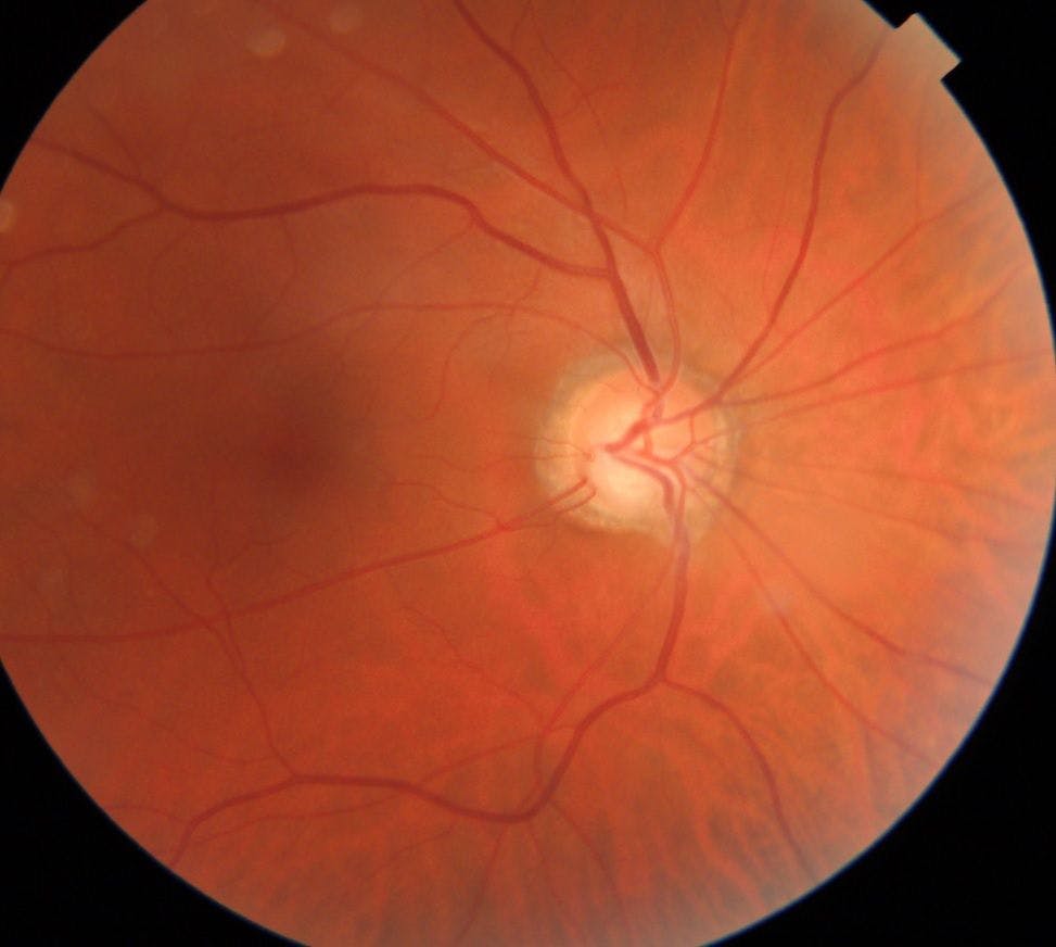
Figure 1: Courtesy of Kristin White, OD.
Cataracts
Low levels of vitamin D were shown in one study to be linked to posterior subcapsular cataracts (PSC). Patients in this study with PSC had levels of vitamin D found to be in the “vitamin D insufficiency” range (24ng/mL [+/-11SD]).
In five patients with early posterior subcapsular water cleft cysts and retro dots who were given 5,000IU of vitamin D daily, their PSC resolved completely. The time frame for this resolution was not mentioned in this study.28
Age-related macular degeneration
Vitamin D receptors and the enzymes that metabolize vitamin D are found in the retina, choroid, and retinal pigment epithelium (RPE). Low levels may be implicated in the pathogenesis of age-related macular degeneration (AMD).
One possible etiology is that vitamin D causes a decrease in amyloid beta proteins that are found in drusen. Another possible method is by reducing inflammation which is thought to be at the root of AMD. Vitamin D can inhibit angiogenesis.29
While there is controversy over whether vitamin D status is related to AMD progression, some studies do show an inverse relationship between vitamin D plasma levels and late atrophic and/or neovascular AMD.29
Diabetic retinopathy
Similarly, vitamin D may impact the development of diabetic retinopathy through its anti-inflammatory role and its effect on new blood vessel growth. Low levels of vitamin D have been thought to lead to inflammation and angiogenesis, which promotes diabetic retinopathy.30
While studies have shown conflicting data, many observational studies have shown lower serum vitamin D values associated with an increased prevalence of diabetic retinopathy and greater severity in patients with both type 1 and type 2 diabetes.30
Figure 2 shows fundus photographs of a patient with severe diabetic retinopathy.

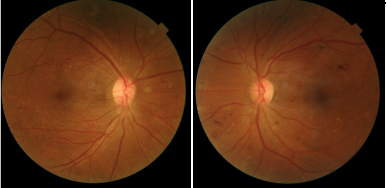
Figure 2: Courtesy of Kristin White, OD.
Dry eye
Vitamin D’s anti-inflammatory role may also impact dry eye. One clinical trial showed improvement in tear osmolarity and tear stability when patients with clinical vitamin D deficiency (<20ng/ml) were treated with vitamin D in addition to routine dry eye therapy.31
Antioxidants
Lutein/Zeaxanthin/Meso-zeaxanthin
While not vitamins, per se, a discussion on nutrition would be remiss without the mention of antioxidants. These free radical scavengers are crucial to preventing damage by oxidative stress, arguably the main driver behind most, if not all, ocular diseases.
The most common non-vitamin A carotenoids that are important for ocular health include lutein, zeaxanthin, meso-zeaxanthin, and astaxanthin. These have been shown to slow the progression of macular degeneration. Further, lutein and zeaxanthin have also been shown to have effects benefitting cognition.33
While lutein and zeaxanthin can be found in foods like leafy green vegetables and egg yolks, it is impossible for those who are showing signs of ocular disease (e.g., macular degeneration) to consume sufficient quantities from diet alone. Meso-zeaxanthin, a metabolite of lutein and zeaxanthin, is not found in food.32
Recommended doses for antioxidants:
- Lutein: 6 to 20mg/day
- Zeaxanthin: 4 to 10mg/day
Astaxanthin
Astaxanthin is an extremely potent antioxidant derived from red algae, krill, and salmon. Astaxanthin has been shown to benefit AMD, cataracts, and glaucoma in addition to dry eye, eye strain, and accommodation.33
Dietary sources of astaxanthin:33
- Red algae
- Salmon
- Krill
- Shrimp
The recommended dose for astaxanthin is 4 to 6mg/day.33
Omega 3s
Fish oil, specifically eicosapentaenoic acid (EPA) and docosahexaenoic acid (DHA), is arguably one of the most important nutrients for ocular and systemic health. We’re all familiar with fish oil for dry eye, but did you know it can benefit retinal disease as well?
Given that over 50% of the lipid content of the photoreceptors is comprised of DHA, it’s no wonder that fish oil can improve the function of the retina. Consuming cold water, fatty fish can improve outcomes in patients with exudative AMD, diabetic retinopathy, and glaucoma.
These include “SMASH” fish, which are typically smaller and have a lower mercury content—sardines, mackerel, anchovies, salmon, and herring. Supplementing with fish, cod liver, or krill oil is also beneficial.34
Studies on omega 3s and ocular diseases
AMD
One study showed reduced levels of vitreal vascular endothelial growth factor (VEGF) in patients with wet AMD receiving intravitreal anti-VEGF injections who also took 1000mg fish oil compared with those receiving injections alone.35
Diabetic retinopathy
Fish oil not only helps increase the sensitivity of the beta cells in the pancreas, but also directly improves capillary permeability and retinal inflammation. It has been shown to reduce the onset of diabetes and improve diabetic retinopathy as well as diabetic macular edema.34
Glaucoma
Fish oil has been shown to increase circulation, which can assist with optic nerve perfusion and improve visual field indices.34 It should be considered an adjunct therapy for our POAG patients.
Table of food sources for vitamins and fatty acids5,6,8,11,12,19,25,34,32,33
| Vitamin/Essential Fatty Acid | Food Sources |
|---|---|
| A | Liver, fish, eggs, dairy products, leafy greens, orange/yellow/red colored fruits and vegetables |
| B1 (Thiamine) | Organ meat, pork, seeds, squash, fish (especially trout, mackerel, salmon, and tuna), and legumes |
| B6 | Wide variety of plant and animal foods, including leafy and root vegetables, fruit such as bananas, red meat, poultry, and seeds (especially sunflower and pumpkin) |
| B9 | Organ meat, green vegetables (both leafy and non-leafy), legumes, beets, avocados, papayas, strawberries, and pomegranates |
| B12 | Fish (especially sardines, salmon, tuna, and cod), shellfish such as shrimp and scallops, organ meat, beef, eggs, poultry, and some fermented soy products like tempeh also contain vitamin B12 |
| C | Broccoli (102mg/cup), red bell peppers (95mg/cup), strawberries (85mg/cup), oranges (70mg in 1 medium orange), all citrus fruits, dark leafy green vegetables, papaya, guava, cantaloupe, and berries |
| D | Sun exposure (greatest source), salmon, tuna, mackerel, wild mushrooms, fish roe, liver, and eggs |
| Omega 3 | Cold water fish such as salmon, sardines, krill, and mackerel |
| Lutein/Xeaxanthin | Leafy green vegetables, egg yolks, pistachios, orange, and yellow-colored vegetables |
| Astaxanthin | Salmon, shrimp, krill, and red algae |
Table 1: Courtesy of Kristin White, OD.
Case report of ocular disease and vitamin deficiency
A 15-year-old caucasian female presented to the optometry department of a community health center with complaints of blurry vision. She was having trouble seeing the tablet she was using for distance learning during the COVID-19 pandemic.
Findings from testing
Upon testing, her best-corrected visual acuity (BCVA) was 20/50 in each eye. Given her age, functional amblyopia was the top differential. The general stress of COVID-19 and family life stressors in an otherwise “healthy” appearing teenager may have been affecting her vision.
The patient returned for an optical coherence tomography (OCT) and visual field testing. The OCT showed full rims (although there are no age-matched controls for teens), and the visual field showed diffuse loss, but not the restricted fields necessary for a definitive diagnosis.
Relevant patient history
Upon further discussion with the family, the patient had been diagnosed a couple of years prior with a stomach disorder that caused her to be very uncomfortable whenever she ate.
At the time of her eye exam, she had been exclusively eating potatoes for several months, as this was the one food that didn’t make her stomach feel upset. When she got tested, it was found that she had multiple vitamin deficiencies.
Treatment recommendations
This is an extreme case and a difficult one to treat as she had a lot of anxiety around food after all the discomfort she had been in. This patient was prescribed vitamins/smoothies by her doctor to slowly incorporate foods and nutrients back into her diet and help restore visual (and other systemic) functions.
In the meantime, the patient was also connected with her school district’s teacher of the visually impaired to get her a larger monitor to use at home as her vision loss was affecting her ability to do her school work.
For more information on vitamins, supplements, nutrition, and the eye, check out Review of Optometry’s wellness guide and this article on nutraceutical medical food.
Conclusion
Testing vitamin status and advocating for vitamin supplementation may not be standard-of-care therapy or practical for every patient. However, a thorough understanding of vitamin status and nutrition can help eyecare providers better educate and care for their patients.
When it comes to the management of ocular disease, adjunct testing and therapy may be warranted, especially when standard-of-care treatments are not fully effective.
Testing a patient’s vitamin status and educating them on the potential benefits of appropriate supplementation may improve treatment outcomes for many common (and not-so-common) eye conditions.
Vitamin deficiencies should always be on the differential diagnosis list when striving to provide good quality patient care.
