


The Diabetic Retinopathy Study (DRS) and Early Treatment Diabetic Retinopathy Study (ETDRS) were two groundbreaking clinical trials that brought a paradigm shift to our management of diabetic retinopathy (DR).1,2 Stemming from these trials, stage-based treatment protocols of non-proliferative diabetic retinopathy (NPDR) and proliferative diabetic retinopathy (PDR) were established.3 Panretinal photocoagulation (PRP) and macular focal laser photocoagulation became critical in preventing vision loss in diabetic macular edema (DME) and PDR.4
PRP still stands as the gold standard for the treatment of PDR.5 These trials further led to the United Kingdom Prospective Diabetes Study (UKPDS), which helped establish that the best treatment for mild to moderate NPDR is through tight glycemic control.6 Although, to counter diabetic macular edema, at any point in the spectrum of DR, intravitreal agents with vascular endothelial growth factor (VEGF) inhibitors are currently used as first-line treatment.7
Modern diagnostic tools serve as a backbone to determining DR staging and have evolved significantly. Staging is essential as most treatment protocols are stage-dependent. In the recent past, disease staging was predominantly dependent on clinical exam, which has limitations due to various patient and physician factors–such as the lighting of the equipment, type of the handheld lenses used, patient’s palpebral fissure size, sensitivity to light, and level of cooperation.8
However, with the advent of Optical Coherence Tomography (OCT), subtle anatomical changes can be detected straightforwardly.9 Intravenous fluorescein angiography (IVFA) is the gold standard test to study the fundus microvasculature;10 many ophthalmologists are trending towards OCT-angiography (OCTA) because of some advantages over IVFA. OCTA is quicker, noninvasive, and harbors no risk of allergic reaction (no intravenous fluorescein infusion) as well as the ability to visualize structural morphology and microvasculature in one test.11
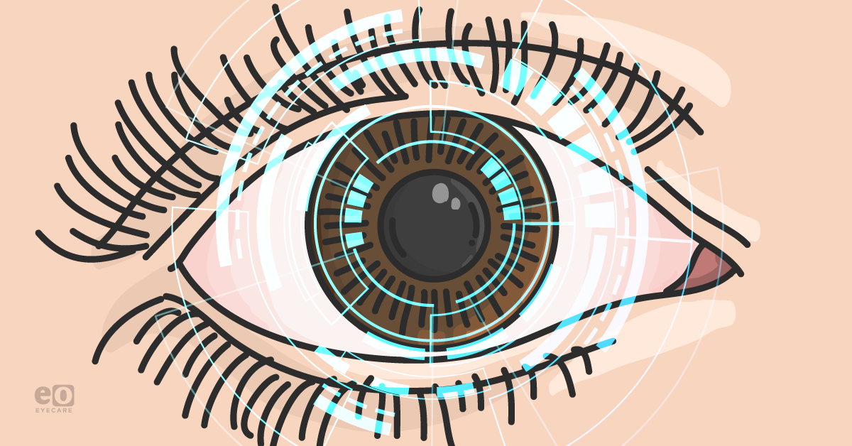
“With evolving medical diagnostics, it was only a matter of time until artificial intelligence (AI) came into play to help diagnose DR more efficiently and accurately.”
Intelligent Evaluation of Diabetic Retinopathy (EviRed) is a European clinical trial that aims to test if AI can be effectively trained to aid ophthalmologists to diagnose stages of DR.12
EviRed Project’s existing details
Overview
The primary objective of the EviRed Project is to develop and validate a system aiding the ophthalmologist by improving the decision-making during DR follow-up.13 It intends to replace the current classification of DR, which provides an insufficient prediction precision, as it involves some degree of subjectivity among ophthalmologists. It anticipates that AI will be able to properly integrate data provided by modern fundus imaging devices with other patient medical data.
A cohort of 5000 diabetic patients has been recruited and followed for an average of two years to collect data to train and validate the new prediction system.
Study description
The study will be recruiting a cohort of 5,000 diabetic patients with different stages of DR and following them for an average of 2 years. Each year, general data as well as ophthalmological data will be collected. Retinal images and videos of both eyes will be obtained using different imaging modalities, including ultra-widefield photography, OCT, and OCTA.
The EviRed cohort will be split into two groups: one group of 1,000 patients (validation cohort) will be randomly selected during the inclusion period by an unbalanced draw to be representative of the general diabetic population.14 Their data will be used for the validation of the algorithms. The results of the remaining 4000 patients (training cohort) will be used to train the algorithms.
The primary objective will be the validation of the prognostic tool and evaluation of how accurately the algorithm can predict progression to severe retinopathy in the following year. Secondary objectives will be to evaluate how accurately the algorithm can assess DR severity and individual components of DR complications as well as to compare prediction by algorithm to that made by ophthalmologists based on the current DR classification.
Study design
Actively recruiting observational prospective cohort study involving 5000 participants. The official title of the study is Intelligent Evaluation of Diabetic Retinopathy, and the commencement date was 21 December 2020, with an estimated primary completion date of 21 May 2024.
Outcome measures
The primary outcome measure is progression to severe diabetic retinopathy within a study duration of twelve months. The secondary outcome measure is algorithm performance within a time frame of three years, where algorithms will be evaluated for their automaticity to assess the components of DR and will be compared with human evaluators.15
At each patient's visit, study investigators will evaluate the risk of DR progression based on the current DR classification and their clinical experience. The clinician will express the risk of DR and its estimated probability of progression as a continuous variable or as a semi-quantitative variable. The performance of the human prediction will be compared to the algorithm using sensitivity, specificity, and area under the curve (AUC).
Study population
The study is targeted towards patients of all sexes who are 18 years or older have either type 1 or type 2 diabetes mellitus (DM) or other rarer forms of DM. The study's goal is to train the algorithms to predict the progression of DR at one year and exclude DM patients with no risk of progression.16 This cohort of patients will include 10% of patients without DR or minimal NPDR and 90% of patients with moderate or severe NPDR.
Subjects will be excluded from the study if they have a history of vitrectomy, ungradable retinal imaging (IVFA/OCT/OCTA), if unwilling or unable to comply with study procedures, and/or considered part of a vulnerable population (minors, legally detained, incarcerated, patients under legal protection). With this study design, it is estimated that all DR stages will be represented; however, there will be a greater proportion of patients with moderate or severe stages of DR who are at higher risk of DR progression.
Results
The results of this trial are pending at the time of this publication.
Importance of the EviRed Project
In 2018, the FDA permitted the marketing of AI-based devices, such as IDx-DR, to detect specific DM-related eye problems.17 The EviRed study can potentially change the future of diagnosis and treatment of DR, similar to the DRS and ETDRS studies. Integration of AI in clinical practice can change the outlook of the ophthalmic practice in the future. Algorithms in the future devices will be fully automated. Human operators with minimal training can be deployed at ophthalmic and primary care practices to make the early diagnosis of DR easier and more efficient.18
References
- Photocoagulation treatment of proliferative diabetic retinopathy. Clinical application of Diabetic Retinopathy Study (DRS) findings, DRS Report Number 8. The Diabetic Retinopathy Study Research Group. Ophthalmology. 1981 Jul;88(7):583-600. PMID: 7196564.
- Early Treatment Diabetic Retinopathy Study design and baseline patient characteristics. ETDRS report number 7. Ophthalmology. 1991 May;98(5 Suppl):741-56. doi: 10.1016/s0161-6420(13)38009-9. PMID: 2062510.
- Grading diabetic retinopathy from stereoscopic color fundus photographs--an extension of the modified Airlie House classification. ETDRS report number 10. Early Treatment Diabetic Retinopathy Study Research Group. Ophthalmology. 1991 May;98(5 Suppl):786-806. PMID: 2062513.
- Techniques for scatter and local photocoagulation treatment of diabetic retinopathy: Early Treatment Diabetic Retinopathy Study Report no. 3. The Early Treatment Diabetic Retinopathy Study Research Group. Int Ophthalmol Clin. 1987 Winter;27(4):254-64. doi: 10.1097/00004397-198702740-00005. PMID: 3692707.
- Photocoagulation treatment of proliferative diabetic retinopathy. Clinical application of Diabetic Retinopathy Study (DRS) findings, DRS Report Number 8. The Diabetic Retinopathy Study Research Group. Ophthalmology. 1981 Jul;88(7):583-600. PMID: 7196564.
- King P, Peacock I, Donnelly R. The UK prospective diabetes study (UKPDS): clinical and therapeutic implications for type 2 diabetes. Br J Clin Pharmacol. 1999 Nov;48(5):643-8. doi: 10.1046/j.1365-2125.1999.00092.x. PMID: 10594464; PMCID: PMC2014359.
- Heier JS, Korobelnik JF, Brown DM, Schmidt-Erfurth U, Do DV, Midena E, Boyer DS, Terasaki H, Kaiser PK, Marcus DM, Nguyen QD, Jaffe GJ, Slakter JS, Simader C, Soo Y, Schmelter T, Vitti R, Berliner AJ, Zeitz O, Metzig C, Holz FG. Intravitreal Aflibercept for Diabetic Macular Edema: 148-Week Results from the VISTA and VIVID Studies. Ophthalmology. 2016 Nov;123(11):2376-2385. doi: 10.1016/j.ophtha.2016.07.032. Epub 2016 Sep 17. PMID: 27651226.
- Schneiderman H. The Funduscopic Examination. In: Walker HK, Hall WD, Hurst JW, editors. Clinical Methods: The History, Physical, and Laboratory Examinations. 3rd edition. Boston: Butterworths; 1990. Chapter 117. Available from: https://www.ncbi.nlm.nih.gov/books/NBK221/
- G Panozzo, B Parolini, E Gusson, A Mercanti, S Pinackatt, G Bertoldo & S Pignatto (2004) Diabetic macular edema: an OCT-based classification, Seminars in Ophthalmology, 19:1-2, 13-20, DOI: 10.1080/08820530490519934
- Classification of diabetic retinopathy from fluorescein angiograms. ETDRS report number 11. Early Treatment Diabetic Retinopathy Study Research Group. Ophthalmology. 1991 May;98(5 Suppl):807-22. PMID: 2062514.
- Tan, A., Tan, G., Denniston, A. et al. An overview of the clinical applications of optical coherence tomography angiography. Eye 32, 262–286 (2018). https://doi.org/10.1038/eye.2017.181
- Clinical projects [Internet]. EviRed. Available from: https://evired.org/about-evired/
- Lanzetta, Paolo et al. “Fundamental principles of an effective diabetic retinopathy screening program.” Acta diabetologica vol. 57,7 (2020): 785-798. doi:10.1007/s00592-020-01506-8
- Ting DSW, Cheung CY, Lim G, Tan GSW, Quang ND, Gan A, Hamzah H, Garcia-Franco R, San Yeo IY, Lee SY, Wong EYM, Sabanayagam C, Baskaran M, Ibrahim F, Tan NC, Finkelstein EA, Lamoureux EL, Wong IY, Bressler NM, Sivaprasad S, Varma R, Jonas JB, He MG, Cheng CY, Cheung GCM, Aung T, Hsu W, Lee ML, Wong TY. Development and Validation of a Deep Learning System for Diabetic Retinopathy and Related Eye Diseases Using Retinal Images From Multiethnic Populations With Diabetes. JAMA. 2017 Dec 12;318(22):2211-2223. doi: 10.1001/jama.2017.18152. PMID: 29234807; PMCID: PMC5820739.
- Zhang A, Zhang Q, Chen CL, Wang RK. Methods and algorithms for optical coherence tomography-based angiography: a review and comparison. J Biomed Opt. 2015 Oct;20(10):100901. doi: 10.1117/1.JBO.20.10.100901. PMID: 26473588; PMCID: PMC4881033.
- Bai P, Barkmeier AJ, Hodge DO, Mohney BG. Ocular Sequelae in a Population-Based Cohort of Youth Diagnosed With Diabetes During a 50-Year Period. JAMA Ophthalmol. 2022;140(1):51–57. doi:10.1001/jamaophthalmol.2021.5052
- Commissioner Oof the. FDA permits marketing of artificial intelligence-based device to detect certain diabetes-related eye problems [Internet]. U.S. Food and Drug Administration. FDA; 2018. Available from: https://www.fda.gov/news-events/press-announcements/fda-permits-marketing-artificial-intelligence-based-device-detect-certain-diabetes-related-eye
- Grzybowski, A., Brona, P., Lim, G. et al. Artificial intelligence for diabetic retinopathy screening: a review. Eye 34, 451–460 (2020). https://doi.org/10.1038/s41433-019-0566-0
