


The SARS-CoV-2 pandemic has taken an enormous and devastating toll on the world’s population, affecting people of all ages, backgrounds, and predisposing medical conditions. With the novel omicron variant on the rise, there are currently upwards of 332 million total cases and 5.5 million deaths worldwide, as per data from Johns Hopkins University.1 A disease that exerts its effects through ACE/ACE2 balance disruption and RAAS activation, SARS-CoV-2 can present with a wide array of symptoms, ranging from generalized muscle weakness, new loss of taste or smell, shortness of breath, clotting, sepsis, and death.
Post-COVID conditions, otherwise known as long-haul COVID, post-acute COVID-19, or chronic COVID, are a series of returning, ongoing, or new symptoms that can occur to those infected by COVID-19, approximately four or more weeks after initially contracting the virus. These symptoms are debilitating and can affect those expressing severe symptoms and those with mild symptoms or those showing no initial signs.
Secondary effects from COVID-19 can present as various combinations of health issues for varying lengths of time.
Symptoms resulting from secondary COVID-19 effects can include:
- Chronic fatigue
- Lightheadedness
- Post-exertional malaise
- Brain fog (difficulty concentrating)
- Exertional chest pain
- Altered taste and smell
- Changes in menstrual period cycles
- Neuropathies (pins-and-needles sensation)
- Difficulty breathing
- Cough
- Sleeping difficulties
- Mood changes
As long-haul COVID has been a relatively new development with less than two years of research under its belt, the CDC and other public health experts across the nation are continuing to learn more about the long-term health effects associated with COVID-19.
Potential long-term effects of COVID-19
In addition to long-haul COVID, COVID-19 has the potential of causing long-term effects throughout multiple organ systems that can linger for months after the time of initial infection.
These effects can manifest through various body systems, some of which include: the cardiovascular system, in which cardiomyopathies, pericarditis and myocarditis, and clotting conditions have been reported; the nervous system, in which strokes, peripheral neuropathies, altered mental status, and multiple intracerebral hemorrhagic episodes; pulmonary, renal, and integumentary systems, and most notably, the immune system, in which COVID-19 can cause a high degree of systemic inflammation and amplify various autoimmune conditions such as HLH, Raynaud’s, and multisystem inflammatory syndrome.
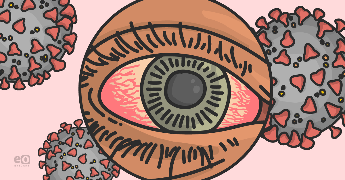
While many of these aforementioned conditions, such as clotting, fatigue, and chronic inflammation are important and relevant, one organ involved in multiple short and long-term conditions related to COVID-19, the eye, has started to garner more attention and excitement in recent months.
At the commencement of the global pandemic, eye-protective gear was a highly relevant topic in the conversation, when face shields were marketed as a safe wearable item to protect against transmission of aerosolized particles through the conjunctiva. The more relevant topics in COVID-19 and ophthalmology are the secondary effects on the eye that can occur due to COVID-19 infection, use of personal protection equipment (PPE) and the COVID-19 vaccine. To date, ocular symptoms have been reported in over a third of COVID-positive patients with variable symptoms.
Ocular manifestations of COVID-19 occur through direct effects due to the virus, altered physical conditions, immune-mediated tissue damage, activation of the coagulation cascade and hypercoagulable state induced by the viral infection, secondary reactivation of dormant viruses, associated comorbidities, and drugs used in disease management.
Some of these effects of COVID-19 are represented as follows:
- Dry eye syndrome
- Mask-induced chalazion
- Conjunctivitis
- Secondary viral infections
- Herpes simplex viruses
- Herpes zoster viruses
- Prone position ventilation-induced glaucoma
- Hypoxemia-induced retinal damage
- Multisystem inflammatory syndrome in children (MIS-C)
- Inflammatory eye conditions
- Uveitis
- Retinitis
- Cranial nerve palsies
- Myasthenia gravis
Direct effects due to coronavirus
In addition to the primary route of COVID-19 transmission through the nasal and oral cavities, direct transmission can also less commonly occur through the conjunctiva and the ocular route. When one coughs, sneezes, talks, or yells, the infected droplets can come in direct contact with the mucosal membranes that line the eyelid, otherwise known as the conjunctiva, through aerosolized transmission.2 The viral particles can then spread through the blood vessels within the conjunctiva and subsequently infect the rest of the body. Patients have been found to have ocular manifestations such as epiphora, conjunctival congestion, chemosis, and most commonly, conjunctivitis.
Face shield protection against ocular transmission has been evidenced by a 2020 Lancet study to reduce the risk of infection from 16% to 6% compared to those without eye protection.2 Face shields provide additional protection when paired with face masks and social distancing for those in high-risk categories for the benefit of ocular protection.2 However, while SARS-CoV-2 viral particles have been detected on the ocular surface and in conjunctival secretions of patients testing positive for COVID-19, the virus is less likely to be transmitted through the ocular route.3
Dry eye syndrome
Dry eye syndrome (DES) is multifactorial and consists of a myriad of ocular symptoms resulting from the chronic inflammation of the ocular surface.4 There is a significant amount of literature to suggest that the overall situation related to DES has likely worsened during the COVID-19 pandemic due to multiple reasons.5
Some contributing factors to dry eye are:
- Lifestyle modifications of lockdown
- Staying indoors
- Increased screen times
- Delays in healthcare delivery due to services rendered only to urgencies and emergencies (particularly during the first wave of the pandemic)
- Prolonged face mask-wearing as a way to reduce viral transmission
- Inflammatory process as a part of multisystem inflammatory syndrome secondary to COVID-19 infection.6
In many surveys, infected patients recovered from COVID-19 reported symptoms of dry eye syndrome.7 Below we will discuss how mask-wearing has contributed to DES.
During the COVID-19 pandemic, the world adapted to an increased face mask-wearing to prevent disease transmission. Although it is essential for infection prevention, it does have ocular health implications. Many ophthalmic practices have observed a corresponding increase in ocular irritation and dryness among regular mask wearers. This finding has not been described in the pre-pandemic literature.
Besides the symptoms of dryness assessed on ocular surface disease indices, many mask-wearing patients reported an awareness of upwards flow of exhaled air into their eyes. This increased airflow can cause an accelerated evaporation of the protective tear film and may produce ocular surface irritation or inflammation.8
Besides, among the healthcare workers who used tape on their cheeks to prevent this air convection and reported DES, it is postulated to have possibly resulted from restricted lower eyelid excursion and possible mechanical ectropion with secondary lagophthalmos. Similar symptoms have been previously reported among patients using devices of mechanical ventilation.9 Another implication of face mask-induced exposure keratopathy could be an increased vulnerability of such eyes, without wearing protective eyewear, towards the pathogen invasion.10
Since the use of face masks is essential in the foreseeable future during the pandemic to help control viral transmission, it is vital to preempt ocular surface implications resulting from the factors discussed above.
Specific suggestions on face masks:
- Use of face masks that fit the face better, such as the ones with flexible metallic nasal wires or respirator masks
- Use of lubricant eye drops
- Use of protective eyewear, particularly in those with prolonged use of face masks and the ones with preexisting DES or its risk factors
We invite our colleagues to conduct further investigations to establish a correlation between wearing face masks and having DES globally to help develop future guidelines for eye protection.
Face mask-induced chalazion
Another important ocular implication secondary to the use of face masks being reported in the literature is the increased incidence of chalazion. There was a retrospective study conducted in California to show this undesirable outcome.11
As discussed previously, facial mask-wearing is a mechanism of accelerated evaporation of tear film to produce DES, which can be a mechanism for anterior blepharitis and Meibomian oil hardening and lead to chalazion formation.12 Increased exhaled airflow into the eyes, as also evident in the fogging of eyeglasses, causes alteration in the eyelid flora. Chalazion, usually a noninfectious process, has been associated with specific bacterial pathogens changes in the gut microbiome.
Staphylococcus aureus, commonly associated with blepharitis, is also often found in oral flora,13 which can be channeled from mouth to the eyes by talking, sneezing, and coughing.14, 15
Additionally, mask wearing is often escorted with frequent manual adjustments promoting bacterial transfer from hands onto face. It is also proposed that during the low infection rates of COVID-19 in California in the summer of 2020, corresponding with increased mask wear, in turn, simultaneously raised the incidence of chalazion further supports the potential of association between mask wear and chalazion development.11
The increased incidence of chalazion formation with mask use does not counterweight the noteworthy benefits of protective facial coverings during the pandemic. However, it is recommended to take preemptive measures to mitigate the multifactorial risk of chalazion formation.11 These measures may improve oral hygiene by using antiseptic mouthwashes containing alcohol, hydrogen peroxide, or povidone iodine. Mouthwashes have shown to reduce bacterial load (as well as the viral load of SARS CoV-2 itself) and may lessen the spread of oral flora onto the eyes and the periorbital region.16
Eyelid hygiene can be improved with the daily use of eyelid scrubs which may act as an antiviral and antibacterial agent, as well as a blepharitis deterrent.
Specific behavioral adaptations can also help minimize transferring of microorganisms onto the eyes, including:
- Frequent washing of cloth face masks with hot water
- Improving hand hygiene habits
- Minimizing face touching
- Refraining from excessive mask adjustment
- Daily face washing
- Reducing air convections from masks onto eyes by using facial quality adhesive tapes on the bridge of the nose of the masks.
Conjunctivitis
Conjunctivitis, otherwise known as pink eye, is the most frequently reported ocular manifestation and can develop at any stage of the disease. It occurs when the virus infects the mucosal membranes that line the eyelid, the conjunctiva and includes symptoms such as swollen, itchy, or red eyes. Patients may present with conjunctival hyperemia, epiphora, chemosis, mild eyelid edema, a foreign body sensation, burning, or pruritis in the eye.
Conjunctival congestion can be an early indicator COVID-19 infection; conjunctivitis was found in 2.26% of patients even before the development of systemic infection. Patients presenting with conjunctivitis should be of high index of suspicion for having COVID-19, and a thorough review of systems should be performed. The more common forms, follicular and viral conjunctivitis as well as the rarer forms, hemorrhagic and pseudomembranous conjunctivitis, have both been reported in patients presenting with COVID-19.17
In two large research studies18, 19 on 586 and 127 patients respectively, follicular conjunctivitis has been reported in multiple COVID-19 positive patients. These patient samples found that respiratory tract symptoms were associated with conjunctival congestion, conjunctivitis typically occurs in the middle phase of the infection, and that the viral shedding in the conjunctiva can persist for some time after a positive nasopharyngeal COVID-19 test.
Two studies confirmed cases of viral keratoconjunctivitis multiple days after presenting with other symptoms. They were successfully treated with moxifloxacin and antiviral therapy,20,21 while another COVID-19 patient presented with hemorrhagic and pseudomembranous conjunctivitis and more severe symptoms was treated with dexamethasone and azithromycin.22
Another study found presentations of conjunctivitis in children presenting with COVID-19.23 Some of these pediatric conjunctivitis cases have been linked to vasculitis and MIS-C and an excessive immunological response (e.g., cytokine storm) associated with COVID-19 and were treated with intravenous immunoglobulin (IVIG), aspirin, and corticosteroids. Episcleritis, a similar self-limiting condition of the episclera, was also reported to be a potential early sign associated with COVID-19 infection.24 This condition occurs in autoimmune phenomena and may demonstrate the exacerbation of COVID-19 on autoimmune conditions.
Treatment for COVID-19 conjunctivitis is mostly supportive care; however, it can generally be managed with cold compresses, antihistamine drops for pruritus, preservative-free artificial tears, or a short course of topical antibiotics to prevent bacterial superinfection. Most conjunctivitis cases associated with COVID-19 resolve within 14-30 days.
The more contagious forms of COVID-19, such as the delta and the newer variants, could also be associated with a higher incidence of conjunctivitis. In contrast, the alpha variant may be related to respiratory tract symptoms.25
Viral Infections
Secondary infections relating to COVID-19 have been receiving increased attention in recent times. Although patients with severe illness are more likely to have secondary bacterial or fungal infections than secondary viral infections, the proportion of all COVID-19 patients with secondary bacterial or fungal infections has been lower than previous viral pandemics.26 The most common secondary viral infections include respiratory syncytial virus (RSV) and Influenza-A.27
Studies have demonstrated that long-haul COVID is associated with inflammatory reactivation of secondary viruses, most often Epstein-Barr Virus (EBV).28 EBV is a class of herpesvirus that lays dormant after the initial infection. Even when asymptomatic, EBV is contagious and remains dormant until a trigger such as a weakened immune system, stress, or hormonal changes are introduced, leading to reactivation. Similarly, reactivation of secondary viruses such as herpes zoster and herpes simplex can lead to ophthalmic pathology.
Herpes Zoster Virus (HZV), otherwise known as shingles, is a reactivation of the Varicella-Zoster virus (VZV) that lies dormant within dorsal root ganglia. HZV commonly presents with pain, itching, or paresthesia along individual dermatomes that is self limiting and typically resolves after 10-15 days. When VZV is reactivated within the ophthalmic division of the trigeminal nerve, the resultant disease is named Herpes Zoster Ophthalmicus (HZO). In addition to a vesicular rash within dermatomes, HZO can present with keratitis, conjunctivitis, uveitis, retinitis, and rarely episcleritis. These findings can lead to chronic inflammation, pain, and eventual vision loss.
A case study confirmed the presence of HZO in four patients ranging in age from 7 to 42 despite them being immunocompetent.29 As none of the other typical reactivating triggers were identified in the patients of this study, and all were confirmed to have COVID-19 using a nasopharyngeal smear test 4-5 days preceding HZO, it was hypothesized that stress from COVID-19 itself can trigger reactivation of HZO.
Managing COVID-19 associated HZO requires timely antiviral intervention to minimize ocular complications. Systemic and topical acyclovir in addition to topical prednisolone acetate are an effective treatment.29
Latent HSV can also be reactivated and spread to the eye. The most common ophthalmic consequence of reactivation of HSV is keratitis, or inflammation of the cornea, and is most frequently caused by HSV-1. Ocular pathology is second only to oral infection in terms of localization of the virus. Symptoms include photophobia, redness, tearing, decreased vision, and pain.
Initial keratitis may result from entry to the surface of the eye by way of droplets or by spreading to the eye from other bodily tissue. Recurrent keratitis can induce scarring, thinning, and neovascularization of the cornea leading to vision loss. Less frequent ocular presentations include uveitis, conjunctivitis, and acute retinal necrosis.
A study found a concomitant increased incidence of herpes keratitis in a period that COVID-19 cases were surging relative to the previous two years.30
Bilateral acute angle closure glaucoma with prone position ventilation
Some ophthalmologists have utilized the prone position as a provocative test for angle closure.31,32 Forward shifting of the lens-iris diaphragm, causing relative pupillary block or blockage of the angle of the anterior chamber, can cause an increase in the intraocular pressure (IOP) during prone position.
Bilateral acute angle-closure glaucoma, previously reported after face down spinal surgery and general anesthesia, is probably associated with the prolonged prone position during surgery.33, 34 Angle-closure glaucoma following prone positioning after vitreoretinal surgery has also been reported.35
Early prone position ventilation for severe COVID-19 pneumonia has been found to reduce mortality and is also part of the protocol for the management of associated acute respiratory distress syndrome (ARDS).36 Prone ventilation has become a standard treatment protocol for managing ARDS associated with COVID-19 pneumonia. Patients may be ventilated for as many as 12 hours a day, which is a risk factor for angle-closure glaucoma.37, 38
Elevated IOPs following prone ventilation can result from orbital compartment syndrome (bilateral periorbital edema, optic disc edema, and retinal hemorrhages with elevated IOPs), or bilateral acute angle-closure glaucoma.39, 40 The extent of this increase in IOP is proportional to the time the patient is in the prone position for ventilation.
Acute rises in IOP may affect the ocular perfusion pressure and risk irreversible vision loss because of ischemic events. It is being proposed that the prone ventilation can also cause primary angle-closure (PAC) and primary angle closure glaucoma (PACG), which are different from the secondary forms of angle closure glaucoma as they do not result from pre-existing ocular risk factors. The following mechanisms may result in primary angle-closure glaucoma in a prolonged prone position: pupillary block, plateau iris, lens-induced glaucoma, and ciliary block glaucoma.41
The ICU staff must look for ocular signs and symptoms, particularly in prone ventilation patients. The altered direction of airflow in mechanical ventilation devices can cause redness in the eyes. But it should not be overlooked as redness in the eyes may also signify acute angle closure. A prompt ophthalmic consult is recommended.
Retinal diseases
Changes to the vasculature due to COVID-19 infection can be seen through changes to the retinal vessels. In a recent study of 59 eyes from 32 COVID-19 patients and 80 eyes from 53 unexposed subjects, retinal arteries and veins of patients with acute COVID-19 were found to be significantly dilated compared to those of subjects unexposed to the virus.42
Retinal damage can occur due to vascular, inflammatory, and neuronal changes in the viral infection.43 Patients may present with a central retinal vein occlusion (CRVO), a central retinal artery occlusion (CRAO), or acute macular neuroretinopathy (AMN), which is believed to occur either due to a hypercoagulable state or hypoxia associated with COVID-19; this can induce the endothelial cells to release tissue factor and trigger the extrinsic coagulation cascade.44, 45 Management for retinal diseases associated with COVID-19 includes high dose steroids to reduce inflammation, anti-vascular endothelial growth factor (anti-VEGF) as well as anticoagulation therapy.
Hypoxia and vision loss
Vision loss can be seen in extreme and severe cases of COVID-19. In COVID-19 infected patients presenting with respiratory distress and low oxygen levels, poor perfusion can eventually lead to ischemia of metabolically active tissues such as the retina or the optic nerve. Additionally, an acute stroke resulting from severe COVID-19 infection can affect the areas of the brain which control various fields of vision. Furthermore, several patients affected with COVID-19 have reported blurred vision, either as an effect of long-haul COVID or as a result of a secondary neurological effect from COVID-19 such as cranial nerve palsy.
Multisystem Inflammatory Syndrome in Children (MIS-C)
Various inflammatory eye conditions have also been observed due to COVID-19, including uveitis and retinitis. One of the most notable and severe conditions, Multisystem Inflammatory syndrome in children (MIS-C), has occurred in various pediatric cases across the nation due to COVID-19 infection.
This fatal condition occurs when the child enters a systemic hyperinflammatory state, affecting organs such as the lungs, stomach, heart, brain, skin, and eyes. The condition most commonly affects school-aged children between the ages of 8-9 but has also been seen in infants and young adults.46 It can occur around four weeks after initial COVID-19 infection and can include fever, stomach pain, diarrhea, bloodshot eyes, presyncope, vomiting, or skin rash. In those diagnosed with MIS-C, severe cases can increase intracranial pressure and vision loss.47
Cranial Nerve (CN) palsies
Cranial nerves are a series of twelve pairs of nerves that have roots in the brainstem or central nervous system, supply the head and neck, and carry out vital sensory, motor, and visceral functions of the human body. Cranial nerve palsies are characterized by a decreased or complete loss of function of one or more cranial nerves due to a congenital or acquired abnormality such as a tumor, trauma, ischemia, or viral infections such as COVID-19.
Cranial Nerve I (CN I) has been linked to one of the more known effects of COVID-19, anosmia, or loss of sense of smell, with some involvement in prolonged anosmia after COVID-19 infection.48 However, several other anomalies in cranial nerve function have also been reported, including those affecting ocular function. Of the four cranial nerves controlling ocular functions (II, III, IV, VI), cranial nerves III (oculomotor), and VI (abducens) have been reported to be affected in multiple patients after COVID-19 infection.
The abducens nerve (CN VI) is the most commonly involved, followed by the oculomotor nerve (CN III). The most commonly reported symptom among those with COVID-associated cranial nerve palsies is diplopia or double vision.49 Patients experiencing abducens nerve palsy cannot abduct (lateral movement) the affected eye due to dysfunction in the innervation of the lateral rectus muscle. These patients present with binocular horizontal diplopia and may attempt to compensate by rotating their heads to allow the eye to look sideways.50
Several cases of abducens nerve palsy have also been reported following the COVID-19 vaccination.51,52 CN III supplies several important extraocular muscles and the ciliary muscle, sphincter pupillae, and levator palpebrae superioris. While there are several established causes of CN III palsy, most notably hypertension, and diabetes mellitus, COVID-19 is also a potential cause. Some symptoms that these patients experienced include unilateral reduction of adduction and elevation of their eye, ptosis, pupil dilation, and diplopia.53
COVID-19 has also been linked with Miller-Fisher Syndrome (MFS),54 a rare, acquired nerve disease and variant of Guillain-Barré syndrome, with reported ophthalmoplegia and blurred vision symptoms. Isolated cases of trochlear (IV) nerve involvements due to COVID-19 infection have also been noted.55
While these neurological manifestations may indicate severe disease, cranial nerve palsies in the setting of COVID-19 infection have been linked to mild viral symptoms and great recovery rates.53 In fact, many cranial nerve palsies associated with COVID-19 disease resolved in 2-6 weeks. However, the prognosis and pathogenesis are still being researched. A misdirected immune system triggered by the viral infection is believed to be the source of these issues.
Myasthenia gravis
COVID-19 symptoms can include neurological etiology, including anosmia, ageusia, and headaches. There have been reports of post-infectious neurological diseases such as Guillain Barre syndrome and Miller Fisher syndrome associated with COVID-19.56 Furthermore, exacerbation of COVID-19 in patients with Myasthenia Gravis (MG) has been reported along with the typical ocular signs such as ptosis, diplopia, and lagophthalmos.
An observational study found that COVID-19 patients with MG who were hospitalized were more likely to be admitted to the ICU or put on mechanical ventilation.57 Inversely, exacerbation of MG has been linked to COVID-19. A large retrospective study of 91 COVID-19 patients showed a high proportion of MG exacerbation.58
Furthermore, a smaller study reported that a quarter of these MG patients went into Myasthenia Crisis, which resulted in respiratory failure. Theoretically, immunosuppressive therapy associated with MG might be involved in the mechanism of COVID-19 exacerbation. However, increasing evidence suggests that this is not the case, with some proposing immunosuppressive therapy might offer protective benefits to the virus.59
Conclusions
COVID-19 has changed our society, and profession, in a myriad of ways. Not only wearing a facial covering and social distancing became the new normal, also our understanding of virology, immunology and infectious diseases has evolved manyfold. Similarly, the complications secondary to the illness from COVID-19, in many cases, have changed lives permanently through their implications on ocular health.
As the novel variants and spiking COVID-19 cases become the new normal across the nation, both eyecare professionals and their patients should be aware of the typical and atypical ocular manifestations. Ophthalmologists and other clinicians may find themselves in the position to be the first providers to detect ocular signs and symptoms of varying etiologies that may raise suspicion for initial COVID-19 diagnosis. As more knowledge becomes available about the novel COVID-19 variants and their associated ocular manifestations, clinicians can be well-equipped to manage their patients’ health conditions and guide their patients to develop a better prognosis.
We invite our colleagues to continue their research on ocular manifestations of COVID-19 to improve the public’s ocular health on a global scale.
References
- Covid-19 map. Johns Hopkins Coronavirus Resource Center. Retrieved January 13, 2022, from https://coronavirus.jhu.edu/map.html
- Chu, Derek K., et al. "Physical distancing, face masks, and eye protection to prevent person-to-person transmission of SARS-CoV-2 and COVID-19: a systematic review and meta-analysis." The lancet 395.10242 (2020): 1973-1987.
- Hemphill, Nancy. “Conjunctivitis Most Common Ocular Sign of COVID-19.” Healio, 25 June 2021, https://www.healio.com/news/optometry/20210625/conjunctivitis-most-common-ocular-sign-of-covid19.
- Boyd, Kierstan. “What Is Dry Eye? Symptoms, Causes and Treatment.” American Academy of Ophthalmology, 5 Jan. 2022, https://www.aao.org/eye-health/diseases/what-is-dry-eye.
- Freni, Francesco, et al. "Symptomatology in head and neck district in coronavirus disease (COVID-19): a possible neuroinvasive action of SARS-CoV-2." American journal of otolaryngology 41.5 (2020): 102612.
- Barabino, S. A Narrative Review of Current Understanding and Classification of Dry Eye Disease with New Insights on the Impact of Dry Eye during the COVID-19 Pandemic. Ophthalmol Ther 10, 495–507 (2021). https://doi.org/10.1007/s40123-021-00373-y
- Asiedu, Kofi. "Rasch analysis of the standard patient evaluation of eye dryness questionnaire." Eye & contact lens 43.6 (2017): 394-398.
- Moshirfar M, West WB Jr, Marx DP. Face Mask-Associated Ocular Irritation and Dryness. Ophthalmol Ther. 2020 Sep;9(3):397-400
- Kousha, Obaid, Zubaid Kousha, and Jonathan Paddle. "Exposure keratopathy: Incidence, risk factors and impact of protocolised care on exposure keratopathy in critically ill adults." Journal of critical care 44 (2018): 413-418.
- Sun C, Wang Y, Liu G, Liu Z. Role of the eye in transmitting human coronavirus: what we know and what we do not know. Front Public Health. 2020;8:155. https://doi.org/10.3389/fpubh.2020.00155/full.
- Silkiss, Rona Z., Michael K. Paap, and Shoaib Ugradar. "Increased incidence of chalazion associated with face mask wear during the COVID-19 pandemic." American Journal of Ophthalmology Case Reports 22 (2021): 101032.
- Nemet, Arie Y., Shlomo Vinker, and Igor Kaiserman. "Associated morbidity of chalazia." Cornea 30.12 (2011): 1376-1381.
- McCormack, M. G., et al. "Staphylococcus aureus and the oral cavity: an overlooked source of carriage and infection?." American journal of infection control 43.1 (2015): 35-37.
- Mégarbane, Bruno, and Ramin Tadayoni. "Cluster of chalazia in nurses using eye protection while caring for critically ill patients with COVID-19 in intensive care." Occupational and environmental medicine 77.8 (2020): 584-585.
- Cui, Yuanyue, et al. "A study of the correlation between chalazion and intestinal flora in chinese children." (2020).
- Vergara-Buenaventura, Andrea, and Carmen Castro-Ruiz. "The Use of mouthwashes against COVID-19 in dentistry." British Journal of Oral and Maxillofacial Surgery (2020).
- Frysh, Paul. “Coronavirus and Your Eyes (Conjunctivitis): Symptoms, Transmission, and More.” WebMD, WebMD, https://www.webmd.com/eye-health/covid-19-and-your-eyes.
- Chen, Lu, et al. "Ocular manifestations of a hospitalised patient with confirmed 2019 novel coronavirus disease." British Journal of Ophthalmology 104.6 (2020): 748-751.
- Sindhuja, K., et al. "Clinical profile and prevalence of conjunctivitis in mild COVID-19 patients in a tertiary care COVID-19 hospital: A retrospective cross-sectional study." Indian Journal of Ophthalmology 68.8 (2020): 1546.
- Cheema, Marvi, et al. "Keratoconjunctivitis as the initial medical presentation of the novel coronavirus disease 2019 (COVID-19)." Canadian journal of ophthalmology 55.4 (2020): e125-e129.
- Guo, Dongyu, et al. "Relapsing viral keratoconjunctivitis in COVID-19: a case report." Virology journal 17.1 (2020): 1-7.
- Navel, Valentin, Frédéric Chiambaretta, and Frédéric Dutheil. "Haemorrhagic conjunctivitis with pseudomembranous related to SARS-CoV-2." American journal of ophthalmology case reports 19 (2020): 100735.
- Danthuluri, V., Grant, M.B. Update and Recommendations for Ocular Manifestations of COVID-19 in Adults and Children: A Narrative Review. Ophthalmol Ther 9, 853–875 (2020). https://doi.org/10.1007/s40123-020-00310-5
- Otaif, Wael, Abdulaziz Ismail Al Somali, and Ahmed Al Habash. "Episcleritis as a possible presenting sign of the novel coronavirus disease: A case report." American Journal of Ophthalmology Case Reports 20 (2020): 100917.
- Singh, Sneha, et al. "SARS-CoV-2 and its beta variant of concern infect human conjunctival epithelial cells and induce differential antiviral innate immune response." The ocular surface (2021).
- Lansbury, Louise, et al. "Co-infections in people with COVID-19: a systematic review and meta-analysis." Journal of Infection 81.2 (2020): 266-275.
- Williams Thomas C., Sinha Ian, Barr Ian G., Zambon Maria. Transmission of paediatric respiratory syncytial virus and influenza in the wake of the COVID-19 pandemic. Euro Surveill. 2021;26(29):pii=2100186. https://doi.org/10.2807/1560-7917.ES.2021.26.29.2100186
- Gold, Jeffrey E., et al. "Investigation of Long COVID Prevalence and Its Relationship to Epstein-Barr Virus Reactivation." Pathogens 10.6 (2021): 763.
- Nofal A, Fawzy MM, Sharaf El Deen SM, El-Hawary EE. Herpes zoster ophthalmicus in COVID-19 patients. Int J Dermatol. 2020 Dec;59(12):1545-1546. doi: 10.1111/ijd.15240.
- Epub 2020 Oct 11. PMID: 33040343; PMCID: PMC7675560. Majtanova, Nora, et al. "Herpes Simplex Keratitis in Patients with SARS-CoV-2 Infection: A Series of Five Cases." Medicina 57.5 (2021): 412.
- Ichioka I.Short-term prone-position test in angle-closure glaucoma. Jpn J Clin Ophthalmol. 2006;60:1619–1623.
- Kim TW, Park KH, Hong C. Dark-room prone-position test for intermittent angle closure. Korean J Ophthalmol. 2007;21:151–154.
- Singer MS, Salim S. Bilateral acute angle-closure glaucoma as a complication of facedown spine surgery. Spine J. 2010;109:7–9.
- Gayat E, Gabison E, Devys JM. Bilateral angle closure glaucoma after general anesthesia. Anesth Analg. 2011;112:126–128.
- Sutter FKP, Smorgon A, McClellan K. Acute angle closure in the fellow eye as a complication of prone positioning after vitreoretinal surgery. Arch Ophthalmol. 2003;121:1057.
- Ghelichkhani P, Esmaeili M. Prone position in management of COVID-19 patients; a commentary. Arch Acad Emerg Med. 2020;8:1–3.
- Soare C, Nowak VA, Osborne S. Eye care in the intensive care unit during the COVID-19 pandemic and beyond. Anaesthesia. 2021;75:1118–1119.
- Hearne BJ, Hearne EG, Montgomery H, et al. . Eye care in the intensive care unit. J Intensive Care Soc. 2018;19:345–350.
- Sun L, Hymowitz M, Pomeranz HD. Eye protection for patients with COVID-19 undergoing prolonged prone-position ventilation. JAMA Ophthalmol. 2021;139:109–112.
- Nerlikar, Roopali R et al. “Bilateral Acute Angle Closure Glaucoma After Prone Position Ventilation for COVID-19 Pneumonia.” Journal of glaucoma vol. 30,8 (2021): e364-e366. doi:10.1097/IJG.0000000000001864
- Weinerb, RN & Friedeman, DS. "Angle closure and angle closure glaucoma-reports and consensus statements of the 3rd global AIGS consesus meeting on angle closure glaucoma. The Hague, The Netherlands". Kugler Publications (2006). 1-20.
- Invernizzi, Alessandro, et al. "Retinal vessel modifications in acute and post-COVID-19." Scientific reports 11.1 (2021): 1-9.
- Sen, Mrittika, et al. "COVID-19 and eye: A review of ophthalmic manifestations of COVID-19." Indian Journal of Ophthalmology 69.3 (2021): 488.
- Dumitrascu OM, Volod O, Bose S, Wang Y, et al. Acute ophthalmic artery occlusion in a COVID-19 patient on apixaban. J Stroke Cerebrovasc Dis. 2020;29:104982.
- Acharya S, Diamond M, Anwar S, Glaser A, Tyagi P. Unique case of central retinal artery occlusion secondary to COVID-19 disease. IDCases. 2020;21:e00867.
- Mis-C and covid-19: Uncommon but serious inflammatory syndrome in kids and teens. Johns Hopkins Medicine. Retrieved January 13, 2022, from https://www.hopkinsmedicine.org/health/conditions-and-diseases/coronavirus/misc-and-covid19-rare-inflammatory-syndrome-in-kids-and-teens
- How covid-19 affects the eyes (updated 8/24/21). USC Roski Eye Institute. (2021, September 2). Retrieved January 13, 2022, from https://eye.keckmedicine.org/ask-the-expert-how-covid-19-affects-the-eyes-2021/
- Butowt, Rafal, and Christopher S. von Bartheld. "<? covid19?> Anosmia in COVID-19: Underlying Mechanisms and Assessment of an Olfactory Route to Brain Infection." The Neuroscientist (2020): 1073858420956905.
- Falcone MM, Rong AJ, Salazar H, Redick DW, Falcone S, Cavuoto KM. Acute abducens nerve palsy in a patient with the novel coronavirus disease (COVID-19). J AAPOS. 2020 Aug;24(4):216-217. doi: 10.1016/j.jaapos.2020.06.001. Epub 2020 Jun 24. PMID: 32592761; PMCID: PMC7311910.
- Greer, Christine E. MD, MS; Bhatt, Jaydeep M. MD; Oliveira, Cristiano A.; Dinkin, Marc J. MD Isolated Cranial Nerve 6 Palsy in 6 Patients With COVID-19 Infection, Journal of Neuro-Ophthalmology: December 2020 - Volume 40 - Issue 4 - p 520-522 doi: 10.1097/WNO.0000000000001146
- Reyes-Capo, Daniela P., Shanlee M. Stevens, and Kara M. Cavuoto. "Acute abducens nerve palsy following COVID-19 vaccination." Journal of American Association for Pediatric Ophthalmology and Strabismus (2021).
- Solomon, R. (2021, August 12). A case of acute abducens nerve palsy after COVID-19 vaccination. American Academy of Ophthalmology. Retrieved January 13, 2022, from aao.org/editors-choice/case-of-acute-abducens-nerve-palsy-after-covid-19
- Douedi, Steven, et al. "Third Cranial Nerve Palsy Due to COVID-19 Infection." Cureus 13.4 (2021).
- Li, Zheng, et al. "Miller Fisher syndrome associated with COVID-19: an up-to-date systematic review." Environmental Science and Pollution Research (2021): 1-6.
- Doblan, Ahmet, et al. "Cranial nerve involvement in COVID-19." American Journal of Otolaryngology 42.5 (2021): 102999.
- Mehta SK, Sunder A. Getting paralysed after COVID: Guillain-Barre syndrome. J Family Med Prim Care. 2021 Jul;10(7):2706-2708. doi: 10.4103/jfmpc.jfmpc_2454_20. Epub 2021 Jul 30. PMID: 34568159; PMCID: PMC8415676.
- Camelo-Filho, Antonio E et al. “Myasthenia Gravis and COVID-19: Clinical Characteristics and Outcomes.” Frontiers in neurology vol. 11 1053. 11 Sep. 2020, doi:10.3389/fneur.2020.01053
- Muppidi, Srikanth, et al. "COVID-19-associated risks and effects in myasthenia gravis (CARE-MG)." The Lancet Neurology 19.12 (2020): 970-971
- Mehta, Puja, et al. "COVID-19: consider cytokine storm syndromes and immunosuppression." The lancet 395.10229 (2020): 1033-1034.

