


Meibomian gland dysfunction (MGD) is an increasingly common condition as screen time increases and blink rate decreases. It is also heavily tied to lid hygiene and a balanced ocular surface microbiome.
This article provides a review of the anatomy of the meibomian glands, the pathophysiology of meibomian gland dysfunction, how it is affected by blepharitis, and treatments for both conditions.
Anatomy of meibomian glands
Meibomian glands reside in the superior and inferior tarsal plates.1 One end of the gland is terminal, while the other opens to the lid margin, where its contents are secreted into the tear meniscus.2 These glands are thought to be regulated by neural and hormone factors as well as the mechanical force of blinking.3
Meibum is the oily secretion produced by meibomian glands and is composed of lipids, proteins, and nucleic acids.4 These secretions are the main component of the lipid layer of the tear film and prevent aqueous evaporation.5
Download the Patient Handout on Lid Hygiene Basics here!

Patient Handout: Lid Hygiene Basics for MGD Patients
These take-home instructions describe basic lid hygiene strategies MGD patients can follow to maintain good eyelid health.

Conditions that impact the meibomian glands: MGD and blepharitis
Meibomian gland dysfunction is typically characterized by decreased secretion, caused by either hyposecretion or obstruction. The believed mechanism of disease is hyperkeratinization of the epithelium, leading to obstruction, decreased meibum flow, dilation, and eventual gland atrophy.6
MGD presents as a spectrum of changes to eyelid morphology, including:7
- Eyelid telangiectasia, thickening, and notching
- Distichiasis
- Obstruction and position change of gland orifices
- Foamy tear film
- Altered secretion viscosity
These changes can be assessed at the slit lamp and with gentle pressure on the lid. Gland atrophy may be assessed with a transilluminator on an everted lid or with infrared photography.
Several internal and external factors contribute to MGD, including age, hormones, environmental stress, diet, topical and systemic medications, and ocular surface microbiome.8 Meibum is partially hydrolyzed by commensal bacteria into free fatty acids and other glycerides.9
This alteration in lipid composition leads to a disruption in the ratio of polar to non-polar lipids, subsequently increasing the risk for hyperkeratinization and decreasing tear film stability.10 Cultures of this bacteria, mostly Staphylococcus aureus, can be seen in meibum from patients with and without blepharitis.11
Etiology of blepharitis
Blepharitis is defined by a pattern of onset, location, and appearance (ulcerative or non-ulcerative). Acute, ulcerative blepharitis is normally caused by staphylococcal bacteria but may also be viral from herpes simplex or zoster. Acute non-ulcerative blepharitis may be associated with allergy or atopy.12
Chronic blepharitis is more often defined by location. Chronic anterior blepharitis is most commonly caused by Staphylococcus but is also caused by seborrheic disease or rosacea.12 Chronic posterior blepharitis is associated with MGD.12
Of note, Demodex folliculorum (anterior) and Demodex brevis (posterior) are mites that also cause blepharitis.12 Demodex mites contribute to blepharitis through direct damage, introducing bacteria, and inducing host hypersensitivity reactions. Mite involvement is still being studied as asymptomatic patients and symptomatic patients have been seen with the same amount of mites.13
The prevalence of blepharitis is not well known but can be estimated to be around 37 to 47% and is more commonly seen in patients over 50 years old.12 Increased age, rosacea, and diabetes are all risk factors. Patients typically present with complaints of itching, burning, dryness, redness, and crusting of the lids.
Signs of blepharitis on clinical exam include:14
- Edema
- Erythema
- Telangiectasia
- Scaling or collarettes
- Collarettes are pathognomonic for Demodex
- Lash anomalies in anterior blepharitis
- Meibomian gland dilation, obstruction, or scarring in posterior blepharitis
In more severe or chronic cases, corneal involvement such as punctate keratitis, recurrent erosions, and infiltrates are possible. There is no further testing indicated in the diagnosis of blepharitis, however, tear break-up time (TBUT) is helpful to document as it is typically decreased in all types of blepharitis.
Connecting blepharitis and MGD
Patients with MGD are much more likely to present with Demodex or blepharitis than healthy patients.15 These patients present with lower-quality meibum and higher concentrations of fatty acids, increasing inflammation and decreasing tear film stability. Chalazia and hordeola are also shown to be sequelae of chronic blepharitis and meibomian gland dysfunction.16
The value of lid hygiene in managing blepharitis and MGD
Good lid hygiene is characterized by a balanced lid and ocular surface microbiome. Maintaining this lid hygiene is the main target for the treatment of blepharitis.
The ideal sequence is 10 minutes of moist heat with a dry eye mask followed by cleansing with an eyelid scrub towelette, spray, or foam. The moist heat breaks down oils and debris, facilitates a deeper clean, and melts meibum to decrease gland obstruction.
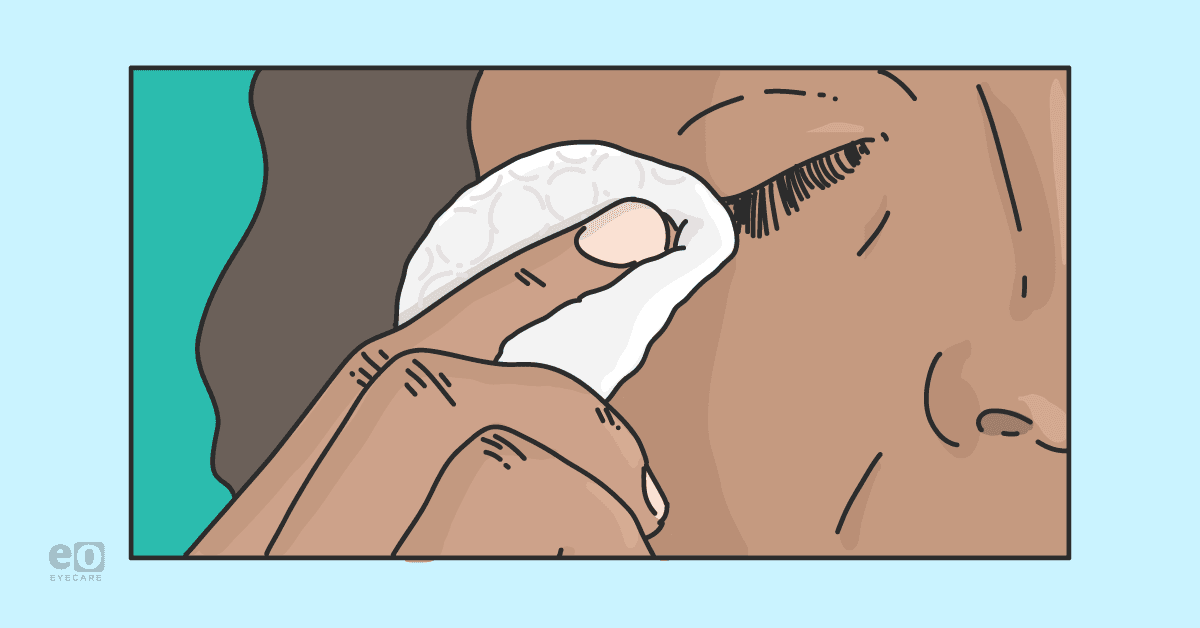
Lid cleansing
A significant reduction in dry eye symptoms, measured by Ocular Surface Disease Index (OSDI) score, in 60 patients with grade 2 MGD was demonstrated with 5 minutes of warm compress followed by 30 to 60 seconds of lid scrubs twice a day.17
There was no statistically significant difference in symptoms between OCuSOFT lid scrubs and dilute baby shampoo. Differences in secondary outcomes such as meibum quality, expressibility, and ocular stain were also insignificant between the two groups.
Comparing the efficacy of baby shampoo and lid cleansers for MGD
To our knowledge, designated lid cleansers should be superior to baby shampoo, however, several other studies have shown similar improvement in clinical signs and symptoms between dilute baby shampoo and lid cleansing scrubs.18,19 Many of these studies were performed involving subjects with mild blepharitis and MGD. Further research on subjects with more advanced ocular surface disease may confirm or deny any superiority regarding designated lid scrubs versus baby shampoo.
In a study of 53 contact lens-wearing patients, using phospholipid-liposome lid-cleaning solution demonstrated superior improvement in OSDI score, non-invasive TBUT, and overall objective clinical lid appearance (based on double-blind evaluation of slit lamp photographs) when compared to baby shampoo.20 Some available phospholipid-liposome lid scrubs include Tears Again and Retaine Liposome Spray.
Ideally, I recommend designated lid scrubs for my patients; however, if patients have financial restraints, I recommend baby shampoo lid scrubs. If recommending baby shampoo, it is important to instruct your patient to dilute the solution. Higher concentrations of 1:1 show greater efficacy against blepharitis; however, they also cause decreased goblet cells, likely from the concentration of detergent.21
Topical hypochlorous acid (0.01%) has also been proven to decrease bacterial load up to 90% without reducing biodiversity in patients with blepharitis.22 This study showed a >99% reduction in staphylococcal load on the surfaces of 71 eyes only 20 minutes after application.
Hypochlorous acid’s antimicrobial activity includes oxidation and halogenation of bacteria. By eliminating bacteria, lipase production is halted, allowing for the production of normal host lipids and subsequent improved tear film stability.23 While hypochlorous acid lid scrubs offer a natural bactericidal effect, they do not break down oils and may be more effective when used directly after a surfactant lid scrub.
Don't forget to download the Patient Handout on Lid Hygiene Basics!
Treatments for Demodex blepharitis
If collarettes are present, treatment of Demodex is indicated. Although there is no universally accepted regimen for treating Demodex blepharitis, current treatment includes lid scrubs with tea tree oil, microblepharoexfoliation, intense pulsed light (IPL), and topical lotilaner solution.
Topical or oral antibiotics
Refractory cases may require topical or oral antibiotics. Anterior staphylococcal blepharitis is often responsive to QD to BID topical treatment for 2 to 8 weeks with bacitracin or erythromycin ointment or azithromycin solution.24 Posterior blepharitis, especially in patients with rosacea, is often responsive to oral doxycycline and cyclical azithromycin.
Dosage has not yet been standardized for doxycycline but is typically prescribed in some combination of an active dose of 100mg BID for 1 to 4 weeks followed by a maintenance dose of 50 to 100mg QD for 1 month to indefinite use.25 Remember to inform patients of sun sensitivity and contraindications in pregnant patients and children under 8 years old.
Dosage for azithromycin is most commonly accepted at 500mg QD for 1 day, followed by 4 days of 250mg (which can be prescribed as a Z-Pak). This course may be isolated or repeated every 1 to 4 weeks, typically for no more than 3 cycles.
Although doxycycline has been a gold standard in ocular surface disease, the efficacy of doxycycline and azithromycin have been shown to be similar. The decreased dosage of azithromycin is making it more appealing to patients and clinicians.
Managing inflammation
Another target for the treatment of blepharitis and MGD is inflammation. An imbalance of omega fatty acid ratios can cause inflammation; supplementation with omega-3 fatty acids containing 2,000mg of EPA/DHA improves meibum quality and patient symptoms.26 Although the effects are very minimal, remind your patient to check with their primary care physician before starting omega-3 supplements because of their natural blood thinning capability.
Antibiotic-steroid combination ointments are helpful in reducing bacterial load and inflammation but are not recommended for long-term use for more than 2 to 3 weeks. Due to ointment’s tendency to blur vision, most patients tolerate QHS-only dosing best. Cyclosporine has also shown a reduction in signs and symptoms from posterior blepharitis; however, further research is indicated.27
Patient education is key to success
At the end of the exam, I find patient education is more effective when they have a visual guide. Click here to download the take-home sheet you may print or use as a guide to create your own.
Patients often interject during education to ask for written instructions, brand suggestions, etc., so I find initiating this conversation by handing them an instruction sheet that outlines the information allows for a more seamless education.
In addition, giving a brief explanation of MGD and blepharitis and how each of these treatments help. Patients feel more confident in their treatment when they understand why each part of the regimen is important.
Dry eye disease is a chronic condition that requires chronic maintenance therapy, an emphasis on consistency, and reassurance that symptoms may not resolve overnight are essential to successful outcomes.
Conclusion
The toolbox to treat meibomian gland dysfunction and blepharitis is constantly evolving.
Using clinical-based evidence and clinical personal experience, optometrists can confidently improve the quality of life in patients suffering from lid disease.

