


Intraocular pressure (IOP) elevations after cataract surgery frustrate patients and providers alike. For the patient, consequences include delay in healing time and potential for the additional need for postoperative eye drops and office visits. To providers, IOP elevations can be a source of stress and need for further follow-up.
Thankfully, IOP elevations after cataract surgery are typically transient. Herein, I will discuss the prevalence and causes of IOP elevations after cataract surgery. Additionally, I will equip you with strategies for management.
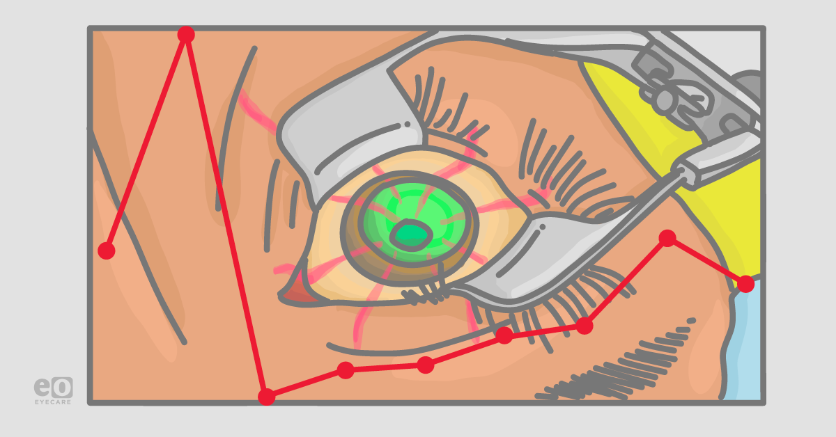
IOP elevations after cataract surgery
First 24 hours after surgery
In studies evaluating IOP in the hours after cataract surgery, significant IOP elevations were observed in the first 24 hours, and these typically normalized by postoperative day 1.1,2 Between 13 and 70% of eyes had a pressure exceeding 30mmHg in the first few hours after routine surgery depending on the study and viscoelastic device used.1,2
First few days after surgery
Of course, in routine clinical care, we rarely measure IOP in the first few hours after surgery, so many of these “spikes” may be missed. Thus, clinically IOP elevations after cataract surgery may be perceived as infrequent, particularly in otherwise healthy eyes.3,4
Weeks to months after surgery
In a study evaluating topical versus intracanalicular corticosteroids for postoperative inflammation after routine cataract surgery, three eyes of 262 had an elevated IOP ≥ 10mmHg above baseline, suggestive of steroid response.5
Even in glaucomatous eyes, IOP elevations in the first weeks are not expected. We can look to the trabecular stent pivotal trials for data in glaucomatous eyes. In the HORIZON trial evaluating the Hydrus microstent, for example, one eye in each group (Hydrus with cataract extraction versus cataract extraction alone) required a paracentesis in the second postoperative week for IOP elevation. Out to 24 months, 0.5% of microstent eyes and 2.7% of cataract surgery-only eyes had IOP elevations ≥ 10mmHg above baseline.6
Similarly, in the iStent Inject pivotal trial, 0.3% of stent eyes and 2.5% of cataract surgery-only eyes had an IOP elevation requiring oral or intravenous medications or surgical intervention beyond the 1-month mark, and 2.1% of stent eyes and 0.8% of cataract surgery only eyes experienced an IOP elevation ≥ 10mmHg above baseline beyond the 1-month mark.7
Causes of elevated IOP after cataract surgery
When eyes do develop post-operative IOP elevations, what are the causes? The table below highlights common and uncommon causes of these IOP elevations and associated risk factors.
Causes of Elevated IOP Post Cataract Surgery
| Common | Uncommon | |
|---|---|---|
| Post-operative Day 1 | Glaucoma Risidual ophthalmic viscoelastic device Inflammation | Vitreous or nuclear material from complex surgery Malignant glaucoma |
| Post-operative Week 1 | Inflammation Glaucoma Steroid response | Retained nuclear material |
| Post-operative Month 1 | Steroid response | Glaucoma Retained nuclear material |
As inferred by the observation of different rates of IOP elevation depending on the viscoelastic device used, retained viscoelastic is a likely offender for IOP spikes in the first 1-2 days after surgery;1,2 elevations from this cause should be truly transient. Additional risks may play a contributing role.
In a very recent retrospective study of 1587 eyes undergoing cataract extraction, male sex, high myopia (axial length ≥ 26.5 mm), a history of glaucoma treatment, poor pupillary dilation, intraoperative floppy iris syndrome, and baseline elevated IOP were risk factors for IOP elevation during the early post-operative period.4 Exfoliation syndrome, even in the absence of glaucoma, may also be a risk for elevated IOP in the earlier post-operative period.3 Additionally, shallower anterior chamber depth, thicker lenses, and shorter axial lengths may increase relative risk.8
By month one, steroid response is likely to play a significant part in observed IOP elevations, and in most patients, steroid taper or cessation is sufficient treatment. Thorough gonioscopy and a high suspicion index can help identify retained nuclear fragments that may rarely be the cause.
Download this flowchart of Causes for Elevated IOP Post Cataract Surgery!
Preventative measures
In eyes at high risk for IOP elevations or for whom IOP elevations would be particularly harmful, multiple studies have demonstrated the benefit of installation of a single IOP lowering eye drop after surgery.3,9 No clear guidance exists regarding the specific patient population who would most benefit from this treatment. In my practice, patients with fixation-threatening visual field loss are the ones I worry most about, and topical drops or even oral acetazolamide can be used during the first 24 hours to mitigate elevations.
Treatment
In glaucomatous eyes, their typical IOP lowering therapy regimen may be on hold after cataract extraction while they wear the patch and await the first post-operative visit. In these patients, resumption of their complete regimen will likely address the IOP elevation.
In patients naïve to therapy, one must decide whether treatment of the IOP elevation is warranted. Generally speaking, non-glaucomatous eyes can withstand a few days of elevated IOP without significant consequence. Still, the presence of an IOP elevation may motivate a shorter interval of follow up than would otherwise be expected in your practice.
If treatment is required, I typically consider a generic fixed-dose combination aqueous suppressant as my first line, given the outstanding efficacy, tolerance, and affordability during the brief post-operative period.
I rarely perform an aqueous paracentesis or wound “burp” for IOP elevations due to the risk of endophthalmitis. Still, I would consider this for IOPs > 40mmHg and unresponsive to oral acetazolamide and drops after 1-2 hours in the office or for eyes with fixation-threatening visual field loss at high risk for severe sequalae.
Baseline glaucoma testing
Glaucomatous eyes are particularly vulnerable to post-operative IOP elevations because of the already compromised outflow system in these eyes. Thus, a post-operative IOP elevation may be the first clue that an eye is a glaucoma suspect. For this reason, once healed from surgery, eyes that experienced a post-operative IOP elevation or a clear steroid response that was not previously under surveillance for glaucoma may benefit from baseline glaucoma testing including static perimetry and optical coherence tomography (OCT).
The benefit of baseline testing is to detect subtle glaucoma that may have gone unrecognized and set a baseline for higher-risk patients moving forward.
Conclusions
IOP elevations after cataract surgery are generally transient and more common in the first hours after surgery. Eyes with glaucoma are particularly vulnerable, and additional risk factors include axial myopia, poor pupillary dilation, elevated baseline IOP, and exfoliation syndrome, among other factors. IOP elevations may be blunted by the installation of a post-operative topical antihypertensive.
Some eyes may require treatment in the post-operative period by initiating or escalating topical anti-hypertensives. Consider baseline glaucoma testing in eyes that unexpectedly experienced post-operative IOP elevations and were not previously under evaluation for glaucoma suspect.
References
- Rainer G, Menapace R, Schmid KE, et al. Natural course of intraocular pressure after cataract surgery with sodium chondroitin sulfate 4%-sodium hyaluronate 3% (Viscoat). Ophthalmology. 2005;112(10):1714-1718. doi:10.1016/j.ophtha.2005.05.011
- Rainer G, Schmid KE, Findl O, et al. Natural course of intraocular pressure after cataract surgery with sodium hyaluronate 1% versus hydroxypropylmethylcellulose 2%. Ophthalmology. 2007;114(6):1089-1093. doi:10.1016/j.ophtha.2006.08.048
- Levkovitch-Verbin H, Habot-Wilner Z, Burla N, et al. Intraocular pressure elevation within the first 24 hours after cataract surgery in patients with glaucoma or exfoliation syndrome. Ophthalmology. 2008;115(1):104-108. doi:10.1016/j.ophtha.2007.03.058
- Oku H, Mori K, Watanabe M, et al. Risk factors for intraocular pressure elevation during the early period post cataract surgery. Jpn J Ophthalmol. 2022;66(4):373-378. doi:10.1007/s10384-022-00918-z
- Lu AQ, Rizk M, O’Rourke T, et al. Safety and Efficacy of Topical versus Intracanalicular Corticosteroids for the Prevention of Postoperative Inflammation after Cataract Surgery. J Cataract Refract Surg. Published online May 9, 2022. doi:10.1097/j.jcrs.0000000000000963
- Samuelson TW, Chang DF, Marquis R, et al. A Schlemm Canal Microstent for Intraocular Pressure Reduction in Primary Open-Angle Glaucoma and Cataract: The HORIZON Study. Ophthalmology. 2019;126(1):29-37. doi:10.1016/j.ophtha.2018.05.012
- Samuelson TW, Sarkisian SR, Lubeck DM, et al. Prospective, Randomized, Controlled Pivotal Trial of an Ab Interno Implanted Trabecular Micro-Bypass in Primary Open-Angle Glaucoma and Cataract: Two-Year Results. Ophthalmology. 2019;126(6):811-821. doi:10.1016/j.ophtha.2019.03.006
- Sanchez FG, Rees J, Gardiner SK, Mansberger SL. Biometric parameters can predict IOP change and IOP spikes after cataract surgery in glaucoma patients. ARVO Annual Meeting, June 2021.
- Barak A, Desatnik H, Ma-Naim T, Ashkenasi I, Neufeld A, Melamed S. Early postoperative intraocular pressure pattern in glaucomatous and nonglaucomatous patients. J Cataract Refract Surg. 1996;22(5):607-611. doi:10.1016/s0886-3350(96)80018-6
