


Dry eye disease (DED), as defined by TFOS DEWS II, “is a multifactorial disease of the ocular surface characterized by a loss of homeostasis of the tear film, and accompanied by ocular symptoms, in which tear film instability and hyperosmolarity, ocular surface inflammation and damage, and neurosensory abnormalities play etiological roles.”1
Up to 35% of all adults suffer from DED, yet the incidence of DED in cataract surgery patients tends to be higher than the general population. Studies by Trattler et al. and Gupta et al. have shown that nearly 50-70% of patients presenting for cataract surgery can have signs of DED on clinical examination and testing. Surprisingly, almost half of these patients are unaware that they have an underlying condition.2,3
Ocular surface disease, particularly DED, can significantly impact measurements used for cataract surgery. Errors in keratometry and biometry can lead to inaccurate intraocular lens (IOL) power selection and ultimately unexpected and unwanted postoperative refractive errors. This can be particularly problematic in patients receiving premium IOL technology, such as toric, trifocal, or extended depth of focus IOLs, where small degrees of refractive inaccuracy can significantly degrade the quality of vision.
Epitropoulous et al. found that patients with hyperosmolar ocular surfaces, often seen in patients with DED, had significant variability in keratometry readings and astigmatism measurements compared with patients without hyperosmolarity.4
How I approach the ocular surface before cataract surgery
I am rigorous about evaluating and treating the ocular surface before cataract surgery in my clinical practice. Before cataract surgery pre-operative evaluations, patients are instructed by my office staff to remain out of contact lenses for at least one week. In patients with long-term, rigid gas permeable (RGP) contact lens use, I often recommended a contact lens holiday for a week per decade of use (i.e., a five-week holiday for a patient who has used RGPs for 50 years).
“All patients obtain in-office meibography, topography, and biometry before eye drops.”
I critically evaluate all imaging and measurements, looking for areas of data dropout or disruption of Placido disc mires on corneal scans, inconsistencies in keratometry, axial length and astigmatism measurements, and any evidence of meibomian gland architecture disruption on meibography.
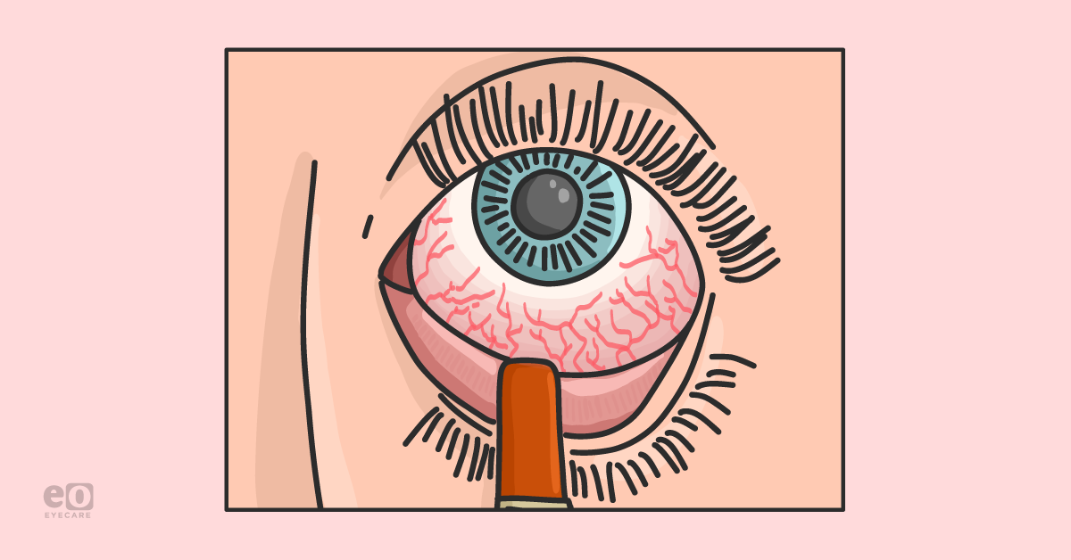
A fluorescein strip is the simplest and most cost-effective in-office test that I use to evaluate DED.
After placing one drop of saline or proparacaine solution on the tip of the strip, I dab fluorescein into the patient’s inferior fornix and allow that small amount of fluorescein to highlight changes on the ocular surface. I prefer using the strip to a fluorescein drop to avoid bathing the entire ocular surface in stain and missing subtle irregularities. I can easily see low tear lakes, rapid tear break-up times, slight conjunctival and corneal staining, and even surface pathology such as anterior basement membrane dystrophy or Salzmann’s nodular degeneration.
I am also pushing, pulling, and everting the upper and lower eyelids to assess meibomian gland dysfunction or eyelid or conjunctival lesions. In patients I suspect neurotrophic corneal disease, I am checking corneal sensation and performing a thorough review of systems to identify any risk factors for neurotrophic keratitis such as prior herpetic disease, medication toxicity, or previous neurological surgery. The American Society of Cataract and Refractive Surgery (ASCRS) has developed a comprehensive algorithm to evaluate ocular surface disease that I recommend surgeons refer to as they begin performing their DED assessments.5
Patient communication is the first step in treatment
Pictures speak a thousand words. I show my patients their topographic scans, pointing out areas of mire dropout or meibomian gland atrophy or tortuosity, and explain to them how necessary the treatment of DED is before pursuing cataract surgery.
“My treatment approach for DED before cataract surgery is multi-pronged.”
I typically start a short course of topical steroids for two weeks to quiet down the ocular surface. In patients with significant aqueous deficiency DED, I also recommend topical prescription therapies such as cyclosporine 0.05% or 0.09% or lifitegrast to increase tear production. I employ punctal occlusion, either with dissolvable or permanent plugs, in the bilateral lower eyelids and upper eyelids depending on the severity of keratopathy. If I see significant meibomian gland dysfunction and concomitant evaporative dry eye, I encourage patients to pursue thermal pulsation therapies to improve the function of their meibomian glands.
In patients with autoimmune-related DED, I advocate for amniotic membrane therapy, autologous serum tears, and punctal cautery to optimize the ocular surface further.
After 4 to 6 weeks of treatment, I re-image patients and compare their pre-operative and post-treatment biometry and keratometry. I have seen nearly 1 to 2 diopter changes in the proposed IOL power and even increases and decreases in the recommended toricity of an IOL after ocular surface optimization. I will repeat imaging until I have two sets of measurements that are consistent and stable.
In cases where I am implanting premium IOL technologies, I will also use intra-operative aberrometry to confirm my IOL power choice further. Scleral or PROSE lenses may be needed in patients whose DED is recalcitrant despite weeks of therapy, and I often counsel these patients that they may not be ideal candidates for premium IOL technology and collaborate with my optometric colleagues to improve their ocular surface.
Caring for the ocular surface continues after surgery
Treatment of DED before cataract surgery requires patience from the surgeon and the patient. For best outcomes, time must ensure that the highest quality data is utilized to determine a patient’s IOL choice. I always counsel my patients that their treatment for DED cannot stop after cataract surgery but must also continue in the post-operative period to ensure they maintain an excellent ocular surface and, thereby, excellent quality of vision.
References
- Craig JP, Nichols KK, Akpek EK, et al. TFOS DEWS II Definition and Classification Report. Ocul Surf. 2017;15(3):276-283.
- Gupta PK, Drinkwater OJ, VanDusen KW, Brissette AR, Starr CE. Prevalence of ocular surface dysfunction in patients presenting for cataract surgery evaluation. J Cataract Refract Surg. 2018;44(9):1090-1096.
- Trattler WB, Majmudar PA, Donnenfeld ED, McDonald MB, Stonecipher KG, Goldberg DF. The Prospective Health Assessment of Cataract Patients' Ocular Surface (PHACO) study: the effect of dry eye. Clin Ophthalmol. 2017;11:1423-1430.
- Epitropoulos AT, Matossian C, Berdy GJ, Malhotra RP, Potvin R. Effect of tear osmolarity on repeatability of keratometry for cataract surgery planning. Journal of Cataract & Refractive Surgery. 2015;41(8).
- Starr CE, Gupta PK, Farid M, et al. An algorithm for the preoperative diagnosis and treatment of ocular surface disorders. Journal of Cataract & Refractive Surgery. 2019;45(5).
