


Diabetic retinopathy is the most common cause of vision loss among working-age adults. Despite continued advancements in diagnosis and management, the prevalence of diabetes in our population continues to rise each year, as does the number of patients with diabetic retinopathy. Early diabetic changes can be asymptomatic, and by the time a patient presents for an eye exam, they may have irreversible damage.
This is a complex issue with many facets, such as poor education, access to healthcare, and loss to follow-up (LTFU). In our experiences in public healthcare and academic medicine, we have cared for countless patients who lost eyesight. This article will focus on changes we can make during the examination, treatment, and follow-up to improve outcomes and decrease rates of LTFU.
Overview of diabetic retinopathy
The Diabetic Retinopathy Severity Scale nicely demonstrates the broad spectrum of diabetic retinal disease. While all patients start with visually insignificant early disease, many will progress at some point. They may only present to their eyecare provider once their condition is advanced and irreversible.
Non-proliferative diabetic retinopathy (NPDR) comprises mild, moderate, and severe disease. Later phases of NPDR may need treatment with laser or intravitreal injections.
Proliferative diabetic retinopathy (PDR) is described as high-risk and non-high-risk and is associated with a higher risk of vision loss and disability. These patients nearly always require treatment. Additional associated complications with later-stage diabetic retinopathy include neovascular glaucoma (NVG) and tractional retinal detachments (TRD) which may require complex surgery.
Diabetic macular edema can present at nearly any stage of the disease and often requires treatment.
Reducing diabetic patient no-shows: the trouble with follow-up
It can be challenging for patients with diabetes to keep up with their eye appointments for several reasons.
They may need to be seen as frequently as once a month. If young and employed, it is difficult to take off significant amounts of time during the workweek. Financially, that lost time off work can add up. Even with insurance, the cost of co-pays, continued travel to appointments, childcare, etc. can be challenging.
Regular visits can be overwhelming, and patients may have other medical appointments to manage diabetic complications, such as nephropathy and neuropathy. Many are also undergoing dialysis, which is usually multiple times per week.
The clinic itself can also be intimidating, especially if the patient requires treatment. Understandably, intravitreal injections sound scary. People picture something out of A Clockwork Orange, but that couldn’t be farther from the truth. The reality is intravitreal injections are very common and essentially pain-free.
People also fear laser procedures or surgery. If they’re asymptomatic, it’s hard to appreciate the risk of blindness associated with skipping visits. Many patients seeing “fine” at the moment may find it hard to accept that they have a condition where blindness is imminent. Patients may also lose or change insurance and get lost in the shuffle of this transition
Unfortunately, for many of these reasons, the LTFU rate in the diabetic population continues to be a problem. It results in many patients losing vision that may have otherwise been saved.
Decreasing diabetic patient no-shows starts with your front office
One of the areas of your clinic that can improve LTFU rates is the front office. Monitoring patients is critical, and an appointment recall system can be beneficial. Better yet, ensure your patients leave with their scheduled appointment and a copy of the date and time handed to the patient.
Text messages, email reminders, and phone calls are very effective in decreasing patient no-shows. These can improve patient retention and compliance with follow-up visits. Reiterate the importance of their visit (i.e., monitoring versus treatment) and get them rescheduled.
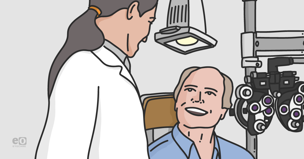
The patient diagnostic experience
History-taking is the most critical time to identify patient barriers to healthcare and follow up. Establish rapport with your patients right from the start. Patients are often not eager to share where they have fallen short in taking care of themselves. They often report lower blood sugars than their actual numbers or a lower HbA1c.
If you can communicate without judgment, patients are more likely to be honest and demonstrate accountability. Understanding what motivates them in life will better equip you to empower them to make positive changes.
Moreover, connect them with resources in the community, like diabetes educators or a diabetes support group at a local hospital, where they may be able to communicate with others to have accountability and motivation.
Educate yourself on tips that can help patients. Discuss how positive changes in diet and exercise can make a big difference. Maintaining a healthy diet and lifestyle can help improve and maintain optimal health levels.
Additionally, if patients are struggling financially, connect them with support programs and social workers to improve healthcare access.
Engage your patients about their diabetes. Ask the following questions:
- How often do you check your blood sugar?
- What was your last blood sugar reading?
- What was your last A1c; when was it taken?
- Who is your PCP?
- What medications do you take for your diabetes?
- Were any medication changes made at your previous visit?
- What is your biggest challenge in managing your blood sugar: Sugar cravings? Life stress? Shift work or other causes of disrupted sleep?
Dilated fundus exam
Most people do not like dilated exams, but this is an essential part of checking a patient’s eyes for signs of diabetic retinopathy. Early disease identification acts as a warning sign that their diabetes may be poorly controlled and provides an opportunity to intervene before irreversible damage takes place.
Explain that their primary care provider has specifically requested the dilated portion of the exam and that you need to do this test to provide them with an accurate report. The additional support of their PCP will make them less apprehensive of a dilated exam. Have patients expect that their eyes will be dilated at each diabetic exam so they can bring a driver if needed.
Optical Coherence Tomography (OCT)
OCT is a quick and painless way to obtain a volumetric scan of the patient’s macula to assess for diabetic macular edema and retinal ischemia. Ensure the proper height of the equipment so patients with neck or back troubles are not uncomfortable during testing. Have a comfortable and adjustable chair for the patient to sit in during image acquisition.
Fluorescein Angiography (FA)
Fluorescein angiography (FA) is an important diagnostic tool for assessing vascular integrity and perfusion in the retina. FA can evaluate the extent of diabetic disease and guide treatment. It can also be intimidating to patients as it is one of our more invasive testing modalities and involves injecting an intravenous dye.
Thorough education, preparation, and technique can make the experience much easier on patients.
Potential risks of FA
- Nausea & vomiting: Patients should be aware of the risk of nausea and vomiting, which can happen in around 1-5% of people. If someone is complaining of nausea, an ice pack to the back of their neck can improve symptoms. Using a lower concentration or volume of dye and injecting the dye more slowly may decrease the risk of nausea. Have a receptacle and tissues nearby if the patient requires it.
- It typically happens and passes quickly, with the patient feeling normal within a few minutes.
- Fainting: People may feel faint and have a vasovagal reaction, especially in the presence of a needle. Watch the patient closely so they don’t fall and encourage them to communicate if anything feels abnormal. Sometimes laying people flat on their back with their legs raised can increase blood pressure and decrease symptoms. Have a blood pressure cuff nearby to monitor the BP.
- Typically, patients will improve quickly.
- Pain: Using small-gauge butterfly needles can make the injection nearly pain-free. Take time to identify the best venipuncture site to avoid multiple attempts. We typically try to locate a vein in the antecubital fossa as sticks are often less painful.
- Bruising: Cosmesis is necessary, and patients dislike leaving their visit with extensive bruising. Using smaller needles, finding a good vein, avoiding multiple sticks, and applying immediate pressure and bandage to the injection site can make a big difference. If patients are on blood thinners, apply pressure for longer than usual.
- Dye extravasation: Fluorescein dye can be painful if extravasation occurs. Patients can have localized burning and discomfort. Confirm you've cannulated a vein by drawing back blood before injecting. Have an icepack nearby to apply pressure to the injection site if extravasation occurs.
Aftercare
Be sure to remind patients that their skin may have an orange/yellowish tint, as will their urine, for 24-48 hours as the dye clears from the body.
Treating your patients with diabetic retinopathy
Intravitreal injections
Although ubiquitous, intravitreal injections (IVI) are a significant source of anxiety for patients. Understandably, the concept of a needle in the eye is unsettling, and injections can significantly contribute to LTFU rates.
As physicians, we can do several things to make injections less intimidating. Start with a discussion about the procedure and what to expect. While there are varying approaches to numbing, I prefer 0.1cc or less of subconjunctival lidocaine, which provides complete anesthesia. Small-gauge needles between 32g-34g also allow for minor discomfort. Subconjunctival hemorrhage (SCH) is painless but still bothersome in cosmesis.
Some people do not like the feeling of an eyelid speculum holding their lids open. Using narrower eyelid speculums or manually keeping the eyelid open can be much more comfortable. Betadine is the most significant source of discomfort with IVI secondary to corneal epitheliopathy. The pain often develops shortly after the injection and can last 12-24 hours.
Additionally, I encourage patients to use artificial tears frequently to help with discomfort.
Panretinal Photocoagulation (PRP)
Panretinal photocoagulation (PRP) is routinely deployed to manage proliferative diabetic retinopathy. With appropriate anesthesia, PRP can be well tolerated. Additionally, staging laser over multiple PRP sessions can make the process much more tolerable for patients.
Surgery
Every surgical signup is different, and taking time preoperatively to address risks, expectations, questions, and outcomes is essential. Sometimes the fear of surgery can be so significant that patients postpone care and have worse outcomes. Communication and education throughout the process are critical tools to ease apprehension.
Detailed post-operative instructions and expectations will reduce perioperative anxiety.
Patient communication: the keystone of successful treatment
The most impactful thing that can be done to improve patient follow-up and outcomes is education. It’s hard for patients to appreciate the potential complications of uncontrolled diabetes when they feel fine. Early management of diabetes can have a massive impact on long-term outcomes. Engaging and empowering patients to take good care of themselves early in the disease process is extremely important.
PEARL: Educate, educate, educate. Get patients involved in understanding diabetes and diabetic retinopathy, the importance of treatment, and the dangers of missing appointments. Their participation is crucial to good long-term health.
Co-managing diabetic retinopathy
Diabetic patients do well with combined optometric and ophthalmic co-management. Working closely with the allied eye health practitioners in conjunction with the patient's PCP minimizes LTFU.
Conclusions
To help preserve our patients’ sight, we need to help them understand the importance of annual dilated eye exams and urgent exams with sudden vision changes.
Additionally, patients must be encouraged to become active participants in their treatment and lifestyle programs to manage their systemic disease successfully. If treatment is needed, the patient must understand the importance of continued follow-up, as sometimes their ocular disease can worsen if treatment is not continued.
