


If you perform enough surgeries, you will likely encounter some very tough cases—and some do not always go as planned.
This article will highlight three common complications in cataract surgery, how to preoperatively identify risk factors, and how to manage each as smoothly as possible.
3 complications associated with cataract surgery
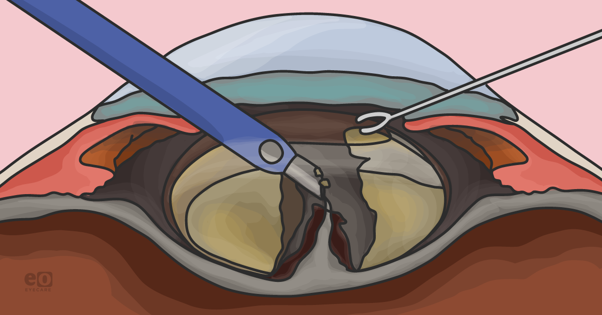
1. Posterior capsule rupture
While some cases of posterior capsule rupture (PCR) may be iatrogenic, it is essential to note that patients with pre-existing trauma (even an aberrant needle from an anti-vascular endothelial growth factor [VEGF] injection grazing the posterior capsule counts!), posterior polar cataracts, and those with very dense lenses tend to be at higher risk.1
Extra care should be taken before starting the case (including consenting the patient regarding their higher risk of vitrectomy and even inability to place a lens at the time of surgery). The operating room (OR) team (nurses, scrub techs) should also be aware of the possible need for vitrectomy so that they are prepared to assist with instrument assembly, etc.
Pre- and intraoperative pearls for treating PCR
Prior to starting the case, the surgeon should assess pupillary dilation, zonular status, and if all appropriate machinery and instruments are available in your OR to handle a possible PCR. Pupillary stretching devices such as iris hooks and rings, capsular tension rings or hooks, and Trypan blue (ThermoFischer Scientific) to help stain the anterior capsule can be incredibly helpful. In addition, extra viscoelastic, which may be used to push vitreous back, viscodissect the lens nucleus, and float nuclear fragments up and away from a posterior capsular tear can be especially helpful, as well.
Unfortunately, even if all steps of a case go as planned, in eyes with preexisting posterior capsule compromise, the surgeon must be prepared (to the best of their ability) to remove all cataract material, place a lens and perform a vitrectomy, if necessary.
If a posterior capsule tear is noted at the time of surgery—DO NOT PANIC—and try your best to keep your instruments where they are, with infusion still running from the phaco probe.1 Removing instruments abruptly from the eye in the presence of a PCR causes a change in pressure gradient and can cause the vitreous to prolapse anteriorly, creating vitreous traction and possibly subsequent retinal detachment.1
With infusion running, the surgeon may instill viscoelastic through the paracentesis incision (preferably dispersive, but cohesive may also be used) before removing the phaco probe.1 Some surgeons may inject triamcinolone into the anterior chamber to help stain any vitreous, if present.
The main treatment goals here are to:
- Acknowledge there is a problem (the posterior capsule rupture).
- Identify what needs to be done (e.g., is there remaining nuclear/cortical material? Is there vitreous entangled in the remaining cataract? Is there enough support to insert an intraocular lens (IOL) or even attempt anterior chamber IOL [ACIOL] implantation?)
- Execute the remainder of the surgery as safely and efficiently as possible (yes—even if that means not implanting a lens or cleaning up as best as possible and referring to your retina colleague for further management).
When to perform an anterior vitrectomy
If vitreous is present in the anterior chamber and/or prolapsing from the wound, this should be handled by performing a thorough anterior vitrectomy. A Weck-cell sponge may also be gently touched to the wound, and scissors should be used to excise any prolapsing vitreous outside the eye (a “Weck-cell vitrectomy”).
If there is remaining lens material, the surgeon may need to alternate between utilizing the phaco/I/A probe and vitrectomy instruments to effectively remove the lens material and vitreous.1 Alternatively, some surgeons have extensive experience and comfort with performing pars plana vitrectomy—in this situation, the vitreous can be removed from a posterior approach—some argue this may allow for less traction on the vitreous during the case, as it is not being pulled anteriorly.
Evaluating the capsular bag for IOL implantation
Once the lens material and vitreous (if present) have been sufficiently removed, the status of the capsular bag must be assessed. If there is only a small round rent in the posterior capsule, one may attempt to insert an intraocular lens into the capsular bag—be aware this comes with a risk of opening the tear in the capsule further and losing the IOL to the posterior segment.
If sufficient anterior capsule support is present, insert a 3-piece IOL in the sulcus. If the status of the capsular bag is very poor, and therefore no support may be offered for an IOL in-the-bag or in the sulcus, the surgeon may consider closing and coming back later for a scleral-fixated or iris-sutured IOL.
“Alternatively, one may consider implanting an ACIOL at the time of surgery.”
Using a miotic, such as Miochol (Bausch + Lomb), at the end of the case is helpful to detect a peaked pupil (which could denote vitreous traction to a wound and will allow for placement of an ACIOL if indicated. All wounds should be meticulously checked for vitreous presence, as well as wound leaks. If there is any concern for leaking incisions, a suture should be placed to ensure the stability of the globe.
Once the surgery is complete, it is important to document the events that occurred in your operative note and if the planned IOL could not be implanted (or any IOL, for that matter). If a retinal consult and/or more surgery is warranted, it is imperative to discuss this with the patient and plan for the next step(s).
2. Corneal edema
Despite significant advances in phacoemulsification instrumentation, femtosecond laser-assisted cataract surgery (FLACS), and advanced viscoelastics, corneal edema can still occur with modern cataract surgery. Fortunately, most cases are transient and mild, resolving independently within a few days. However, some cases may require treatment—both medical and/or surgical. It is important to identify eyes at high risk for recalcitrant corneal edema, as well as how to appropriately counsel patients and treat this condition.
Often, patients with very dense lenses (thereby requiring high levels of ultrasound during phacoemulsification) are at risk for increased corneal edema postoperatively.2-4 These patients may benefit from FLACS, as well as certain phacoemulsification techniques that do not utilize as much ultrasound energy (e.g., a chopping technique vs. divide and conquer).
Intraoperative pearls for treating corneal edema
Unfortunately, iatrogenic injury to the cornea may occur during surgery, such as damage to the endothelium due to instrumentation or accidental scrolling or detachment of Descemet’s membrane during insertion and removal of the phaco probe.2-4 These cases can often be managed medically. However, there have been some successful cases documenting the use of an air bubble to help reattach a large area of detached Descemet’s membrane (much in the same way air is used in endothelial keratoplasty cases).2-4
Patients with preexisting corneal weakness—such as those with endothelial dystrophy (e.g., Fuch's endothelial corneal dystrophy and posterior polymorphous dystrophy) and those post-keratoplasty (both endothelial and penetrating) are at higher risk of endothelial dysfunction and corneal edema postoperatively.2-4 These patients must be counseled regarding the risk of corneal swelling following cataract surgery, including the possible need for topical medications (in addition to their routine postoperative care) and even surgery.
Follow-up care for corneal edema
Typically, if corneal edema is not clearing adequately within a few days, surgeons may recommend using hypertonic saline drops (sodium chloride 2% or 5%) two to four times per day and ointment at night. Often, this will help to clear the edema completely, although sometimes, an additional topical steroid is required.
Should several months pass without adequate clearing of corneal edema, the patient should be referred to a cornea specialist to discuss a corneal transplant. Many of these patients are excellent candidates for endothelial keratoplasty (Descemet endothelial membrane keratoplasty [DMEK] and Descemet stripping automated endothelial keratoplasty [DSAEK]), and can do incredibly well following this surgery.
3. Iris trauma
Try we as might, we can’t blame every case of iris trauma on Flomax (Tamsulosin, Sanofi)—or its related compounds. Though floppy iris syndrome can lead to iatrogenic iris injury at the time of surgery, other causes of iris damage can occur before, during, and after phacoemulsification—and that being said, some patients on Flomax, or other medications in that class, have no evidence of floppy iris syndrome whatsoever.
Small pupils
Cataract surgery among eyes with small pupils is a known risk factor for complications both intraoperatively and postoperatively.5-7 Though pupil stretching devices are very helpful for these eyes (ex., hooks and rings), they may be associated with iris sphincter tears and even iris disinsertion in less experienced hands (as well as resultant bleeding in the anterior chamber).5-7
Surgeons should take their time while utilizing these devices to avoid iatrogenic iris injury, hyphema, and issues with IOP postoperatively.
Iris prolapse
The most common cause of iris prolapse is due to a difference (higher) in pressure gradient posterior to the iris versus anterior—this may be due to overaggressive hydrodissection (trapped saline behind the lens), trapped viscoelastic behind the lens/IOL, and intraoperative floppy iris syndrome (IFIS).5-7
Additionally, iris prolapse may result from hydrated vitreous secondary to a posterior capsule tear or balanced saline solution (BSS) getting pushed through the zonules, a poorly constructed or leaking incision, patient moving/coughing/sneezing during surgery, a suprachoroidal hemorrhage, as well as a shallow anterior chamber.5-7
Treating and preventing iris prolapse
While treating iris prolapse, forcefully pushing the iris back into the eye is not the best strategy. This can lead to iris tears, bleeding, and more iris tissue prolapsing out of the wound (especially if manipulation is done through the main incision).5-7
It is important for the surgeon to identify the cause of iris prolapse and treat it—e.g., if there is trapped BSS behind the lens, gently rocking or tapping on the lens can release the BSS and create a more normal pressure gradient.5-7 Also, instilling a small amount of viscoelastic through the paracentesis incision (over the iris) before inserting any instrument may allow for more gentle manipulation of the ocular tissue(s).5-7
Prevention of iris prolapse is key—although iris prolapse does not usually cause significant postoperative visual problems, excessive manipulation can lead to iris damage, holes, atrophy, and chronic visual disturbances (glare, monocular diplopia, among others).5-7 Secure, well-constructed wounds are the first steps in preventing iris prolapse (and wound leaks, for that matter); surgeons should avoid over-inflating the eye during hydrodissection and even during instillation of viscoelastic prior to IOL implantation.5-7
If prolapse occurs after phaco, hydrating the primary and paracentesis incisions may decrease wound leakage.5-7 The surgeon may also adjust their fluidics—decreasing flow rate and vacuum can minimize the pressure gradient.5-7
Conclusion
Thankfully, most cataract surgeries are straightforward and provide our patients with improved vision and quality of life. It is essential to identify patient and ocular risk factors for complications during surgery, as well as be able to discuss these with patients and their families.
Having a plan and knowing the correct steps during tough cases will allow your patients to have good visual outcomes and maintain their trust in your care.
