


Meet the OCT Queen
Full disclosure, if you ask my Athens staff about me, they’d tell you I was the OCT queen. And I wouldn’t deny it.
Optical Coherence Tomography (OCT) is an incredibly useful, non-invasive imaging tool that helps us pinpoint a diagnosis or shed light on management strategy. With this technology, infrared light records the path of scattered photons; this in turn provides high resolution cross-sections of ocular structures. OCT scans give us a detailed anatomical understanding of numerous ocular pathophysiologies. Review ocular conditions with their corresponding OCT structures here.

As optometric technology evolves, we explore how we can better utilize our OCT practices to maximize our patient care.
Current OCT Protocols for Ocular Diseases
Distinguishing anatomical variations in retinal layers or abnormal thickness values can enhance monitoring and screening measures of various ocular diseases. OCT testing plays an instrumental role by highlighting structural components and quantifying irregularities.
Prepare your OCT analysis knowledge by brushing up on differential diagnoses with our OCT Retinal Bootcamp Course.
After successfully spotting and diagnosing an ocular condition, tracking progression is integral to optimize our patient’s visual and pathological outcomes.
Screen, Diagnose, and Monitor

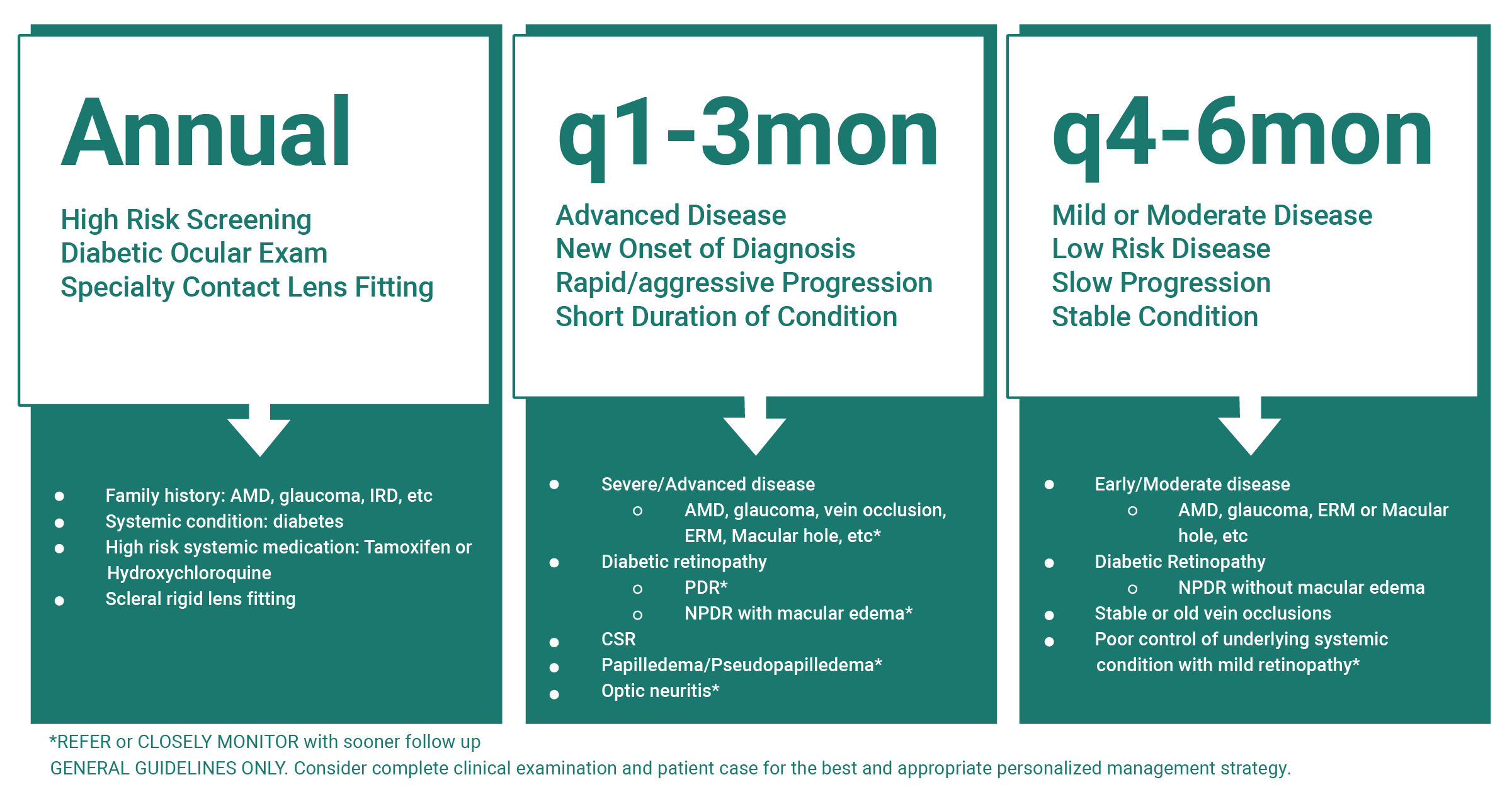
With your clinical assessment, performing sequential OCT imaging tests when indicated can generate an in-depth evaluation for respective management. It provides ease of classification, helps identify severity and pick up concomitant pathology, and discern factors of progression.
Disease Tracking Tips
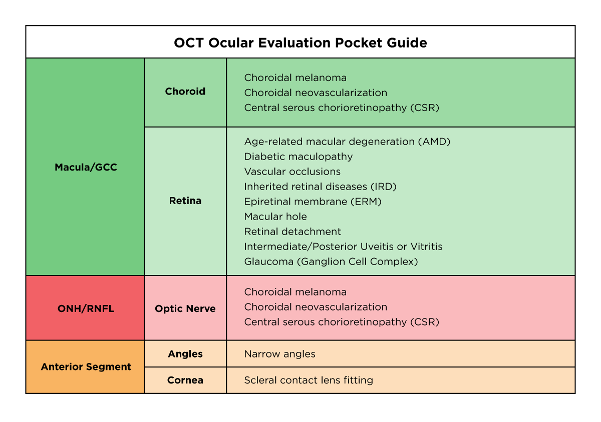
OCT-Macula
It is evident that monitoring progression with OCT analysis will help you identify any detrimental progression—certainly for serious conditions that cause permanent vision loss. In AMD, be vigilant for the accumulation of retinal fluid, and CNVM. For diabetes, also be vigilant for the accumulation of retinal fluid as well as macular edema.
With a macular cube, minor but clinically significant changes can be noted as well. Macular pucker distortions, macular hole stages, and vitreo-macular adhesions can be distinctly visualized and monitored.
In fact, OCT imaging is more affluent in uveitis cases, proving beneficial in monitoring of Vogt-Koyanagi-Harada disease, Behçet’s disease, and birdshot chorioretinopathy.2
Interestingly, as pictured below in Figure 1, I had an 11-year-old female pediatric case with BVCA 20/25- OU. Her irregular macula appearances caused me to investigate with dilation and OCT macula. She was ultimately diagnosed with primary iridocylcitic and intermediate uveitis with CME.

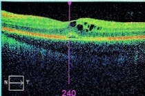
Figure 1
OCT-ONH and RNFL
We know that for glaucoma patients, an optic nerve head (ONH) and retinal nerve fiber layer (RNFL) analysis is essential for careful management by tracking serial measurements. Performing a GCC scan in glaucoma suspects and early disease is foundational to note any preliminary changes. We can also use these high resolution images to consider other optic nerve anomalies such as optic neuritis, papilledema, and pseudopapilledema.
Discerning the abnormal structural contour in the right eye of this 50yo female with an OCT-ONH provided corroborating optic disc edema evidence. A relatively homogenous elevation and slight “lazy V” pattern in the photoreceptor and RPE layer can be seen in Figure 2. With the clinical exam, I suspected optic neuritis—which soon after led to a confirmation in multiple sclerosis diagnosis.


Figure 2
OCT-Anterior Seg
Better and more precise specialty contact lens fitting of sclerals can be done with the OCT anterior segment add-on by quantifying and visualizing optimal clearance and landing zones. This course provides in-depth ocular disease indications for use of this specialty lens.
Refer and co-manage
Identifying severe disease or one that requires immediate attention will lead to urgent referrals. A questionable choroidal structure can be scanned to uncover a mass that is suspicious for melanoma. This integral finding may potentially confirm a possibly alarming diagnosis, especially if malignant.
Although careful clinical assessment of retinal detachments are necessary, a quick OCT scan can help if the status of macula-on or macula-off seems questionable. In addition, a patient case that presents as possible retinitis pigmentosa can benefit from providing corroborating evidence of a missing or severely abnormal photoreceptor layer.
Suspicion for narrow angles can be precisely viewed with anterior segment OCT; then it can be referred for an appropriate LPI procedure.
Guidelines to utilizing OCT for COVID complications
It’s well known that conjunctivitis and keratitis can result in COVID patients. However many more ophthalmic associations have been reported, especially in those with severe disease and extensive systemic comorbidities.
OCT-Macula
Posterior segment manifestations include a variety of conditions, occurring about 12 days after diagnosis.1 Among the common findings of retinal hemorrhages, exudates, and cotton wool spots are vascular deformities, macular defects, and neuro-ophthalmic alterations—some leading to permanent vision loss.
COVID links to cerebrovascular accidents or cerebral venous thrombosis that can trigger acute vision loss. In immunocompromised patients, acute retinal necrosis presentations have increased risk. Retinal vascular occlusions can indicate a macula OCT to detect edematous findings in CRVO or BRVOs. Central retinal artery occlusions have been reported as well, where an OCT would highlight outer retinal layer and inner nuclear layer hyperreflectivity.
Additional macula OCT findings can be localized with COVID-associated acute macular neuroretinopathy or macular neuroretinitis. These would display hyperreflective outer retinal disruptions and lesions as well as possible neurosensory detachments.
Infectious and inflammatory cascades have led to substantial vision threatening complications such as endophthalmitis, vitritis, and reactivation of serpiginous choroiditis. For instance, we can take advantage of discovering vitreous quality using OCT by appreciating hyperreflective vitreous cells.
Neuro-ophthalmic consequences of COVID
OCT-ONH and RNFL
Neurology related alterations in COVID patients have a broad and extensive presence, appearing in more than one-third of cases around day 5 after symptoms present.1 Consider a visual field and OCT when indications point to neuro-ophthalmic findings such as optic neuritis, papilledema, and pseudopapilledema.
Virus-caused mechanisms also instigate autoimmune disorders such as Guillain-Barre syndrome and associated Miller-Fisher syndrome. Cranial nerve palsies incidents have occurred, most commonly affecting cranial nerve 6.
The intricate workings of COVID present abnormal coagulation factors, complex inflammatory reactions, and pathways that are still unknown. Careful consideration of reported manifestations and investigation with clinical exam and OCT testing can provide insight and guidance on these novel cases.
OCT angiography versus fluorescein angiography
Fluorescein angiography is a diagnostic test that reconstructs the circulation of blood flow in the retina and choroid by fluorescent dye injection. OCT Angiography (OCT-A) is the non-invasive counterpart that takes sequential scans of blood vessel movements based on light reflectance.
With OCT-A, the advancement in technology and procedural simplicity displays signs of retinal edema, microaneurysms, microvascular abnormalities, or perfusion deficiencies. Appropriate screening of progressive or severe AMD with OCT-A can highlight measurable structural impairments, whether drusen, geographic atrophy or leakage.
Diabetic OCT-A protocol considerations
In diabetics, early detection OCT-A testing is valuable with preclinical changes and unexplained visual acuity deterioration--typically due to ischemic correlations of enlarged avascular zones and significant capillary dropout. Early proliferative diabetic retinopathy is readily distinguished by notable retinal blood vessel growths. In addition, differentiating types of vein occlusions can become more apparent based on the area of non-perfusion.
This is an exceptional OCT modality that we can conduct for managing wet and dry AMD, monitoring diabetic retinopathy progression, retinal vascular complications, and ischemic conditions such as giant cell arteritis and ocular ischemic syndrome.
Although fluorescein angiography is the gold standard for accuracy of these retinal microvascular changes, OCT-A does define minute vascular inconsistencies and otherwise inconspicuous telangiectasia or subtle neovascularization. Here is a course to assist visualization of ocular pathologies on OCT-A.
Presenting OCT testing objectives
Clinical applications of OCT are endless. Be intentional about why performing it is necessary. Develop an understanding with appropriate patient education and discussion of diagnosis and findings.
Patient script
Introduce what OCT is and why it benefits the patient: “This instrument takes a scan right through the macula and optic nerve. The photo will show all detailed layers and will provide critical information for me to accurately diagnose and manage your conditions. Not only is this a proactive measure, but it will help me provide the best care to help reduce progression rate, delay damage, and subsequent vision loss.”
Sharing these advantages sheds light on the significance of imaging technology. Discuss that baseline testing for suspected disease patterns or family history can identify elements that can indicate disease markers. It is also important to educate patients about appropriate follow up schedules on OCTs to monitor the severity or progression of their condition. Stress that these are measures that will lead to better prognosis and treatment options.
Now, why was I the OCT Queen?
Because I recognized the power of this tool.
OCT provides for:
- Early detection of a myriad of ocular conditions.
- Baseline testing for suspicion or associated family history.
- Careful monitoring of progressive eye diseases.
- Forming the best management strategy and referral timeline.
Utilizing the various facets that it offers, OCT technology is invaluable for eye care physicians. Performing this special testing will expand our scope for early detection, diagnosis, management, and referrals.
Most know that a queen is the most powerful piece on the chessboard. But you still have to execute moves with skill and intention for short-term goals and long-term strategies. Utilize OCT technology by the same token for your disease management. Your outstanding care will build patient confidence and differentiate your expertise as a clinician.
References
- Honavar SG, Sen M, Sharma N, Sachdev MS. Covid-19 and eye: A review of ophthalmic manifestations of covid-19. Indian Journal of Ophthalmology. 2021;69(3):488. doi:10.4103/ijo.ijo_297_21
- Thomas AS. How Oct is aiding uveitis management. Retina Specialist. https://www.retina-specialist.com/article/how-oct-is-aiding-uveitis-management. Published June 4, 2019. Accessed October 15, 2021.
- Sowka J. Imaging Technology in Glaucoma Diagnosis and Management. Lecture presented: 2018.
