


What is OCT angiography?
Optical coherence tomography angiography (OCT-A) is a novel technology that provides a dyeless, three-dimensional view of chorioretinal microvasculature. OCT-A adds a layer of dynamic and functional vascular data onto the structural anatomical information recognizable in conventional OCT imaging.
Highly vascular ocular conditions, such as age-related macular degeneration (AMD) and diabetic retinopathy (DR), are applications in which OCT-A looks to be promising as adjuncts to or alternatives to traditional imaging techniques.
How does OCT angiography Work?
OCT-A uses laser light reflectance on the surface of moving red blood cells, imaging blood vessels through various areas of the eye and creating a three-dimensional view.
During OCT-A imaging:
- Multiple sequential OCT-B scans are captured at the same cross-sectional location, stationary objects are filtered out, and the remaining movement of red blood cells is left.
- A three-dimensional rendering of B scans creates a three-dimensional map of blood flow and visualization of the retinal tissue.
- As moving particles in the blood create the most variations in phases and intensity of backscattered light, blood vessels appear bright while static tissue appears dark, similar to fluorescein angiography (FA).
Segmentations
OCT-A software typically divides retinal layers into the following vascular segmentations:

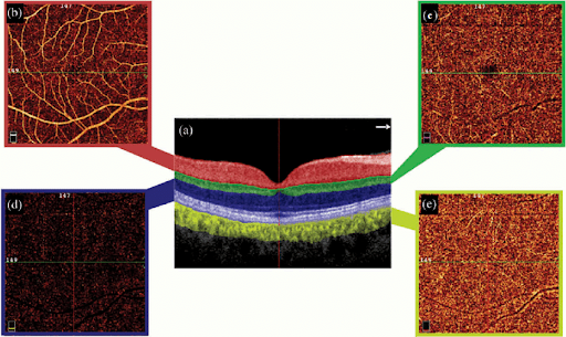
A: Vitreous
B: Superficial vascular plexus: this vascular area consists of large retinal vessels and capillaries. It is the area from the internal limiting membrane to the inner plexiform layer.
C: Deep vascular plexus: area between inner nuclear layer to the outer plexiform layer
D: Avascular area: from outer nuclear layer to RPE. In healthy eyes, this area is void of vasculature. However, if neovascularization forms (ex in wet macular degeneration), the source of the neovascular membrane starts in the choroid and proliferates through a break in the Bruch’s membrane.
E: Choriocapillaris: including Bruch’s membrane and 20um below. Neovascular membranes from exudative macular degeneration are isolated in this layer of OCT-A.
Clinical Applications of OCT-A
These are just some examples:
- Nonproliferative Diabetic Retinopathy (NPDR)
NPDR affects the inner retinal vasculature. The enhanced imaging of retinal capillaries using OCT-A detects DR even before clinical signs appear. However, leakage will require using fluorescein angiography.

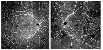
Figure 1.0: Widefield OCT-A of diabetic eyes without clinically significant diabetic retinopathy. Despite no outward signs of diabetic retinopathy, the patient exhibits regions of capillary nonperfusion.
- Retinal Venous Diseases
Similar to diabetic patients, areas of nonperfusion can be seen in patients with retinal venous occlusive disease. Some machines also have an angioplex software that even detects neovascularization on the surface (Zeiss Cirrus OCT).

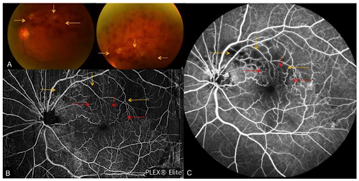
Figure 2.0: Superior temporal branch retinal vein occlusion with tortuous venous segments with flame-shaped, dot and blot hemorrhages, and cotton wool spots. A) Fundus photo, B) OCT-A montage, C) FA showing collateral vessel formation (red arrows) which are blocked by dense hemorrhages (as seen on the fundus photo). There are also areas of non perfusion (yellow arrows) obscured by extensive retinal hemorrhage on the fundus photos.
- Age-Related Macular Degeneration (ARMD)
The choriocapillaris slab of OCT-A helps distinguish small choroidal neovascular membranes from drusen, scars, or nonvascularized pigment epithelial detachments.

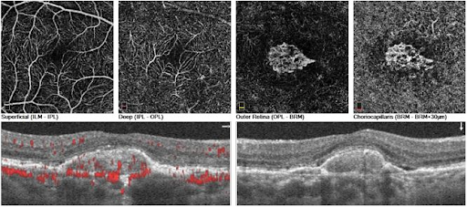
Figure 3.0: Active CNV lesion composed of loops, and peripheral anastomoses. The superficial, deep retinal plexuses, outer retina (normally avascular), and the choriocapillaris are depicted.
Check out this course for more great OCT-A case studies!
Advantages of OCT-A
In comparison with traditional imaging techniques, such as intravenous fluorescein angiography (FA) and indocyanine green angiography(IGA), the biggest advantages of OCT-A include that it is:
- Non-invasive.
- Dyeless (no systemic adverse events to dyes used in FA and IGA).
- Shorter in acquisition time.
- Capable of providing a three-dimensional view of retinal vessels, combining dynamic. blood flow and structural imaging without compromising vision.
Limitations of OCT-A
It is important to keep in mind that OCT-A has a few limitations, as with any other imaging technology. FA still remains the gold standard in modern retinal angiography.
Limitations of OCT-A include:
- It is light source dependent, therefore some ocular conditions (dry eye, cataract, etc.) will negatively affect the quality of the image.
- It is extremely motion-sensitive; patient movements, blinks, or saccades can result in image artifacts.
- Blood flow motion can sometimes be too fast or too slow to detect, and dye angiography is required to confirm areas of presumed nonperfusion. It offers a limited field of view and does not image well into the periphery, the image resolution decreases with area increases to maintain acquisition times.
- Dye leakage and staining are not visible, as in other imaging techniques.
Learn more about OCT-A interpretation and functionality with the ZEISS CIRRUS.
OCT-A in the optometry practice: practicalities and opportunities
OCT-A became commercially available in 2015 but has not yet been fully adopted in clinical settings. While OCT-A is a non-invasive modality that is useful for any condition that has to do with vasculature, it currently lacks algorithms for normative data. However, the number one barrier to OCT-A is the relative lack of education and training available for ODs looking to add this technology to their practice.
However, as companies expand on the research and development of OCT-A and add further continuing education and resources to train eyecare practitioners on this technology, there is ample room to grow OCT-A and its utilization in the optometry practice.
Coding and billing OCT-A
There is not a specific code for OCT-A. CPT code 92134 is a broad ophthalmic imaging code that applies to OCT-A as well. Unfortunately, the reimbursement rate is much lower than dye-based angiography.
However, OCT-A has the advantage over dye-based procedures for patients who are pregnant, those with a history of renal failure, and allergies to dyes. Some conditions are suspicious, and clinicians often feel the need to obtain fluorescein angiography, which involves consulting with retina specialists. OCT-A can help avoid unnecessary referrals to retina practices.
In short
OCT-A is a powerful and emerging tool in the eyecare field, which provides excellent information on chorioretinal structure, dynamic blood flow, and perfusion—albeit with limitations. Though FA still remains the gold standard, there is a newly evolving place for OCT-A in optometry, as new studies emerge and practitioners continue to collaborate to push the boundaries of this technology and its applications.
References:
- De Carlo, Talisa E., et al. "A review of optical coherence tomography angiography (OCTA)." International journal of retina and vitreous 1.1 (2015): 5.
- Gao, Simon S., et al. "Optical coherence tomography angiography." Investigative ophthalmology & visual science57.9 (2016): OCT27-OCT36.
- Koustenis A, Harris A, Gross J, Januleviciene I, Shah A, Siesky B. Optical coherence tomography angiography: an overview of the technology and an assessment of applications for clinical research. Br J Ophthalmol. 2017 Jan;101(1): 16-20.
- Landry, Darrin A. “What OCTA Shows Us.” Optometry Times, 8 June 2019, www.optometrytimes.com/view/what-octa-shows-us.
- Landry, Darrin A. Ophthalmologymanagement.com, 1 June 2019, www.ophthalmologymanagement.com/issues/2019/june-2019/the-state-of-octa.
- Marques, João, and Rufino Silva. Optical Coherence Tomography Angiography in AMD, Jan. 2018, amdbook.org/book/export/html/509.
- Rodman, Julie. “Is OCT-A Right For My Practice?” Review of Optometry, 15 Sept. 2020, www.reviewofoptometry.com/article/is-octa-right-for-my-practice.
- Saraf, Steven S. “How OCT Angiography Is Improving Our View of Diabetic Retinopathy.” Retina Specialist, 5 Apr. 2018, www.retina-specialist.com/article/how-oct-angiography-is-improving-our-view-of-diabetic-retinopathy.
- Stephenson, Michelle. “How to Get the Most from OCT-A.” Review of Ophthalmology, 7 Aug. 2017, www.reviewofophthalmology.com/article/how-to-get-the-most-from-octa.
- Wong, Claire L., et al. “Clinical Applications of Optical Coherence Angiography Imaging in Ocular Vascular Diseases.” MDPI, Multidisciplinary Digital Publishing Institute, 25 June 2019, www.mdpi.com/2076-3417/9/12/2577/htm.


