


Neurotrophic keratitis (NK) is a degenerative corneal disease caused by impairment of trigeminal corneal innervation#, most often involving the ophthalmic branch of the trigeminal nerve.
The resulting reduction or loss of corneal sensitivity disrupts epithelial maintenance and wound healing, allowing progressive epithelial breakdown and loss of corneal integrity. Because symptoms may be muted, the cornea can become vulnerable to trauma, infection, ulceration, stromal melting, perforation, scarring, and vision loss.1-3
Pathophysiology
The cornea is one of the most densely innervated tissues in the body. Sensory fibers detect mechanical, thermal, and chemical stimuli and help drive protective blink and lacrimation reflexes to help support tear film stability, epithelial hydration, and ocular surface protection.2,4,5,6
Corneal nerves also provide trophic support through bidirectional signaling between nerves and epithelial cells.
Key mediators in this neuro-epithelial communication include:
- Nerve growth factor (NGF)
- Brain-derived neurotrophic factor (BDNF)
- Glial cell-derived neurotrophic factor (GDNF)
- Epidermal growth factor (EGF)
- Substance P
- Calcitonin gene-related peptide (CGRP)
Together, these neurotrophins, neuropeptides, and growth factors help regulate epithelial proliferation, migration, adhesion, differentiation, wound healing, and limbal epithelial function. When this neuro-epithelial axis is disrupted, the result may be corneal hypoesthesia or anesthesia, impaired epithelial repair, persistent epithelial defects, ulceration, stromal degradation, secondary infection, and perforation.2,4,5,6
Incidence and prevalence of neurotrophic keratitis
The epidemiology of neurotrophic keratitis remains incompletely defined. Earlier estimates placed its prevalence at approximately 16 to 42 cases per 100,000 individuals.7
An analysis of the American Academy of Ophthalmology IRIS Registry identified 31,915 affected eyes in 27,483 patients and reported an average prevalence of 21.34 cases per 100,000 patients between 2013 and 2018. The mean age at diagnosis was 68 years; herpetic keratitis and diabetes mellitus were among the most common associated conditions, present in 33.7% and 31.6% of patients, respectively.8
The true burden of NK may nevertheless be underestimated, particularly because early disease can resemble dry eye disease or other ocular surface disorders and reduced corneal sensation may delay recognition.1,9
Incidence data are limited. In a German tertiary referral center, Roth et al. estimated an annual population incidence of approximately 1 to 3 new cases per 100,000 residents for moderate-to-severe NK.9 Because that study primarily captured more advanced disease and its population estimate was extrapolated from a single center, these findings may not reflect the incidence of all stages of NK.9
Download the cheat sheet here!

Neurotrophic Keratitis Management by Stage
Use this flowchart to help identify the optimal intervention for patients with neurotrophic keratitis based on the severity of disease.

Etiology and risk factor identification
A careful history is essential because NK can result from damage anywhere along the trigeminal sensory pathway, from the corneal nerve endings to the brainstem. This disruption impairs corneal sensation and epithelial healing.1,2
Relevant causes and risk factors can be grouped as follows:1,2
- Ocular surface disease, injury, and toxicity
- Herpetic eye disease
- Chronic ocular surface disease
- Chemical or thermal injury
- Topical medication toxicity or topical anesthetic abuse
- Contact lens–related trauma
- Surgical and procedure-related factors
- Refractive surgery
- Corneal transplantation
- Cataract or glaucoma surgery
- Retinal detachment repair
- Corneal collagen cross-linking
- Neurosurgical procedures involving the trigeminal nerve
- Neurologic, systemic, and congenital conditions
- Diabetes mellitus
- Multiple sclerosis
- Intracranial tumors, including acoustic neuroma
- Stroke
- Vitamin A deficiency
- Congenital corneal anesthesia
Clinical evaluation: Do not skip corneal sensitivity
Corneal sensitivity testing is essential whenever NK is suspected, examination findings exceed reported symptoms, or an epithelial defect heals poorly despite appropriate therapy. A history of herpetic eye disease, diabetes, ocular surgery, or other established risk factors should lower the threshold for testing.2,3
Testing should be performed before topical anesthetic drops are instilled. Assess both eyes and document sensation centrally and in the peripheral quadrants.
Available methods include:2,10
- Qualitative screening: A cotton wisp provides a readily available assessment of corneal sensation. Record the response as normal, reduced, or absent and compare corresponding areas between eyes. This method does not quantify the degree of sensory loss.2
- Contact esthesiometry: The Cochet-Bonnet esthesiometer uses an adjustable nylon filament to measure mechanical sensitivity. Its limitations include operator dependence, potential epithelial injury, and a restricted stimulus range that can limit assessment when sensitivity is markedly reduced. Testing commonly uses 5-mm changes in filament length, which also constrain measurement resolution.2,10
- Non-contact esthesiometry: The portable Brill esthesiometer delivers controlled air pulses to assess sensitivity without touching the corneal epithelium. It offers an additional option for quantitative testing, although its measurements are not interchangeable with Cochet-Bonnet values. Belmonte-type gas-jet instruments are used primarily in research settings.10,11
- In vivo confocal microscopy: Can provide supplementary information about corneal nerve density and morphology and may help monitor nerve regeneration. Routine diagnosis relies on clinical examination and corneal sensitivity assessment.2,3
Mackie staging: A practical framework
Once neurotrophic keratitis is recognized, the next step is staging disease severity. The three-stage Mackie classification, described in 1995, remains a widely used clinical framework for guiding treatment and follow-up.2,12
| Stage | Clinical Findings | Typical Symptoms |
|---|---|---|
| Stage 1 | Punctate epithelial keratopathy, irregular epithelium, tear film instability, reduced corneal sensitivity | Mild irritation, blur, or absent symptoms |
| Stage 2 | Persistent or recurrent epithelial defect, often with smooth or rolled edges and limited inflammation | Often surprisingly little discomfort |
| Stage 3 | Stromal ulceration, thinning, melting, descemetocele, or perforation risk | Pain may still be less than expected |
The best opportunity to reverse ocular surface damage is in the earliest stage of NK, before a persistent epithelial defect develops.3 By stage 2 or stage 3, the diagnosis may be more obvious, but the risk of scarring, infection, stromal melt, and perforation is higher.2,3
Stage 1: Protect the epithelium early
Stage 1 NK may present with punctate epithelial keratopathy, epithelial irregularity, tear film instability, reduced corneal sensitivity, and mild blur or irritation. Because symptoms may be limited, corneal sensitivity testing should be considered when staining appears disproportionate to patient complaints.2,3
Figure 1: Slit lamp image of stage 1 neurotrophic keratitis with punctate epithelial staining.


Figure 1: Courtesy of Cory J. Lappin, OD, MS, FAAO.
Preservative-free artificial tears may be used frequently throughout the day, with lubricating ointment at bedtime. Punctal occlusion may also be considered as an adjunct to preservative-free lubrication in stage 1 NK.3 Preserved or potentially toxic topical medications should be reduced or eliminated whenever possible, particularly in patients using multiple glaucoma drops.2,3
Patients with suspected herpetic eye disease may require antiviral therapy. Exposure keratopathy, lid malposition, incomplete blink, and significant meibomian gland dysfunction should be addressed to promote epithelial healing.2,3 In selected patients with recurrent epithelial breakdown, a therapeutic bandage contact lens may be considered with close follow-up because reduced corneal sensation can mask early complications.2
Short on time? Download the Neurotrophic Keratitis Management by Stage cheat sheet!
Stage 2: Persistent epithelial defect requires escalation
Stage 2 NK is characterized by a persistent or recurrent epithelial defect, often with smooth or rolled edges and limited inflammation. Despite the seriousness of the epithelial breakdown, patients may report surprisingly little discomfort because of reduced corneal sensation.1-3,6,13
Figure 2: Slit lamp image of stage 2 neurotrophic keratitis with persistent epithelial defect.


Figure 2: Courtesy of Cory J. Lappin, OD, MS, FAAO.
Treatment focuses on promoting epithelial closure and protecting the cornea from further damage.
Options include:2,3
- Topical cenegermin (OXERVATE): Cenegermin-bkbj 0.002% is a recombinant human nerve growth factor and the only FDA-approved treatment for NK. Pivotal trials and the 2021 expert consensus support its use in stages 2 and 3, although FDA approval is not restricted to these stages.3,4,13,14
- The uncontrolled DEFENDO trial suggests potential benefit in stage 1.15 The recommended regimen is one drop in the affected eye(s) six times daily, at 2-hour intervals, for 8 weeks.14
- Autologous serum tears (AST) or platelet-rich plasma (PRP): These blood-derived therapies contain growth factors and other biologically active components that may support epithelial repair in NK-associated persistent epithelial defects.2
- Therapeutic soft bandage contact lenses: A bandage lens can often be placed at the presenting visit to protect the healing epithelium from mechanical trauma caused by blinking.2,3
- Scleral lenses: These lenses vault the cornea and maintain a fluid reservoir, providing protection and hydration. Treatment and corneal protection should continue while fitting and dispensing are arranged.2,5
- PROSE treatment: Prosthetic replacement of the ocular surface ecosystem (PROSE) uses custom scleral prosthetic devices and is available through BostonSight PROSE providers.6
- Amniotic membrane therapy: Self-retained or sutured amniotic membranes provide a biologic scaffold that supports epithelial healing and may reduce inflammation and scarring.2 Prokera uses a cryopreserved membrane mounted on a retaining ring. CAM360 AmnioGraft is a ringless, shelf-stable cryopreserved membrane.15 OptiCover is a ringless, lyophilized (freeze-dried) membrane.
- Both CAM360 and OptiCover can be retained with a bandage lens or collagen shield.16,17 Amniotic membrane may be used at different points during treatment, with or without cenegermin (OXERVATE), according to epithelial healing and clinical response.18
Close follow-up is essential to assess healing and adjust treatment. Patients wearing therapeutic lenses require monitoring for infectious keratitis, hypoxia, and fit-related epithelial injury because reduced corneal sensation can mask developing complications.2,5
An epithelial defect that is not improving warrants reassessment of treatment and consideration of co-management. Treatment and corneal protection should continue while additional care is arranged.2,3
Stage 3: Recognize the emergency
Stage 3 NK is characterized by stromal ulceration, thinning, melting, descemetocele formation, or perforation.
Figure 3: Slit lamp image of stage 3 neurotrophic keratitis featuring corneal ulceration with stromal involvement.
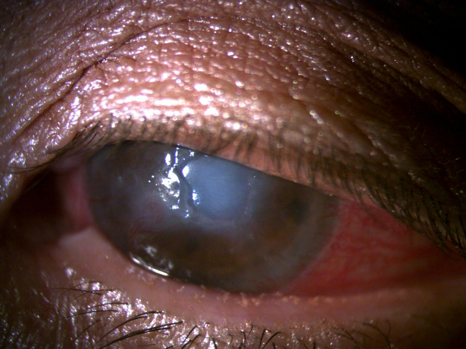
Figure 3: Courtesy of Cory J Lappin, OD, MS, FAAO.
Reduced corneal sensation can mask the severity of these findings, so examination findings should determine the urgency of care:2,3
- Urgent cornea assessment: Measurable progression of stromal thinning between visits, corneal melt, or suspected microbial superinfection warrants same-day cornea consultation. An enlarging defect, new corneal infiltrate, or anterior chamber reaction should raise concern for progression or infection. A descemetocele, positive Seidel test, or suspected perforation requires immediate surgical evaluation.2,3
- Surgical stabilization: Depending on the extent of tissue damage, treatment may include tissue adhesive with a bandage contact lens, temporary or permanent tarsorrhaphy, multilayer amniotic membrane transplantation, conjunctival flap surgery, or tectonic lamellar or penetrating keratoplasty. Care should be coordinated with a service able to provide the required intervention within the time dictated by the clinical findings.2,3
- Corneal neurotization: After acute stabilization, selected patients may benefit from corneal neurotization to address the underlying loss of innervation. This procedure directs a functioning sensory nerve to the cornea, either directly or through a nerve graft. Font and Cortina report return of corneal sensation in approximately 80% of cases, beginning around 5 to 6 months after surgery, although recovery may remain incomplete. Corneal protection remains necessary while reinnervation develops.2
When to consider MRI or neuroimaging
NK may be the presenting clue to trigeminal nerve dysfunction caused by a compressive, inflammatory, post-surgical, traumatic, or neoplastic process.2,19
MRI should be considered when any of the following are present:2,19
- Unilateral NK without an identifiable cause
- Facial numbness or reduced facial sensation
- Findings involving multiple cranial nerves
- New neurologic symptoms
- Orbital signs
- Hearing or vestibular symptoms
- A history concerning for intracranial or orbital pathology
Key takeaways
- When the cornea looks worse than the patient feels, consider neurotrophic keratitis.2,3
- Assess corneal sensation whenever staining, persistent epithelial defects, or corneal ulcers are out of proportion to the patient’s symptoms.2,3
- Important risk factors include herpetic eye disease, diabetes, vitreoretinal and refractive surgery, trigeminal nerve injury, contact lens-related trauma, and chronic topical medication toxicity.2,3
- Mackie staging guides treatment intensity. Escalation and co-management should reflect disease progression, treatment response, and access to needed care.2,3,12
- Stage 3 NK requires urgent cornea specialist involvement, particularly when progressive stromal thinning, corneal melt, descemetocele, perforation, or suspected secondary infection is present.2,3
Conclusion
Neurotrophic keratitis is frequently overlooked because symptoms often underestimate the severity of corneal disease. For optometrists, one simple observation can change the course of care: when the cornea looks worse than the patient feels, assess corneal sensation. Recognizing this clinical mismatch early can lead to timely diagnosis, appropriate intervention, and ultimately help prevent irreversible vision loss.1-3,15

