


In the US alone, more than 37 million people have diabetes,1 and are at risk of developing serious complications, including vision-threatening diabetic retinopathy (VTDR) and diabetic macular edema (DME), a primary cause of vision loss in this population that can occur at any DR stage.2
As such, it is imperative that eyecare providers (ECPs) remain vigilant when monitoring patients with diabetes.
The role of the optometrist in monitoring diabetic patients
Indeed, new research published in JAMA Ophthalmology illuminates just how crucial it is that primary eyecare providers closely monitor these patients. Specifically, the report noted that a staggering 26% of patients with diabetes have DR, and 5% of patients with diabetes develop VTDR.2 “This places significant responsibility at the feet of optometrists and general ophthalmologists—this is not just a problem for retina specialists,” notes Paul Chous, MA, OD, FAAO.
“Research clearly points to the fact that level and duration of metabolic control, diabetes duration, and baseline severity at initial eye examination all influence the risk of VTDR,3,4 which means we need to be involved early in the course of disease onset if we want to have the most impact on the prevention of vision loss,” he added.
But just how soon should you start adding additional, more sophisticated, and predictive testing when examining patients with diabetes? And what specific tests are most useful? Findings from recent studies are challenging diagnostic thresholds with clear indications that it may be useful to begin adding tests, such as optical coherence tomography angiography (OCT-A) and electroretinography (ERG), when a patient presents with prediabetes.5
Current diagnostic protocols
For diseases where early intervention prevents bad outcomes, early identification is ideal. Whether it’s the initial onset of myopia or the very earliest signs of age-related macular degeneration (AMD), primary eyecare providers must be alert to changes suggesting disease onset and progression.
This same level of vigilance has long been advocated for patients with diabetes because earlier good systemic management, surveillance, and appropriate intervention have been proven to reduce the risk of vision loss and other complications unequivocally.
Without treatment, patients with high-risk proliferative DR have a 50% chance of becoming blind within 5 years.2 To that end, the American Diabetes Association's (ADA) 2021 Standards of Diabetes Care recommends that patients with diabetes undergo a comprehensive dilated eye exam at least once per year, with longer or shorter intervals depending on individualized risk factors.6
OCT-A imaging as structural testing in diabetic patients
Of note, “A dilated fundus exam is a cornerstone of any comprehensive appointment, but in patients with diabetes, it’s non-negotiable and essential because it greatly enhances the detection and accurate grading of any retinal disease,” says Frances Bynum, OD. “However, as an added precaution, I also rely on retinal imaging, including OCT and OCT-A, because it establishes a baseline for future comparison.”
OCT-A is another structural testing option that’s gaining increased attention as a supplemental measure for assessing the integrity of retinal microvasculature. “Research shows that OCT-A imaging helps detect DR lesions earlier, including abnormal retinal perfusion and capillary dropout that wouldn’t otherwise even be visible with traditional exam techniques,” says Dr. Chous.
“We need to watch patients with subtle OCT-A abnormalities more carefully, especially those with non-perfusion that is a set-up for ischemia, hypoxia, release of vascular endothelial growth factor (VEGF), and worsening disease. It is likely that improved glycemic control, including avoidance of both hyperglycemia and hypoglycemia, will benefit patients at these earlier stages of disease.”
The benefits of OCT-A imaging for structural testing
“One of the greatest benefits of OCT-A is that it helps you get to the bottom of unexplained vision loss in patients with diabetes by revealing foveal ischemia,” adds Steven Ferrucci, OD, FAAO. “Ideally, it would be standard of care in all patients with DR, but from a practical standpoint—in terms of cost and time—that’s likely not realistic just yet.”
“In addition to these structural tests, visual acuity measurement is the mainstay of assessing visual function—but it has significant limitations in many patients, including those with diabetes,” says Dr. Bynum. “We need to know as much about function as we do about structure in patients with diabetes, but both visual acuity testing and visual fields are subjective tests,” he added.
Utilizing electroretinography
An electroretinogram is an alternative functional test that is entirely objective, but due to size, cost, testing time, corneal contact, and more, it used to be impractical for use in typical private practice.
“Still, this electrophysiological test has long been recognized as being to the retina what the electrocardiogram (ECG/EKG) is to the heart,” says Dr. Ferrucci. “In the same way an ECG is crucial to diagnosing heart problems and monitoring heart function, ERG helps detect retinal dysfunction.”
Recognizing this unmet need for a quick, easy, and repeatable measurement of objective retinal function, a handheld full-field, flicker ERG (ffERG) device has quickly become a must-have in many primary care practices.
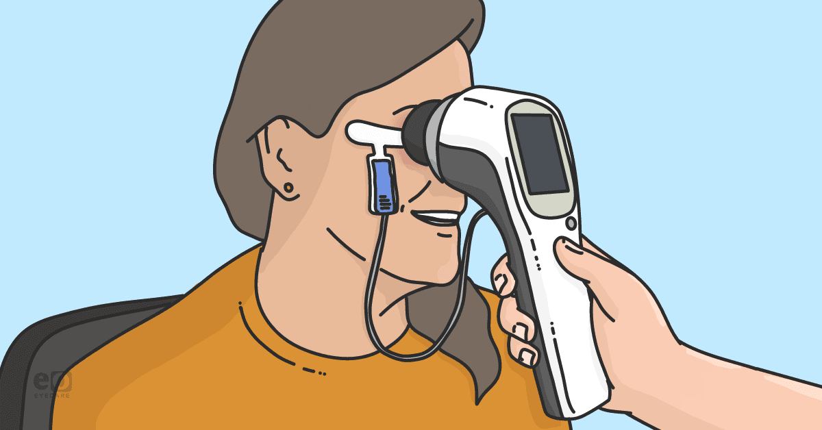
What is the RETeval device?
Currently, there is one FDA-cleared, handheld, battery-operated, non-mydriatic ERG testing instrument on the market. Known as the RETeval device, this ERG allows you to test both eyes in minutes using skin rather than corneal electrodes.
It also adjusts for pupil size in real-time, doesn’t require dilation, and features a DR assessment protocol that generates a DR Score that helps clinicians monitor patients over time and spot trouble right away. For example, a score of 23.4 or higher indicates an 11-fold risk of requiring medical or surgical intervention within 3 years.7
“By combining a retina cell stress measure and a pupil light response, you can rapidly and non-invasively assess all patients with or at risk for DR at every visit to determine current and future risk of disease progression over time as that risk changes,” says Timothy Earley, OD.
Indeed, some ECPs now use the DR protocol to screen all patients with diabetes, whether or not they’ve been diagnosed with retinopathy. “New research supports this as a progressive versus aggressive approach and asserts that we may want to start using this protocol even earlier, in patients with prediabetes,” adds Dr. Earley.
New diagnostic thresholds for diabetic retinopathy
In fact, new findings challenge current diagnostic thresholds for diabetes that were based on increased rates of DR at and above those thresholds. Specifically, researchers in the United Kingdom explored the extent to which non-invasive diagnostic technologies could identify early (pre-clinical) retinal and neurovascular pathology in prediabetes.5
What they found may change how and when patients are diagnosed with early DR. At present, retinopathy on fundus examination or conventional fundus imaging is requisite for diagnosing diabetic retinal disease.
However, in this study of 75 patients, both abnormal retinal neurovascular structure on OCT-A (Heidelberg Spectralis OCT2/OCT-A) and function on ERG (LKC Technologies RETeval device) preceded clinically observable retinopathy in prediabetes.
The authors note that handheld ERG and OCT-A can alert clinicians to abnormalities, indicating earlier, pre-clinical retinal damage has occurred even before retinopathy is visible, which contradicts earlier beliefs.5
Study findings on non-invasive tests for prediabetic patients
In short, the authors concluded that this means non-invasive tests, such as ERG, can be performed in patients with prediabetes in an effort to rapidly identify early retinal neurovascular damage and institute protective strategies against progression to clinical disease.
These findings are consistent with earlier research using both multispectral imaging and adaptive optics to identify structural retinal abnormalities in subjects with insulin resistance and prediabetes.8,9
“The question becomes, what does this mean for primary eyecare providers?” asks Dr. Bynum. “What can we do differently that’s both practical for clinicians and makes a meaningful difference in patients’ lives?” The report offers some guidance in this regard as well.5
Making the case for new protocols
The paper states that OCT-A and ERG can be used for screening purposes in patients who have prediabetes.5 “It makes sense,” explains Dr. Chous. “Structure and function are both very important, and more and more evidence is showing that advanced tools, such as OCT-A and ERG, offer us earlier insight and diagnostic confidence that will help eye care providers given rising rates of diabetes.”
He adds, “More importantly, with modern, objective technology, detecting functional stress can anticipate structural damage and help us offer and reinforce protective strategies against progression much earlier than before.”
ERG vs. structural imaging to assess sight-threatening DR
In studies comparing the ability of ERG and structural imaging to evaluate sight-threatening DR, ERG outperformed traditional imaging at predicting which patients would likely need subsequent medical/surgical intervention and those most likely to progress to VTDR.7,10
Dr. Earley uses a weather analogy to explain why the ability to predict is so important. “Most of the tests we use help us understand what’s happening now, which is like looking out your front door to see whether it’s raining. ERG is more like checking out the weather forecast before you pack for a trip. It helps predict what tomorrow will be like.”
Conclusion: It’s a matter of sink or swim
Projections indicate that, by 2060, some 60.6 million US adults (17.9%) will have diabetes.11 That’s nearly 18% of the US population. The number of people with DR is likewise expected to nearly triple from 5.5 million to 16.0 million by 2050.12 It is anticipated that 3.4 million patients will have VTDR by that time.12
The study authors acknowledge that, although OCT-A is readily available, there are barriers to using it for DR screening. For example, the device itself may be cost-prohibitive, and performing the test requires both pupil dilation and a well-trained technician.
There are fewer barriers to ERG, the authors note. Specifically, they emphasize that ERG is now relatively inexpensive, does not require dilation, and is portable, fast, and easy to use, making it a practical option in most clinical settings.5
“We need to think long and hard about how closely—and how carefully—we evaluate patients who have diabetes,” says Dr. Chous. “Recognizing that a quarter of our diabetes patients have diabetic retinopathy,2 and knowing we can perform a fast, patient-friendly test to assess risk should provide a spark to educate patients and recommend early interventions to reduce retinal damage and vision loss.”
