


Ophthalmic surgery has the potential to exacerbate a patient's pre-existing ocular surface disease (OSD), which, in turn, may result in poor visual outcomes.1
Moreover, a compromised ocular surface can significantly influence intraocular lens choices, limiting patients from obtaining superb visual results and spectacle independence.
Surgeons should carefully consider the underlying causes of their patients' vision loss and ocular discomfort to optimize cataract outcomes as well as their patients' comfort and vision.
Ocular surface disease and refractive surgery
OSD is not an age-dependent diagnosis. Physicians—and especially refractive surgeons—should keep OSD in mind when determining good candidates for different types of cornea-, refractive-, or lens-based refractive surgery.
At our practice, we screen each of our patients for OSD. When a patient is diagnosed with ocular surface or lid margin disease, my primary goal is to actively treat the underlying cause before we discuss surgical options.
Many of our patients are referred to our practice for cataract evaluation, but a large portion leave my office with an additional diagnosis of OSD.
Prevalence of OSD in pre-surgical patients
The prevalence of OSD is high in patients scheduled for cataract surgery—about 55% to 80%.2,3 In a study assessing the rate of undiagnosed dry eye disease (DED) in patients who were scheduled to undergo cataract surgery, less than 25% of them had a prior diagnosis of DED, but a majority (>60%) had asymptomatic DED.4
In another study, 85% of patients with no previously reported ocular surface dysfunction had at least one abnormal tear test with either matrix metalloproteinase-9 (MMP-9) or osmolarity.3
Overview of OSD and DED
OSD is used to describe a broad scope of immune and non-immune conditions that cause a disruption in the function or structure of the surface layers of the eye, including the cornea, conjunctiva, limbus, tear film, and eyelids.1 Dry eye disease is a form of OSD that is characterized by a persistently unstable or deficient tear film that causes discomfort, visual impairment, inflammation, and altered integrity of the ocular surface.5
The most common etiologies of OSD that I see in my practice stem from chronic disease processes such as ocular rosacea, Demodex blepharitis, epithelial basement membrane disease, and mechanical DED. Other common conditions that cause OSD include meibomian gland dysfunction (MGD), eyelid laxity, patients treated with chemotherapy and radiation to the neck or head, uncontrolled diabetes, and neurotrophic keratitis.
Many patients with OSD are treated for years without relief, but identifying the root cause of their OSD can help to improve patient satisfaction.
Treating ocular surface disease
To begin healing the ocular surface, it is important to gather a full diagnostic history of the patient and identify the cause(s) of their OSD. Treating the ocular surface can involve a lengthy period of trial and error with various pharmacologic treatments. Though it takes time and close attention to assess and treat complex cases of OSD, helping patients achieve a healthy ocular surface often leads to improved patient care and excellent surgical outcomes.
Initial treatment for OSD depends on the symptoms and underlying cause of the ocular surface dysfunction. For example, a patient with ocular rosacea, which is almost always accompanied by MGD, will often have improved symptoms after being treated with intense pulse light therapy.
Recently, Tarsus received FDA approval for lotilaner 0.25% ophthalmic solution (Xdemvy), which targets the Demodex mite,6 and eradicates the infestation, which can improve and even resolve MGD, lid inflammation, and poor tear film stability, helping to improve vision.
In my practice, few patients have only one cause of OSD; most have multifactorial disease that requires several treatment modalities to achieve a healthy ocular surface.
Establishing OSD treatment protocols
With any of the OSD therapies, I start conservatively by prescribing over-the-counter (OTC) medications, and then I evaluate the efficacy of treatment at follow-up. For patients who have uncontrolled inflammation, I may combine their OTC medications with an antibiotic and steroid ointment.
If these conservative therapies do not adequately relieve a patient's OSD, then I move on to more intensive ocular treatments, such as amniotic membranes.
The American Society of Cataract and Refractive Surgery Committee created a useful algorithm and questionnaire to aid ocular surgeons in diagnosing and treating visually significant OSD prior to any refractive surgery (download it here).7
When to treat OSD with amniotic membranes
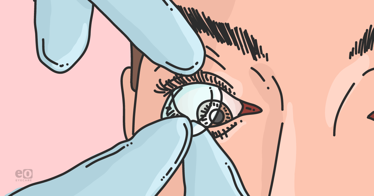
Amniotic membranes are highly beneficial in patients with OSD who do not respond to conservative OTC treatments. Cryopreserved amniotic membrane (CAM)—one such example is Prokera; BioTissue—is especially useful in healing patients with OSD if they have dysregulated corneal surfaces due to basement membrane disease, a superficial corneal abrasion, chemical insult, prior cornea surgery, or severe ocular inflammation. To date, Prokera is the only amniotic membrane that is FDA-cleared with an FDA designation as being anti-inflammatory, anti-fibrotic, and anti-angiogenic along with supporting epithelial adhesion and differentiation.
CAM may also be used in early OSD, for patients with mild DED, to stabilize their prescription and their ocular surface if they are a candidate for surgery. CAM is easy to administer and may be placed on the surface of the eye for 3 to 5 days,8 depending on OSD severity, for patients who require a short duration of regenerative therapy.
For CAM aftercare, I place a tape splint tarsorrhaphy to stabilize the eyelid, and I instruct my patients that they may experience some stinging from the amniotic membrane due to the regenerative properties of CAM. I will also hand my patients a detailed brochure that outlines key counseling points and what to expect with CAM treatment.9
Clinical benefits of treatment with amniotic membranes
Initiating CAM therapy is a case-by-case decision, but early intervention with regenerative therapy in OSD can have profound effects on the ocular surface. Amniotic membranes supply a rich source of biologically active factors that support epithelialization and exhibit anti-inflammatory, anti-microbial, low immunogenicity, anti-scarring, and anti-angiogenesis properties.8
In ophthalmology, amniotic membranes have been shown to accelerate ocular surface reconstruction and reduce the symptoms of DED.8,10 There are two types of amniotic membranes available to treat the ocular surface: cryopreserved and dehydrated amniotic membranes.
Both amniotic membranes can be used for healing ocular dystrophies and for post-surgery wound dressing, but there are important differences.
Dehydrated vs. cryopreserved amniotic membranes
Dehydrated amniotic membranes have a useful lyophilization and drying process that allows for easier storage of the membranes. However, the dehydration process compromises the structural integrity of the tissue and depletes a key biologic factor—heavy-chain, high molecular weight hyaluronic acid/pentraxin 3 (HC-HA/PTX 3)—that contributes to the anti-inflammatory and immune response of amniotic tissue.11,12
Conversely, CAMs have structural integrity that is similar to fresh tissue and are enriched with key biologic signals that are essential for the healing process.
These essential compounds include:13
- HC-HA/PTX 3
- Growth factors
- Collagen (types I, III, IV, V, and VI)
- Fibronectin
- Laminin
- Hyaluronic acid
- Proteoglycans
Patients with OSD have a complicated prognosis due to their impaired ability to heal the ocular surface. Thus, selecting a regenerative therapy should be based on optimizing patient healing outcomes. Because of the differences in amniotic membrane integrity and associated healing benefits, I prefer CAM for my patients with chronic OSD.
Final tips on best OSD treatment practices
When addressing ocular surface disease, remember the following key aspects:
- Ophthalmic surgery can exacerbate or worsen a patient's underlying OSD.
- OSD is often underdiagnosed and undertreated, which leads to suboptimal visual and surgical outcomes.
- Treatment can involve a considerable amount of time spent counseling the patient and healing the ocular surface.
- Using CAM as part of the treatment algorithm for patients with OSD can accelerate their healing, lower inflammation, and reduce a patient's chance of ocular scarring.
- Helping patients achieve a healthy ocular surface can optimize surgical outcomes.
