


Learning to read corneal topography and tomography maps is an essential skill for those in training and practicing ophthalmologists. Identifying corneal irregularities and perhaps even saving a patient from needing an invasive surgery by allowing early diagnosis provided in these images is incredibly important.
This article will review corneal topography and tomography basics, as well as utilization in practice. A “cheat sheet” will be provided as a reminder for when to consider topography and tomography, as well as a quick reference on how to interpret corneal elevation maps on the go in a busy office/clinic.
Corneal topography
Corneal topography is a photographic diagnostic technique that diagrams the cornea's surface. It may identify and evaluate the anatomic and astigmatic impact of entities such as corneal scarring, growths (such as pterygia or Salzman nodules), keratoconus, and other ectatic diseases as well as aid in contact lens fitting. Corneal imaging is especially helpful in diagnosing early keratoconus as the cornea may appear normal on a slit lamp exam.1-4
Topography is typically performed using Placido disc patterns or concentric mires that are reflected off the tear film of the anterior cornea. These patterns are converted to color scales—and imaging for interpretation. More recently, technological advancements have added scanning slit methods to better assess elevation data. Ocular surface disease can play a role here; tearing film's irregularities can impact the image quality.1-4
Placido disc imaging
A Placido disc is a device made of concentric rings (generally white rings on a black background).1-4 The air-tear film interface acts as a convex mirror and reflects light back in a pattern dependent on the corneal elevations. Contemporary videokeratoscopy topographers use mathematical formulae to provide a point-to-point quantitative gradient of subtle topographic changes, which can illuminate corneal curvature irregularities and alterations in the tear film.
Devices that use Placido disc systems include the Keratron (Optikon, Rome, Italy), Keratograph (Oculus, Arlington WA), Atlas (Zeiss, Dublin CA), and ReSeeVit (Veatch Ophthalmic Instruments, Tempe AZ).4
An example of a Placido disc is shown in Figure 1.

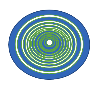
Figure 1. Placido disc
Topography is a must before any refractive surgery, such as LASIK or PRK—not only for screening for abnormalities but also to help map out treatment plans. Corneal topography can also assist in preoperative planning for cataract surgery. Detection of ectasia or even a decentered ablation post-LASIK is important to note if a patient is considering a presbyopia-correcting, or even toric intraocular lens, and counseling regarding candidacy for these technologies can then be done.1-4
Topography is also quite useful post-keratoplasty (usually post-penetrating keratoplasty (PKP) or deep anterior lamellar keratoplasty (DALK). Here, topographic images can elucidate steep areas (usually due to tight/loose sutures) and assist in mapping out suture removal in the postoperative period to best optimize vision and manage postoperative astigmatism.1-3
Finally, serial topography scans can be invaluable in diagnosing progressive keratoconus. If diagnosed, this helps open discussions regarding corneal crosslinking to help halt disease progression.1-3
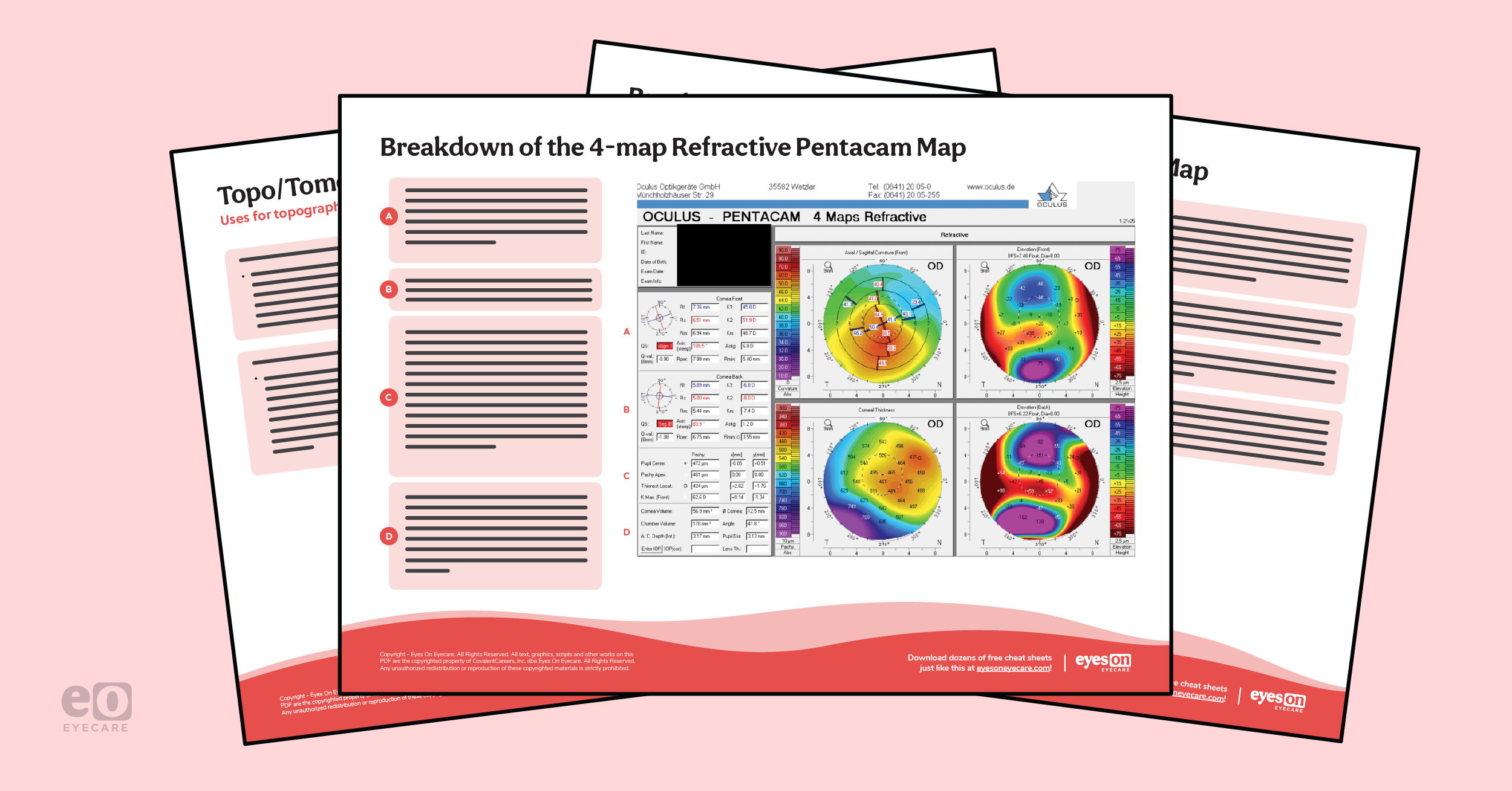
Download the cheat sheet now!

Corneal Topography & Tomography Cheat Sheet
This quick reference covers when and how to interpret corneal elevation maps on the go.

Interpretation of topography mapping
As with any diagnostic imaging, a stepwise approach is taken here.
One of the best places to start is by looking at the quad/multimap screen, which gives them an excellent visual comparison of data. Next, look to the color scale and identify the range and gradient of values given—each scan will have a color-coded scale for the measured indices. For the topographic map, these indices will reflect the dioptric value of the cornea. Hotter colors (orange and red) generally indicate steeper cornea values. Try to identify patterns—look for bow tie astigmatism, asymmetry, and focal changes in color maps.1-4
The next step is to look at the numbers on the maps and in the statistics boxes. Numerical overlays will confirm steepening and thinning and demonstrate central corneal thickness, maximum keratometry, and anterior and posterior corneal elevations. In the statistics boxes, there is useful data on minimal corneal thickness, angle kappa, pupil diameter, 3mm, 5mm, and 7mm zone irregularity as well as white to white diameter (please note that some of these values may be present in only some brands or versions of topographers).1-4
Topography maps and their function
- The axial map will give a quick overview of corneal power, however, it is not as accurate as the tangential map. As this map uses averaging algorithms, central data is more accurate than peripheral data.1-4
- The tangential display map is the most sensitive power map; it measures power and curvature at individual points on the cornea.1-4
- The elevation display map conveys the true shape of the cornea about ‘best-fit sphere’ 1-4 The system(s) measuring the cornea calculate areas of relative elevation and depression based on the deviation from the best-fit sphere, with deviation values displayed in microns. In Placido-disc systems only the anterior surface is measured; Scheimpflug systems measure both anterior and posterior elevations.1-4
- The corneal thickness display maps are only available on scanning slit and Scheimpflug topographers. This display can stage disease conditions like keratoconus and monitor corneal thickness changes due to corneal dystrophy (e.g., Fuchs), as well as contact lens wear.1-4
Finally, you should compare your imaging with slit lamp findings. Remember that topography can be affected by corneal artifacts and ocular surface disease/dry eye.1-4 A patient with severe ocular surface disease may have an ectatic-appearing topography scan that, once treated, may resolve and scans will show normal topography.
Corneal tomography
Compared to topography, which analyses anterior corneal curvature and power, tomography can capture overall corneal shape. This is made possible by scanning slit technology, Scheimpflug-based imaging, and anterior segment OCT, which can provide data on both anterior and posterior corneal surfaces.1-4
Scanning slit technology
Scanning slit is one of the elevation-based methods for assessing corneal curvature/power. Here, several slits from the nasal and temporal side are projected onto the cornea and assessed at several points on each slit. Triangulation between the reference slit beam surface and the reflected beam captured by the camera can be used to analyze the anterior and posterior curvature and corneal thickness. The Orbscan (Bausch+Lomb, Rochester NY) utilizes this technology.1-4
Scheimpflug-based assessment
This imaging modality allows for assessing corneal shape via manipulation of the image plane and drawing an oblique tangent from the image, object, and lens planes. The point of intersection of these planes is called the Scheimpflug intersection. Mathematical manipulation of the image plane and lens plane can create a focused image of the anterior and posterior cornea, as well as allow for the calculation of corneal thickness.1-4
An example of a system that utilizes Scheimpflug imaging is the Pentacam (Oculus, Arlington WA).
Figure 2 offers a guide on how to interpret the most commonly used diagnostic screen on the Pentacam

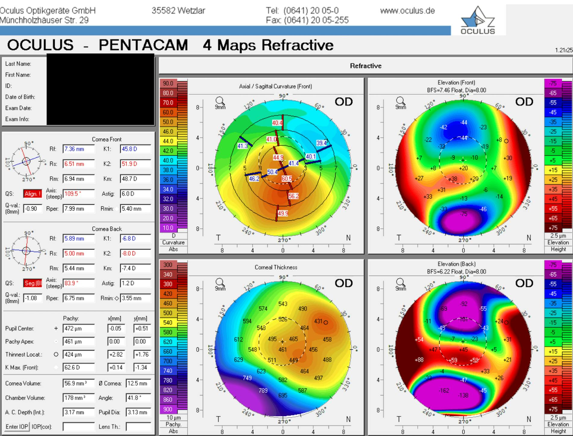
Figure 2: Pentacam(4-Map Refractive Print Out)
Conclusions
Corneal topography is an established and important technology for measuring the shape and power of the cornea—it allows practitioners to assess disease states, follow the course of conditions and help best guide treatments medically and surgically.
Don't forget to download the Guide to Corneal Topography Cheat Sheet!
Correction March 8, 2023: The original version of this cheat sheet erroneously labeled the blue line as the steep axis and the red line as the flat axis. This has been corrected.
