


Over the last few decades, many technological advances have been made in diagnosing and treating ophthalmic disorders. One such advancement was the introduction of corneal collagen cross-linking (CXL) to treat keratoconus. Before cross-linking, management was focused solely on attempting to correct the visual disturbances brought on by keratoconus.
However, the advent of corneal collagen cross-linking finally created a method in which the progressive disease process could be halted or minimized. Today, clinicians further push the bounds of medicine by utilizing this technology in different ways to provide better care for their patients.
Overview of keratoconus
Keratoconus is an ectatic disorder characterized by progressive thinning of the central or paracentral cornea resulting in anterior protrusion. Of all the ectatic diseases, keratoconus is the most common, with an incidence of approximately 1 in 2000.1
There are currently no specific genes or hereditary patterns associated with keratoconus. However, some risk factors have been identified with its development.
Keratoconus risk factors include:1
- Eye rubbing
- Down syndrome
- Vernal conjunctivitis
- Leber’s Congenital Amaurosis (LCA)
- Retinitis pigmentosa
- Floppy eyelid syndrome
- Connective tissue disorders (i.e., Ehlers-Danlos syndrome)
While the pathogenesis of keratoconus is not completely clear, these risk factors help us postulate some potential causes of the disease. For instance, conditions such as Ehlers-Danlos syndrome disrupt connective tissue formation leading to the loss of integrity of collagen throughout the body, including the cornea.
The other conditions mentioned have been associated with increased mechanical forces on the cornea. For example, individuals with Down syndrome and LCA have an increased incidence of eye-rubbing. In contrast, in floppy eyelid syndrome, there is a decrease in the protective function of the eyelids and an increased incidence of corneal exposure and rubbing, particularly during sleep.
Patients typically experience blurred vision, glare, or halos, due to progressively increasing irregular astigmatism. In rare instances, the thinning can become severe enough to cause tears in Descemet’s membrane resulting in rapid corneal edema and acute hydrops. This presents as episodes of sudden decreased vision and pain.
Critical features for diagnosing keratoconus include:
- Progressively increasing astigmatic refractive error
- Scissoring of the red reflex on retinoscopy
- V-shaped deformation of the lower lid on downgaze (Munson's sign)
- Conical deformation of light on the nasal cornea with a temporal light (Rizzuti's sign)
- Apical thinning on slit-lamp examination or presence of iron lines (Fleischer ring)
Ancillary testing, such as keratometry and corneal topography/tomography, can also help identify abnormal corneal parameters and monitor the progression of keratoconus.
Treatment of keratoconus
Treatment of keratoconus initially involves using spectacles and soft contact lenses to improve vision. When the degree of irregular astigmatism is severe, rigid gas permeable contact lenses are used. In cases of progressive thinning, corneal collagen cross-linking can be performed.
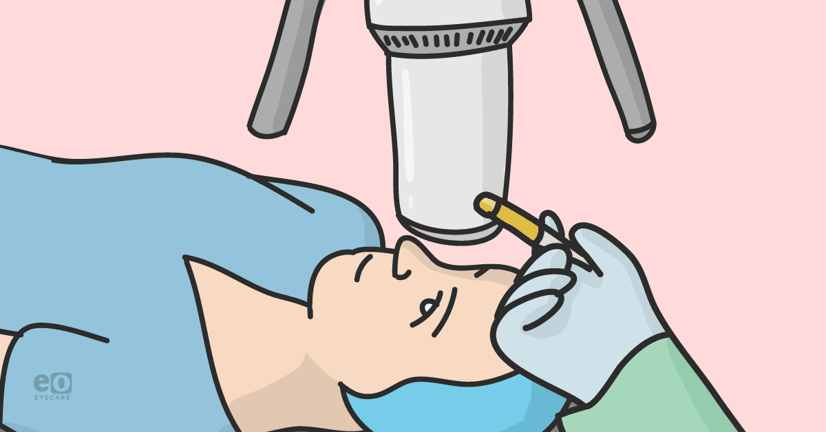
This treatment modality was approved by the FDA in 2016 and involves the application of riboflavin (vitamin B2) eye drops onto the cornea, followed by UVA light exposure to increase collagen bonding to stabilize the cornea.
There are also surgical considerations for patients who suffer from keratoconus. Intracorneal corneal ring segments (ICRS) are alloplastic arcs that can be placed into the corneal stroma to induce flattening of the central cornea. This can be done for mild to moderate cases where patients are intolerant of contact lenses. Advanced topics could require transplantation in the form of deep anterior lamellar keratoplasty (DALK) or total thickness penetrating keratoplasty (PK).
Corneal cross-linking
Corneal cross-linking was first described in 1997 by Spörl Seiler and colleagues as the “Dresden protocol.”1 The treatment begins with administering riboflavin drops to the cornea, followed by exposure to UVA light. The riboflavin serves as a source for free radicals, generated when the UVA light interacts with its ring structure.1
These free radicals lead to oxidation of the corneal stroma and cause collagen fibrils to form stronger chemical bonds with each other. Ultimately, this results in increased stiffening and stabilization of the cornea.
Epithelium-off technique
When corneal cross-linking was first introduced, the standard technique used (and the one that is currently FDA-approved) was the Dresden protocol. It is still the most widely used method of corneal cross-linking. Here, the eye is anesthetized, and the central 7-9mm of the epithelium is debrided using a blade. This is followed by administering riboflavin solution every 2 minutes for a total of 30 minutes and then 30 minutes of UVA light exposure.3
Postoperatively patients are treated with an antibiotic until epithelialization occurs. Today, surgeons can preferentially debride the epithelium using ethyl alcohol, mechanical rubbing with a cellulose sponge, or phototherapeutic keratectomy (PTK) laser scrape.2
One of the significant benefits of this technique is its tried-and-true nature. Debridement of the epithelium allows for maximal riboflavin penetration into the corneal stroma and ultimately better collagen cross-linkage.
Studies of this procedure have shown a halt in cases of rapidly progressive keratoconus along with flattening of the cornea and significant improvement in visual acuity.1,3 More recent studies have also shown that the effects can last up to 10 years.4
However, it is essential to consider the complications of epi-off cross-linking. Debridement of the epithelium can result in intense pain following the procedure and leaves the cornea’s inner layers vulnerable to the external environment. A study performed by Evangelista and Hatch (2018) reported adverse effects associated with epi-off cross-linking, including pain, infectious and sterile keratitis, persistent epithelial defects, corneal melt, stromal haze, and possible damage to the corneal endothelium.
It is important to consider patients and their circumstances when considering epi-off cross-linking. For instance, patients with Down syndrome may have extreme difficulty with post-operative pain or administration of post-operative drops. This could lead to intense eye rubbing, which may increase the possibility of keratitis or a protracted postoperative course.
Similarly, without the surgical intervention of the eyelids, patients with floppy eyelid syndrome may continue to have the potential for mechanical rubbing of their eyes and be predisposed to infection or prolonged epithelial defects.
Epithelium-on technique
Epi-on or transepithelial corneal cross-linking methods allow riboflavin penetration into the stroma without epithelial debridement before UVA exposure.
This has been attempted in different ways, including:2
- Altering the riboflavin solution
- Increasing riboflavin concentration
- Duration of administration
- Use of topical anesthetics and
- Iontophoresis
Iontophoresis refers to electrodes on the corneal surface to alter the electrochemical gradient and theoretically allow riboflavin to be driven across the intact epithelium.
Large metanalyses have been conducted to compare epi-on results to epi-off CXL studies. However, it must be considered that there was no uniform method followed in the epi-on CXL studies. These studies assessed changes in uncorrected and corrected distance visual acuity and changes in keratometry.
The analysis showed no statistically significant differences in visual acuity between the two procedures at one year. Still, more extensive studies have shown improvement in visual acuity at three-year follow-up in the epi-off group.6,7
Also, while studies on the effect of epi-on CXL did show no change in average keratometry reading between 3 and 12 months, a larger metanalysis comparing the two procedures showed a statistically significant difference in the decrease of Ks with epi-off as compared to epi-on.6,8
Lastly, it was demonstrated that epi-on CXL causes less pain and risk of infection, scarring, and haze than the epi-off technique.9,10
Conclusions on corneal cross-linking
Keratoconus is the most common corneal ectasia and causes central or paracentral thinning of the cornea. While early treatments focused on improving visual quality, CXL was developed to halt the progression of keratoconus. This technology is the first to focus on treating the disease process by increasing the bondage of collagen within the corneal stroma and thus stiffening the cornea.
When first described, the procedure involved the debridement of the corneal epithelium to allow for increased riboflavin penetration into the corneal stroma. While this is still the most used technique today, the debridement of the epithelium has some disadvantages, including pain, the potential for infection, and corneal haze. As physicians, it is essential to consider how this could affect our patients and to discuss these possibilities with them.
It is equally as important to consider their comorbid conditions and their potential to affect the postoperative course, which may increase the likelihood of these adverse events.
An alternative method
More recently, the literature has described an alternative method of leaving the corneal epithelium intact to decrease these complications without compromising the effectiveness of cross-linking. These methods include altering riboflavin concentration, chemical additives, and iontophoresis. Studies have shown that these methods do decrease the occurrence of postoperative complications as well as improvement in both visual acuity and stabilization of keratometry.
However, long-term analysis has shown minor improvement in both categories compared to the epi-off technique. So, while being practical, it has been suggested that the procedure may not be as effective as the original procedure.
An important caveat to this is that these head-to-head comparisons did not follow a standard protocol of the epi-on technique. This could be a confounding variable affecting the results of epi-on cross-linking as some protocols (i.e., use of chemical additives) have been reported to be as effective when divided into subgroups.6
In closing
Ultimately, corneal cross-linking is a breakthrough in the treatment of keratoconus. While we further our understanding of the procedure, it is essential to consider the different protocols. This can allow us to assess our patients, their degree of keratoconus, and comorbidities to provide informed recommendations as we collaborate with them in tailoring a treatment plan.
References
- Weisenthal, Robert W, et al. “Corneal Dystrophies and Ectasias.” 2021-2022 Basic and Clinical Science Course, Section 08 External Disease and Cornea, AMERICAN ACADEMY OF OPHTHALMOLOGY, 2021.
- Feldman, Brad H, and Clinton Jordan. “Techniques for Corneal Collagen Crosslinking: Epi-off vs Epi-On.” EyeWiki, 13 Feb. 2022, https://eyewiki.aao.org/Techniques_for_Corneal_Collagen_Crosslinking:_Epi-off_vs_Epi-on#cite_ref-20.
- Wollensak G, Spoerl E, Seiler T. Riboflavin/ultraviolet-a-induced collagen crosslinking for the treatment of keratoconus. Am J Ophthalmol. 2003 May;135(5):620-7. doi: 10.1016/s0002-9394(02)02220-1. PMID: 12719068.
- Raiskup F, Theuring A, Pillunat LE, Spoerl E. Corneal collagen crosslinking with riboflavin and ultraviolet-A light in progressive keratoconus: ten-year results. J Cataract Refract Surg. 2015 Jan;41(1):41-6. doi: 10.1016/j.jcrs.2014.09.033. PMID: 25532633.
- Evangelista CB, Hatch KM. Corneal Collagen Cross-Linking Complications. Semin Ophthalmol. 2018;33(1):29-35. doi: 10.1080/08820538.2017.1353809. Epub 2017 Sep 6. PMID: 28876968.
- Wen D, Song B; Li Q et al. Comparison of Epithelium-Off Versus Transepithelial Corneal Collagen Cross-Linking for Keratoconus: A Systematic Review and Meta-Analysis. Cornea 2018. 37:8 1018-1024.
- Buzzonetti L, Petrocelli G, Valente P. Iontophoretic transepithelial collagen cross-linking versus epithelium-off collagen cross-linking in pediatric patients 3-year follow-up. Cornea. 2019:38 (7) 859-863.
- Meiri Z, Keren S, Rosenblatt A, et al. Efficacy of corneal collagen cross-linking for the treatment of keratoconus: a systematic review and meta-analysis. Cornea. 2016:35(3)417–428.
- Shalchi Z, Wang X, Nanavaty MA. Safety and efficacy of epithelium removal and transepithelial corneal collagen crosslinking for keratoconus. Eye (Lond). 2015;29(1):15–29. doi:10.1038/eye.2014.230.
- Al Fayez MF, Alfayez S, Alfayez Y. Transepithelial versus epithelium-off corneal collagen cross-linking for progressive keratoconus: A prospective randomized controlled trial. Cornea 2015: 34, 10:S53-6.
