


Phototherapy, also known as Syntonic Light Therapy, is an advanced non-invasive treatment that has been around for over 100 years. Phototherapy is known to affect behavior, mood, and physiological functions. That’s why it’s commonly used in the medical community to treat conditions such as sleep disorders,1 Seasonal Affective Disorder,2 neonatal jaundice, and more.
Syntonic light therapy has been used by Optometrists since the 1920s, but became a more common optometric practice by the 1940s. It is an effective treatment of chronic headaches, stress, trauma, brain injuries, and concussions.3 Many case reports4-7 have also shown significant expansion of functional visual fields8 (not physical visual field defects relating to retinal or optic nerve disease) after treatment. Since 80% of learning is through vision, inefficient visual functions can affect academic achievement, athletic performance, proficiency and work, and home life activities. Therefore, optometrists have also applied syntonic light therapy in treating visual disorders affecting learning abilities in children.
Before delving more into syntonics, it is critically important to note that because syntonic light therapy affects the brain, the use of this treatment should be well understood and done with care to prevent any negative effects/reactions. Syntonics should be taken seriously and must be responsibly applied with the proper understanding and knowledge.
The College of Syntonic Optometry (CSO) works to promote and expand the therapeutic use of light in clinical optometric practice through post-graduate education and research to this day. The CSO provides annual conferences with up-to-date continuing education lectures for optometrists to share knowledge on phototherapy. The CSO conference is an excellent resource for one to learn more about phototherapy as clinical trials may not be readily available in the medical literature.
What is phototherapy and how does it work?
Phototherapy uses specific light colors, frequencies, and wavelengths to improve regulatory centers in the brain that influence chemical, hormonal, and electrical balances. These regulatory centers affect body functions, including vision.
Syntonic light therapy uses frequencies of colored light applied to the eyes to access these regulatory centers to help restore balance in the body’s sympathetic and parasympathetic nervous systems.
Here is a friendly reminder of how these nervous systems work:
| Sympathetic Nervous System | Parasympathetic Nervous System |
|---|---|
| Fight or flight response | Controls homeostasis |
| Pupil dilation | Pupil constriction |
| ↓ Accommodation | ↑ Accommodation |
| ↓ Convergence | ↑ Convergence |
| ↑ Pulse, blood pressure, and blood sugar levels | ↓ Pulse, blood pressure, and blood sugar levels |
Light is used to trigger photoreceptor cells in our eyes, which stimulates photoreceptive areas in the brain through the optic nerve via rod and cone pigments. Dr. Harry Riley Spitler explained these pathways in The Syntonic Principle, where he discusses how ocular light stimulation through retinal fibers results in changing the physiology of the thalamus, hypothalamus, and pituitary gland.9
Photoreceptor cells can also stimulate photosensitive elements within the blood through vascular beds, and our eyes are the only organ in the body where the vascular system is directly exposed to light. So application of light therapy through the eyes can access our regulatory centers in more than one way.
Research has shown that certain frequencies of colored light will stimulate different aspects of the nervous system3:
- Higher energy or shorter wavelength colors like blue or violet stimulate the parasympathetic nervous system. These colors can help to reduce adrenaline production, which helps with anxiety and stress.
- Lower energy or longer wavelength colors like red and orange stimulate the sympathetic nervous system to improve fusion and binocularity. The theory behind this is that long wavelengths build an electrical charge within cell membranes and break through synaptic resistance to overcome binocular suppression.
- Wavelengths in the middle frequencies like green and yellow act as physiological stabilizers and detoxifiers for chronic or degenerative health issues.
Syntonics can easily be implemented into any office setting as the equipment is minimal, and the patient is instructed to do most of the treatment at home. Patients wear certain colored filter goggles that have been prescribed by their doctor and look at a light source (either a syntonizer instrument or penlight) for roughly 15-20 minutes per session, about 3-4 times per week.
Visual conditions that may benefit from phototherapy
Syntonic light therapy can be used as the primary or supportive treatment for a plethora of visual syndromes and dysfunctions10, such as:
- Strabismus
- Amblyopia
- Accommodative dysfunctions
- Vergence dysfunctions
- Oculomotor dysfunction
- Nystagmus
- Visual attention deficit
- Vision-related learning and behavioral challenges11,12
- Delayed visual processing speed
- Visual discrimination and other visual information processing challenges
- Constricted functional visual fields associated with visual stress, emotional or physical trauma, brain injuries, binocular suppression, and some degenerative ocular disorders11,12
Considering the visual syndromes previously listed, syntonics could be a great option for patients who experience the following symptoms:3,10
- Fluctuating blurry vision
- Eye turn or lazy eye
- Double vision
- Asthenopia or general eye fatigue
- Glare, light sensitivity, and difficulty with night vision
- Reduced depth perception
- Headaches
- Poor concentration or short attention span
- Reading challenges and reduced academic performance
- Reduced athletic or occupational performance
- Reduced functional peripheral vision
- History of head trauma, fevers, or ear infections
- Stress trauma (physical, mental, or emotional), chronic or severe illness, and chronic allergies
How to identify patients for phototherapy
Syntonics phototherapy courses train optometrists on how to identify patients who would benefit from the treatment based on their pupil response and functional visual fields.
The most common method of identifying patients with an imbalanced autonomic nervous system is the Alpha-Omega pupil.13 Normally, when light is shone directly towards the pupil, swift pupil constriction is expected until the light is removed. In a patient with a disrupted autonomic nervous system, direct illumination with a penlight a few inches away from the eye will show a release in pupil constriction within a few seconds. This release in constriction is the Alpha-Omega pupil and suggests an imbalance in the SNS and PSNS.
Functional visual fields are measured monocularly at a near viewing distance with a campimeter. Optometrists will use white and other colored targets to identify any constricted fields.
Case study
Below is an example of a 27-year-old female with an intermittent left-to-alternating exotropia of high frequency and large magnitude. She has reduced depth perception and intermittent left eye suppression, along with convergence and accommodative insufficiency in both eyes. Her campimeter results show constricted visual fields, more in the left eye than the right. After roughly six sessions of vision therapy, her visual fields noticeably expanded. Unfortunately, she ended her therapy early due to financial restrictions.
Image 1 represents the visual fields of the right eye. The picture on the left is the initial field prior to vision therapy. The picture on the right is the expanded field after roughly six sessions of therapy.

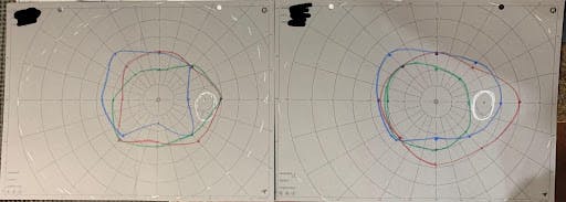
Image 1
Image 2 represents visual fields of the left eye. The picture on the left is the initial field prior to vision therapy, and is more constricted compared to the right eye. The picture on the right is the expanded field after roughly six sessions of therapy.

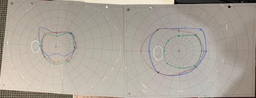
Image 2
Syntonic phototherapy educational courses dive into the four main syndromes that can be successfully treated with phototherapy. When deciding if a patient is eligible for phototherapy, review the four syndromes3 and see what treatment option fits best.
Four main syndromes to treat with phototherapy
Acute Syndrome
Symptoms
History of symptoms from:
- Recent infection
- Head trauma
- Stroke
- Fever
- Headaches
Signs
- Exo posture
- Convergence insufficiency
- Enlarged blind spot
- Visual field defects
- Oculomotor dysfunction
- Conjunctivitis / uveitis
- Cataracts
- Macular disease
Treatment
Blue-green filters
- Reduces cortical or retinal swelling, inflammation, and pain
Chronic Syndrome
Symptoms
Symptoms from chronic or degenerative diseases:
- Fatigue
- Loss of visual stamina
- Asthenopia
- Headaches
- Photophobia
- Transient blur
Signs
- Eso posture
- Accommodative insufficiency
- Low vergence recovery
- Constricted fields
Treatment
Yellow-green filters
- Physiological stabilizer and detoxifier
Indigo-red filters
- Add for any emotional instability
Emotional Fatigue
Symptoms
- Emotional exhaustion
- Depression, mood swings
- Asthenopia
- Headaches
- Transient blur
- Allergies, asthma, or fluid retention
Signs
- Exo posture with fatigue
- Significant Alpha-Omega pupil
- Low vergence break and recovery
Treatment
Red and indigo filters
- Balance out the SNS / PSNS systems
Lazy Eye
Symptoms
- Asthenopia
- Double vision
- Transient blur
- Poor eye-hand coordination
- Reading or learning challenges
- Short attention span
Signs
- Esotropia
- Amblyopia
- Reduced depth perception
- Poor fusion
Treatment
Red-orange filters
- Breaks synaptic resistance in cell membranes to overcome binocular suppression
Implementing phototherapy in your practice
Syntonics is an excellent treatment option in all optometric practice settings, but it takes a trained professional to understand when it can be, or may not be, beneficial to the patient. It is vital for any eye care professional interested in offering syntonics phototherapy in their office to take the educational courses provided by the College of Syntonic Optometry (CSO) and obtain an in-depth knowledge of its application. The CSO was established in 1933 and continues to educate eyecare professionals on the benefits of using phototherapy to treat visual conditions. The organization offers annual conferences with continuing-education courses and tons of resources to learn more!
