


Thyroid eye disease (TED) presents a unique clinical challenge due to its variable presentation, unpredictable course, and potential for significant visual morbidity. Because disease activity and severity can change over time, careful assessment and consistent monitoring are essential components of effective management.
Optometrists are often the first point of ocular care for these patients and play a critical role in identifying disease progression, stratifying risk, and coordinating timely referrals. This article reviews practical clinical scoring systems and follow-up strategies that support accurate documentation, informed decision-making, and collaborative care in patients with thyroid eye disease.
Brief overview of TED
TED is a chronic, immune-mediated orbitopathy most commonly associated with Graves’ disease, an autoimmune disorder that can cause hyperthyroidism.1 It represents the leading cause of unilateral and bilateral proptosis in adults.
Thyroid eye disease is characterized by autoimmune-driven inflammation of the orbital fibroblasts, adipocytes, and extraocular muscles, resulting in tissue expansion, remodeling, and fibrosis.1,2
Approximately 25 to 50% of patients with Graves’ disease develop TED, though the condition may also occur in euthyroid individuals or those with autoimmune hypothyroidism.2
The disease course typically progresses from an active inflammatory phase to a quiescent, inactive phase, with severity ranging from mild ocular surface irritation and cosmetic changes to sight-threatening complications such as exposure keratopathy and dysthyroid optic neuropathy.1,2
Pathophysiology
The pathophysiology of TED is complex and involves aberrant immune activation within the orbit. Orbital fibroblasts overexpress thyroid-stimulating hormone receptors and insulin-like growth factor-1 receptors, which are targeted by circulating autoantibodies, leading to fibroblast proliferation, adipogenesis, cytokine release, and hyaluronan production.1,2
Symptoms and risk factors
This inflammatory cascade recruits T cells and B cells, perpetuating orbital inflammation and driving either fat-predominant or muscle-predominant disease phenotypes. Progressive inflammation may result in extraocular muscle fibrosis, restrictive strabismus, and persistent diplopia.1,2
Clinical manifestations of TED include:1,2
- Eyelid retraction
- Proptosis
- Conjunctival injection
- Periorbital edema
- Chemosis
- Elevated intraocular pressure
- Ocular surface disease
Risk factors for TED include female sex, advancing age, smoking, thyroid dysfunction, genetic predisposition, radioactive iodine therapy, and coexisting autoimmune disease.1,2 Although men are less frequently affected, they often experience more severe disease. Early recognition, accurate assessment of disease activity and severity, and prompt achievement of euthyroid status are critical components of management.1,2
Figure 1: (A) External color photograph of chronic moderate thyroid eye disease with asymmetric proptosis left > right. (B) Coronal CT of orbits with thickening of the recti muscles left > right. (C) Sagittal cut of the same CT scan.


Figure 1: Thyroid Eye Disease© Courtesy of Ramy Rashad et al. Image used under CC BY 4.0.
Figure 2: Image of a TED patient with strabismus. The right eye is slightly elevated and abducted in comparison to the left eye, as noted by the position of the corneal light reflex. A slight left head tilt is also taken to help combat the diplopia. Right upper eyelid retraction is also noted.


Figure 2: Thyroid Eye Disease© Courtesy of Ramy Rashad et al. Image used under CC BY 4.0.
Management
Most patients with mild disease are managed conservatively with lubrication, risk factor modification, and observation, while active moderate-to-severe or sight-threatening disease requires systemic immunosuppression and, in selected cases, surgical intervention.1,2
Optometrists play a key role in early detection, longitudinal monitoring, identification of red flags, and timely referral within a multidisciplinary care model to optimize visual outcomes and long-term patient quality of life.2
Figures 3 and 4: Close-ups of a patient (Fig 1) before receiving TED treatment with a medical history of radioactive iodine (RAI) therapy and persistent ocular grittiness that advanced to diplopia, and (Fig 2) following TED treatment composed of infusions of monoclonal antibody therapy.

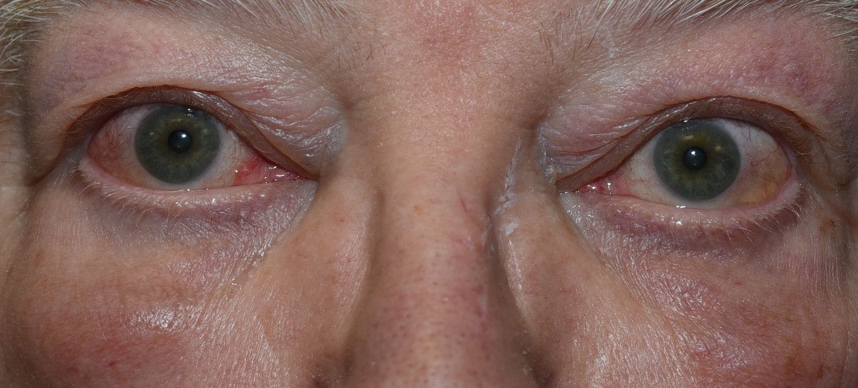
Figure 3: Courtesy of Audrey Ko, MD.

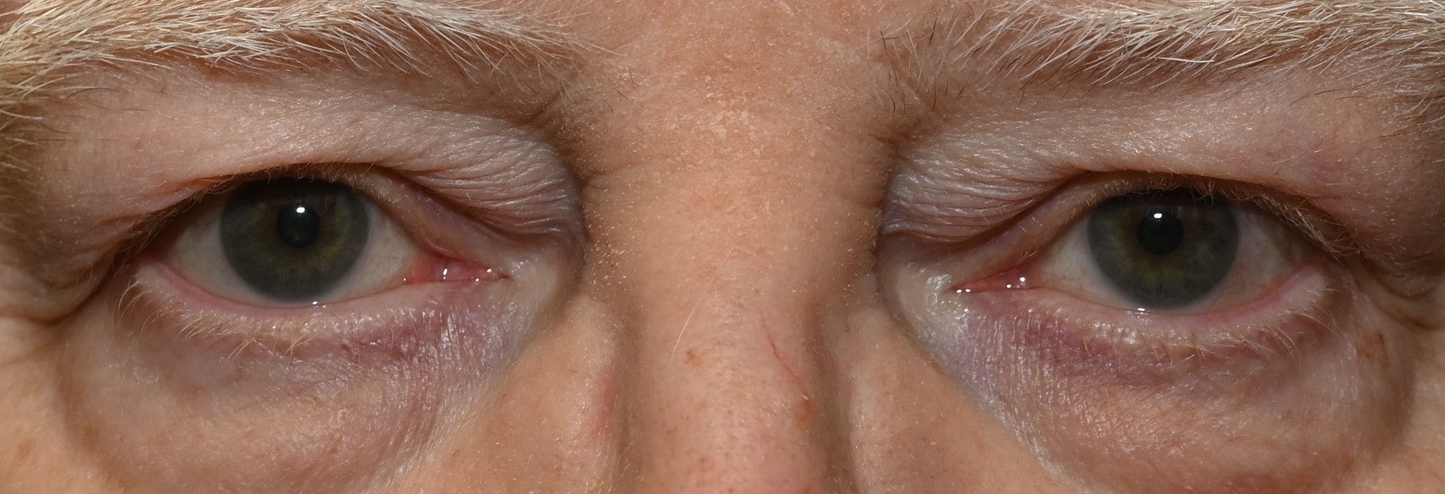
Figure 4: Courtesy of Audrey Ko, MD.
Importance of monitoring TED activity
Underlying pathogenic factors lead to the significant symptomatic challenges in patients with thyroid eye disease, as autoantibodies target orbital fibroblasts. The confined retro-orbital space becomes compromised by an inflammatory process leading to congestion from abnormal fat deposit, irregular orbital tissue, and fibrosis.1
Pearl: Monitoring subtle and often overlooked clinical presentations can streamline appropriate management for these patients.
Active versus inactive TED
Facial disfigurement presents in active TED or elevated thyroid-stimulating hormone (TSI). This occurs due to swelling of the extraocular muscles and adipose tissue, which can entrap and compromise ocular health and vision. Proptosis, diplopia, eye pain, eyelid and ocular adnexa edema, and light sensitivity are among some of the notable active signs and symptoms.3
Inactive TED patients have low, improved, or no inflammatory presentation but may retain some variation of chronic symptoms, such as eyelid retraction, dry eye, and scarring. Unfortunately, fibrosis can continue to cause detrimental progression in signs and symptoms, namely ocular motility, proptosis, and diplopia.3
TED severity grading systems
Treatments and management are dependent on mild, moderate, or severe TED. In 1969, Werner introduced the NO SPECS classification system—an acronym for No physical signs or symptoms, Only signs, Soft tissue involvement, Proptosis, Extraocular muscle signs, Corneal involvement, and Sight loss—with a modified version published in 1977.4
This system was designed to grade the severity of thyroid eye disease based on clinical findings. Importantly, NO SPECS does not assess disease activity; rather, it categorizes the extent of structural and functional involvement at a given point in time.4
Prior recording of TED clinical severity utilized the NO SPECS system with the following parameters:4,5,6
- No signs or symptoms
- Only Signs
- Soft tissues involvement (graded as absent, minimal, moderate, or marked)
- Proptosis (measured as absent, 3 to 4mm over upper normal limit, 5 to 7mm increase, or 8mm increase
- Extraocular muscle signs (graded as absent, limited with extreme gazes, restricted, or fixation of globe)
- Corneal involvement (graded as absent, stippling, ulceration, clouding/necrosis/perforation)
- Sight loss vision impact from optic nerve involvement and is broken into grades 0 to c
- 0 (Absent): no evidence of sight loss.
- a: Optic nerve involvement signs may be present (e.g., disc pallor or “choking”/disc swelling and/or a visual field defect), with visual acuity still relatively preserved at 20/20 to 20/60.
- b: Similar optic nerve involvement findings, but visual acuity is reduced to 20/70 to 20/200.
- c: Severe sight loss (“blindness”) with visual acuity worse than 20/200.
In addition to grading soft tissue involvement, recognizing and measuring diplopia helps classify the severity of TED.
Other pertinent guidelines for TED severity levels include:7,8
- Mild
- Eyelid retraction: <2mm
- Proptosis: <3mm above the reference range
- Corneal exposure: None
- Diplopia: Transient or no diplopia
- Moderate-to-severe
- Eyelid retraction: >2mm
- Proptosis: >3mm above the reference range
- Corneal exposure: Mild
- Diplopia: Inconstant or constant diplopia
- Sight-threatening
- Dysthyroid optic neuropathy
- Corneal exposure: Corneal breakdown
Clinical features of TED to consider when grading severity
Eyelid retraction is most commonly quantified in primary gaze by measuring the distance from the central corneal light reflex to the upper eyelid margin (margin reflex distance 1 [MRD1]). In adults, typical MRD1 values are about 4 to 5 mm; larger measurements are consistent with upper lid retraction.7,8
Proptosis is most commonly quantified clinically with Hertel exophthalmometry, which measures anterior globe position (forward protrusion) in millimeters relative to the lateral orbital rim.8,9
Corneal exposure is primarily assessed on clinical examination by documenting eyelid closure and exposure-related anatomic findings—especially the presence/degree of lagophthalmos (often recorded as the vertical palpebral fissure height during incomplete closure) and associated scleral show (commonly measured in millimeters in lid malposition/retraction evaluations).10
Determining the status of severe corneal exposure and optic nerve compression is critical to ensure an appropriate plan to avoid sight-threatening complications.
Standardized scoring systems for TED
Keeping consistent and accurate documentation of disease activity can be simplified with quantitative scoring systems: not only does it provide standardization, but it also gives a clear trend of the difference between disease activity and changes in severity.
Common clinical scoring systems allow for better reproducibility in scoring of signs and symptoms, playing a significant role in determining progression and management plans. Scoring provides clarity in essential multidisciplinary communication with endocrinologists and primary care physicians.
CAS, VISA, and EUGOGO scoring systems provide more diagnostic value for management.
Clinical Activity Score (CAS)
Years after establishing the NO SPECS classification system, a major conceptual shift occurred in 1989 with the introduction of the Clinical Activity Score (CAS), developed to help distinguish between the active inflammatory phase and the stable or inactive phase of the disease.4
Unlike NO SPECS, CAS focuses on the classic signs of acute inflammation. A score of ≥3 out of 7 at initial evaluation is considered indicative of active disease, helping guide decisions regarding immunosuppressive treatment.4
The CAS is attractive in day-to-day optometric practice because it’s simple, quick, validated, and widely used to define “active” disease (≥3) and to track change over time with the expanded 10-item version.
Its role in evaluating active disease and inflammation is equal weighting, point based on each of the following areas:11
- Spontaneous orbital pain
- Gaze evolved orbital pain
- Eyelid swelling
- Eyelid erythema
- Conjunctival injection
- Chemosis
- Inflammation of the caruncle or plica
Follow-up appointments (1 to 3 months) have additional factors that are added to the CAS score above:
- Increased proptosis of ≥ 2mm
- Reduced eye movements >8° in any direction
- Decrease visual acuity ≥ to 1 Snellen line
Active TED is categorized as CAS ≥ 3 at the initial visit or ≥ 4 at follow-up visits.12 Comprehensive evaluation may reveal other ocular findings, but severity of each also play a significant role in management.11
Limitations of the CAS
The tradeoff is reliability: inter-observer variability can be substantial, meaning two clinicians may legitimately score the same patient differently—sometimes enough to swing a treatment decision toward or away from immunosuppression.13,14
That variability is compounded by the CAS design itself: it’s binary (0/1) per item and gives equal weight to very different clinical realities (for example, eyelid redness and worsening visual function).
While CAS is the most common diagnostic tool that provides a standardized TED characterization score, it’s worth remembering that serious complications like dysthyroid optic neuropathy (DON) can present with low CAS scores, reinforcing that CAS should be used cautiously and ideally alongside other clinical and/or lab parameters.13,14
Alternatives to CAS: VISA and EUGOGO
When you want a system that organizes the exam and management priorities, the VISA and EUGOGO classifications offer advantages beyond CAS alone.
VISA
VISA follows a clinically intuitive flow—Vision, Inflammation, Strabismus, Appearance—and grades each domain independently, which can be particularly helpful when you’re monitoring targeted outcomes (e.g., motility vs. exposure) or communicating what is changing.13,15
Its inflammation scoring can be more nuanced than CAS because it allows graded severity (0 to 2) for edema/chemosis and it includes diurnal variation, a symptom many active-phase patients report and CAS does not capture.13,15
VISA also incorporates progression across its four parameters rather than relying on inflammation alone to define activity, which can feel more aligned with how optometrists follow disease evolution in the clinic. The downside is that VISA is more data-heavy, and some thresholds differ from CAS/EUGOGO (ex., the motility progression cutoff), so clinics need consistency in how measurements are taken.13,15
VISA scoring entails criteria where patients scoring above 5 are considered for aggressive therapeutic options. It has higher point values for more severe presentations, such as eyelid edema and chemosis.
VISA includes the following criteria:15
- Vision: Visual acuity and visual field; signs of optic nerve compression
- Inflammation: Eyelid swelling, conjunctival injection, chemosis, eyelid edema, eye pain
- Strabismus: Restricted motility, degree of misalignment
- Appearance: Proptosis, eyelid retraction, corneal exposure
Table 1: Outline of inflammatory scoring in the VISA system.15
| Criteria | Scoring |
|---|---|
| Retrobulbar ache (at rest of with gaze) | 1 |
| Eyelid edema | 1: Without redundant tissues 2: Redundant tissues, bulging of palpebral skin |
| Eyelid erythema | 1 |
| Conjunctival injection | 1 |
| Chemosis | 1: Conjunctiva behind grey lid line 2: Conjunctiva extends anterior to grey lid line |
| Caruncle edema | 1 |
| Diurnal variation | 1 |
Table 1: Courtesy of Jesús Barrio-Barrio et al.
EUGOGO
EUGOGO (European Group of Graves’ Orbitopathy) tends to shine when the goal is a practical, management-oriented severity framework: it classifies disease as mild, moderate-to-severe, or sight-threatening in a way that maps directly to how aggressively you treat and how urgently you refer. It also formalizes activity assessment through the modified CAS and supports soft-tissue grading with an atlas, which can improve standardization for some findings.13,15
Limitations still exist, as EUGOGO does not clearly record “change in severity” over time, and it groups moderate and severe into the same management category.13,15
EUGOGO distinguishes and is structured around severity. Among the evaluation of corneal integrity and optic neuropathy are the soft tissue changes, which include eyelid swelling, eyelid erythema, conjunctival redness, chemosis, and inflammation of caruncle or plica, eyelid retraction, and lagophthalmos.4 Proptosis and ocular motility are also measured.15
EUGOGO classification is as follows:13,15
- Mild (minimal quality of life [QOL] implications)
- Minor eyelid retraction <2mm
- Mild soft tissue involvement
- Exophthalmos <3mm above normal range
- Absent or Transient diplopia
- Moderate to Severe (QOL affected, justifying risk of immunosuppression, surgical intervention as applicable) with no sight-threatening Graves’ orbitopathy
- Eyelid retraction >2mm
- Moderate-severe soft tissue involvement
- Exophthalmos ≥3mm above normal range
- Diplopia (inconstant or constant)
- Sight-threatening (immediate intervention required)
- Optic neuropathy
- Corneal breakdown d/t severe exposure
- Globe subluxation
- Frozen Eye
- Choroidal fold
- Postural visual darkenings
Choosing a classification
In practice, an eyecare professional might prefer CAS for speed and routine activity tracking, VISA for a structured, domain-based approach that better captures symptom nuance and progression, or EUGOGO for a severity-driven framework that’s especially useful for triage and co-management.
Just keep in mind that VISA and EUGOGO aren’t interchangeable, so consistency within a practice (and across co-managing providers) matters.13,15
Treating thyroid eye disease: From mild to sight-threatening
Thyroid eye disease spans a wide clinical spectrum—ranging from mild, quality-of-life–limiting symptoms to true ocular emergencies with risk of irreversible vision loss. Management hinges on carefully weighing risk factors and severity while determining whether the disease is active or inactive, since therapy and follow-up cadence change meaningfully across phases.16,17
TED most often follows a biphasic course, with an active inflammatory period (often 1 to 3 years) followed by an inactive phase where residual fibrosis and tissue remodeling may persist.18,19
Non-surgical management of TED
Mild TED
For mild disease, the goal is to improve quality of life, promote remission, and prevent progression. Many mild TED patients do not experience symptoms severe enough to justify systemic pharmacologic or surgical intervention and can often be managed with watchful waiting.
When used, a single course of sodium selenite 100μg twice daily for 6 months may be considered, with duration limited to 6 months due to evidence limitations and toxicity risk; though the benefit in selenium-sufficient areas remains unclear.20
Moderate-to-severe TED (shorten the active phase and achieve remission)
When TED becomes moderate-to-severe—particularly in the active phase—medical therapy is aimed at reducing inflammatory activity and limiting downstream remodeling.
The following interventions can be used in managing moderate-to-severe TED:
- IV glucocorticoids are described as a first-line approach for moderate-to-severe active TED without major soft-tissue involvement (e.g., marked proptosis/diplopia).18,19,20
- A commonly cited regimen is IV methylprednisolone 500mg weekly for 6 weeks, then 250mg weekly for 6 weeks, with improvement in CAS reported in many patients. At the same time, the response of proptosis and diplopia is less predictable, and adverse effects increase with higher cumulative dosing.18,19,20
- Mycophenolate mofetil (MMF)—alone or in combination with IV glucocorticoids—has been studied in randomized trials, with reported benefits including CAS reduction/inactivation and, in some studies, improvements in proptosis and motility, with a tendency toward fewer adverse effects compared with IV glucocorticoids alone.18,20
- Practice recommendations differ: EUGOGO supports MMF + IV glucocorticoids as a first-line option, while other consensus groups have been less convinced by available data.18,20
- Teprotumumab (TEPEZZA, Amgen), an IGF-1R inhibitor targeting orbital fibroblast signaling, has reshaped the medical management conversation—especially for patients with active moderate-to-severe TED with soft-tissue involvement such as proptosis and diplopia.18,19,20
- In pivotal trial data, 83% of treated patients achieved ≥2mm reduction in proptosis compared with 10% on placebo, with additional improvements in CAS, diplopia, and quality of life.18,19,20
- Retreatment can be beneficial for some patients who flare after an initial course. Real-world durability is a key consideration, with long-term response noted as limited for some and a portion of patients requiring retreatment.18,19,20
- Adverse effects of TEPEZZA include GI symptoms (including worsening inflammatory bowel disease), muscle cramps/pain, alopecia, fatigue, hyperglycemia, and hearing loss.18
- Because of hearing loss risk, baseline audiogram is recommended prior to initiation.20 Teprotumumab should be avoided in pregnancy, and in patients aged 18 and younger due to insufficient safety data.
- Tocilizumab (IL-6 receptor blockade) is positioned as an option for patients who are intolerant of or unresponsive to IV glucocorticoids.
- While one randomized trial did not show improvement in soft-tissue involvement, real-world outcomes cited include improvements in proptosis and diplopia in a substantial portion of patients. Risks include severe infection, hepatotoxicity, and anaphylaxis.18,20
- Rituximab (anti-CD20) may be considered for corticosteroid-refractory cases; however, it is also described here as having little to no effect on proptosis or diplopia, making it less appropriate when soft-tissue manifestations dominate.20
- Orbital radiotherapy (OR) is presented as an established adjunct in active moderate-to-severe TED, especially for ocular motility/diplopia.18,19 It has been used for decades, potentially through depletion of lymphocytes and orbital fibrocytes.
- A commonly referenced regimen is 20 Gy in 10 daily fractions, though modified regimens may perform similarly.
- OR is noted to be avoided in patients younger than 35 and relatively contraindicated in severe hypertension or diabetes due to retinal vascular risk; transient symptom worsening may occur.18,19
Figure 5: (A) External color photograph of a patient with acute moderate thyroid eye disease beginning 2 months after radioactive iodine therapy. (B) One month later, with progression to severe thyroid eye disease. The exam showed increased conjunctival injection and chemosis. Sixty milligrams of oral prednisone with adjunctive radiotherapy was started. (C) Significant improvement in conjunctival and periorbital edema after 1 month. Steroids were slowly tapered over 8 months.


Figure 5: Thyroid Eye Disease© Courtesy of Ramy Rashad et al. Image used under CC BY 4.0.
To review drugs currently in the FDA pipeline for thyroid eye disease, check out the Clinical Trials Tracker!
Sight-threatening TED: A medical emergency
Sight-threatening TED can stem from dysthyroid optic neuropathy (DON), severe corneal breakdown, or rarely, globe subluxation. This presentation requires immediate recognition and escalation because vision loss may be acute or evolve over weeks to months.20
For DON due to optic nerve compression, IV glucocorticoids are described as first-line therapy, with a regimen of IV methylprednisolone 500 to 1,000mg daily for 3 consecutive or alternate days.20 If the response is inadequate, urgent orbital decompression is recommended.
If DON is primarily due to optic nerve stretching from proptosis, surgical decompression is emphasized as the appropriate next step, since this mechanism rarely responds to medical therapy.20 Additionally, severe corneal involvement warrants urgent referral to corneal specialists to initiate treatment, given the risk of permanent morbidity.
Surgical interventions for TED
Surgical interventions—including orbital decompression, strabismus surgery, and eyelid procedures—continue to play a critical role once inflammatory activity has stabilized, as well as in urgent scenarios such as compressive optic neuropathy.19
Although refinements in technique, including endoscopic approaches, have lowered complication rates and enhanced aesthetic results, surgical management primarily corrects the structural effects of tissue expansion and fibrosis rather than treating the underlying disease process itself.19
Orbital decompression is indicated urgently when DON fails to respond to IV steroids. Decompression is also listed among oculoplastic surgical options used for proptosis management.19,20
Importantly, surgical decision-making is not isolated from medical therapy. Patients requiring decompression for DON may also need adjuvant medical therapy or orbital radiotherapy to inactivate the disease. Referral to oculoplastics is also appropriate for consultation on the timing and sequencing of decompression, strabismus correction, and eyelid procedures.20
Figure 6: (A) Photograph of acute moderate TED with periorbital edema, upper and lower eyelid retraction, and chemosis. (B) Same patient in chronic phase. (C) Patient status post-bilateral orbital fat decompression. No further procedures were needed.


Figure 6: Thyroid Eye Disease© Courtesy of Ramy Rashad et al. Image used under CC BY 4.0.
Co-management of thyroid eye disease
TED is best managed as a multidisciplinary condition. Ophthalmic care benefits from tight coordination with endocrinology for systemic disease control and oculoplastics (and, as needed, neuro-ophthalmology and cornea) for escalation pathways and definitive interventions.
Follow-up strategies in TED
Follow-up intervals should be tailored to risk factors, severity, and whether TED is active versus inactive. Notably, 50% of patients diagnosed with Graves’ disease develop TED within the first 18 months,1,2 and this early window may justify shorter follow-up intervals—especially when compounding risk factors are present.21
Follow-up periods for TED patients:21
- Inactive TED: Follow-up every 6 to 12 months based on findings. Even for inactive or mildly active patients, it can be worthwhile to reassess 1 to 3 months after the initial examination to track changes in CAS and identify early shifts.21
- Moderate-to-severe active TED: follow sooner as indicated. Worsening findings may prompt closer monitoring or treatment adjustment. If active TED appears stable and there is no optic nerve compression, monitoring every 3 to 6 months is reasonable.21
Urgent referrals for sight-threatening TED
Vision-compromising TED requires emergent action. Dysthyroid optic neuropathy and severe corneal involvement warrant urgent referral to the appropriate specialists. Corneal specialists may need to initiate treatment for necrosis or perforation, and timely referral to neuro-ophthalmology and oculoplastics is critical to prevent permanent vision loss.
Because TED can evolve, referral communication should be dynamic and explicit:
- Referral notes to endocrinology should clearly state the TED diagnosis and provide meaningful updates on activity/severity changes. Requesting endocrinology notes helps identify changes in thyroid status that may influence overall risk and stability.19,20
- When available, obtaining a full summary of current and past treatments and any CT/MRI imaging is helpful for longitudinal care and to contextualize changes in exam findings.20
- Referral to oculoplastics supports consultation on proptosis, strabismus, eyelid retraction, and decompression planning.
- Co-management should also recognize that TED treatment is not limited to surgical approaches; non-surgical options discussed here include high-dose systemic glucocorticoids and IV infusion treatments such as teprotumumab, with immunosuppressive agents considered in selected moderate-to-severe cases.19,20
In practice, strong co-management means aligning on phase (active vs. inactive), documenting objective changes (e.g., CAS trends, optic nerve findings, corneal status), and maintaining a shared escalation plan—so mild disease is monitored thoughtfully and sight-threatening disease is treated without delay.20
Closing
Due to multifactorial conditions, with various and varying signs and symptoms, TED is often misdiagnosed or overlooked. The entire process can be incredibly frustrating as its physical ramifications can take an emotional toll.
Stay one step ahead of TED by analysing your clinical findings in patients with underlying thyroid dysfunction. It can help redefine early diagnosis in these patients, potentially preserving the patient’s TED prognosis.
