


Pseudoexfoliation syndrome (PXS)—also referred to as exfoliation syndrome—is a systemic condition characterized by its deposition of white dandruff-like exfoliative material in various organ systems.
In terms of ocular manifestations, these deposits can clog the trabecular meshwork, leading to an elevation in intraocular pressure (IOP).
If this elevated IOP goes unmanaged or uncontrolled, then it can lead to glaucomatous optic nerve thinning and visual field defects, converting into pseudoexfoliation glaucoma.
Overview of pseudoexfoliation glaucoma
Pseudoexfoliation glaucoma (PXG) is a secondary open-angle glaucoma derived from PXS that has eventually led to a diagnosis of glaucoma.
This condition has been linked to the Lysol-oxidate-like 1 (LOXL1) gene in Scandinavian populations.1 This fibrillar, dandruff-like tissue is typically deposited on various ocular tissues, such as the lens capsule, ciliary body, corneal endothelium, zonules, iris, and the trabecular meshwork.1,2
Deposition of fibrillar material occurs in various systemic organ systems including ocular tissues, leading to downstream effects such as obstruction of the trabecular meshwork and weakening of zonular attachments. The presence of deposits in the juxtacanalicular layer of the trabecular meshwork demonstrated in transmission electron microscopy indicates that there is a local production of the material.2
The combination of local production and deposition of fibrillar material causes obstruction of the trabecular meshwork and elevated intraocular pressure. The risk of conversion from pseudoexfoliation syndrome to pseudoexfoliation glaucoma increases with age.
Clinical presentation and diagnosis of PXG
Pseudoexfoliation syndrome and glaucoma are clinical diagnoses. PXS can present monocularly or binocularly; when bilateral, it can be asymmetric. Conversion of PXS to PXG occurs when elevated IOP, glaucomatous nerve damage, and visual field loss are seen.
Exam findings in pseudoexfoliation glaucoma:
- Moth-eaten pupil: Characteristic transillumination defects of the iris at the pupillary margin.
- Bulls eye anterior lens capsule: Pseudoexfoliative material on the anterior capsule is deposited in a bulls-eye pattern due to iris movement/rubbing off material, which leaves a clear intermediate zone between a relatively opacified central and peripheral zone
- Optic nerve cupping: Can be visualized on fundus exam
- Deposition of pseudoexfoliative material: Can be visualized on the pupillary margin
Imaging for pseudoexfoliation glaucoma:
- Visual field: Glaucomatous visual field defects present
- Optical coherence tomography (OCT) retinal nerve fiber layer (RNFL) imaging: Glaucomatous optic nerve thinning and atrophy
Gonioscopy could show:
- Open angle
- Narrow angle due to weakened zonules
- Increased pigmentation of trabecular meshwork
- Sampaolesi line—a pigmented line anterior to Schwalbe’s line
- Settling of pseudoexfoliative material in the inferior angle
Management of PXG
Medical management of pseudoexfoliation glaucoma
Standard medical therapy with aqueous suppression and agents that increase outflow is employed initially. In comparison to primary open-angle glaucoma (POAG), PXG usually follows a more aggressive clinical course, with an increased possibility of surgical intervention.
Surgical management of pseudoexfoliation glaucoma
Selective laser trabeculoplasty for pXG
Laser surgery in PXG has been shown to lead to an IOP decrease of up to 31.6%. To have a more significant response with selective laser trabeculoplasty (SLT), doing 360° of treatment (compared to 90 or 180°) can lead to better IOP reduction.3
In a study done on Japanese patients, it was shown that the baseline IOP at the time of SLT can affect the magnitude of reduction, with IOP >21 mmHg leading to a more significant reduction.3 In addition to significant IOP reduction, a decrease in eyedrop burden in patients has also been observed.
Incisional surgery for PXG
Studies have shown that patients with PXG had similar outcomes as POAG with incisional surgery (e.g., trabeculectomy).2,4,5
Surgical outcomes in terms of post-operative medical treatment, surgical complications, IOP reduction, and the use of antifibrotic agents (mitomycin C or 5-fluorouracil) were not statistically different when measured between patients with PXG versus POAG.4
Cataract surgery considerations for patients with PXG and PXS
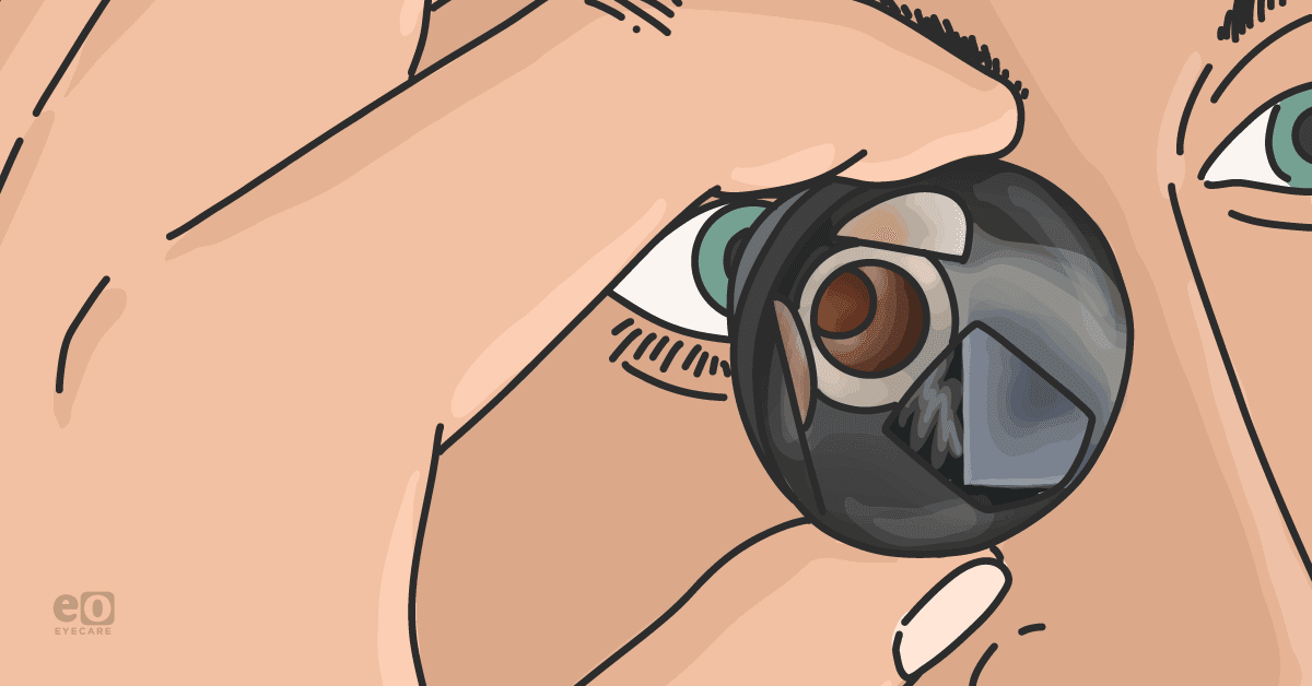
Patients with PXG and PXS have an increased risk for complications during cataract surgery due to their unique pathology.
1. Dilation
The deposition of exfoliative material onto the iris vasculature and iris stroma leads to poor dilation.
When evaluating these patients for cataract surgery, the surgeon should take note of dilation, anticipate intraoperative miosis, and prepare to use iris expanders such as iris hooks or a pupillary expansion ring.
2. Zonular instability
Weakened zonules increase the risk of intraoperative zonular dehiscence. On cataract evaluation, phacodonesis, shallow anterior chamber depth, and narrow angles indicate that zonular instability is likely present.
Operative planning should include consideration of capsular tension hooks, capsular tension rings, and possible anterior vitrectomy.
3. Lens removal
With the increased risk for zonular dehiscence, techniques utilizing less phacoemulsification energy decrease the amount of intraoperative zonular stress, thus potentially mitigating surgical complications.
Creating a larger capsulorhexis, prolapsing the lens out of the bag for removal, and performing chopping techniques (horizontal or vertical chopping) are all strategies that would help reduce the zonular stress.
In conclusion
Between 44 to 50% of patients with PXS are ultimately diagnosed with glaucoma, making it crucial that an eyecare professional performs an annual assessment on these individuals.5
This is especially true for those who develop PFG, as they tend to have more rapid progression than those with POAG.5,6 In addition, patients with PXS more often require surgical intervention.
