While learning about these conditions in my
first year of ophthalmology residency, one of my attendings brought up a point that has stuck with me since. He pointed out that along with the emergent conditions, it is important to also build one’s physical exams skills to evaluate for diseases which can be easily overlooked. The first disease we discussed was pseudoexfoliation syndrome.
He explained that due to its clinically subtle signs it can be easily missed, particularly by new trainees. This could prove to be detrimental, due to the complications that pseudoexfoliation syndrome could cause, such as
glaucoma or lens subluxation, which could then otherwise be considered as primary disease states.
He emphasized that by making this diagnosis early we could optimize our treatment plans for our patients, select treatment modalities proven to be more effective, and adequately prepare for surgical interventions when necessary.
Definition and clinical manifestations of pseudoexfoliation syndrome
Pseudoexfoliation Syndrome (PSX) is a systemic condition in which flaky fibrillar material is deposited throughout various organ systems such as the lungs, liver, kidneys and gallbladder.1 In the eyes it can be found on any of the structures within the anterior and posterior chamber.
PSX is commonly found in individuals over the age of 50 with a prevalence of 5% in people 75-85 years of age.2 Interestingly, it has been found to have the highest prevalence in the Scandinavian population.2
The development of PSX has been linked to mutations of the LOXL1 gene. This gene encodes for proteins, such as lysyl oxidase, which are involved in the formation and maintenance of extracellular matrix and crosslinking of elastin and collagen.2 The compromise of elastic structures and presence of this flaky material are the cause of the pathologic changes and clinical manifestations associated with PSX.
Changes in the eye with PSX, from anterior to posterior
Cornea
Pseudoexfoliative material can deposit on the endothelial surface of the cornea. Studies have shown that there is an increased association of lower corneal endothelial cell count in patients with PSX.3 Like pigment dispersion syndrome, a Krukenberg spindle (vertically oriented streak of pigment) can sometimes be seen in PSX, although it is less common.
Trabecular meshwork
The deposition of fibrillar material into the trabecular meshwork results in mechanical obstruction of the aqueous humor drainage pathway. This is the most common cause of secondary open angle glaucoma. The incidence has been reported to be as high as 15-30% in patients with pseudoexfoliation syndrome.2 The prognosis of glaucoma is also worse than in primary open angle glaucoma. This is due to the commonly higher intraocular pressure and fluctuations that can occur with fibrillar material liberation.2
Also, like pigment dispersion syndrome, a Sampaolesi line (pigmented Schwalbe’s line) may be appreciated on gonioscopy.
Iris
On close examination of the pupillary margin, deposition of fibrillar material can also be seen. Transillumination can reveal atrophy of the iris close to the pupil margin. It is believed this is related to infiltration of pseudoexfoliative material within the iris stroma. This is also linked to reduced function of the pupillary dilator muscle resulting in poor dilation.
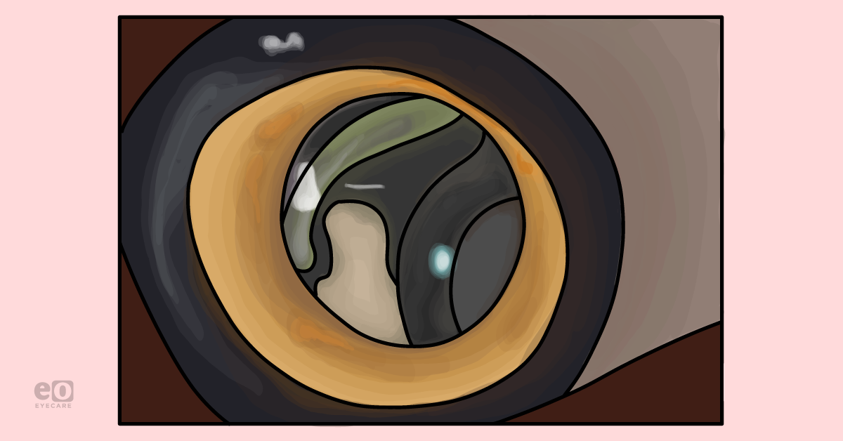
Lens
Flaky white material typically deposits on the anterior lens surface. A common “bulls-eye” appearance can be seen with a central area of flaky material, clear intermediate zone and peripheral deposition. This is thought to occur due to mechanical rubbing of the posterior iris on the anterior lens capsule creating the clear zone. Compromise of zonular fiber elasticity also results in laxity of the crystalline lens, which may be appreciated as phacodenesis on examination.
This can increase the risk of pupillary block and cause angle closure glaucoma. This can also lead to subluxation of the lens from minor trauma and instability of the capsular bag during intraocular surgery. Pseudoexfoliative material can be appreciated on the zonules in cases of subluxed lenses.
PSX treatment and considerations
Treatment of pseudoexfoliation syndrome involves the management of the secondary complications it causes. When
assessing for glaucoma it is important to consider the potential mixed mechanism etiology from pupillary block and trabecular meshwork obstruction. This can alter the approach taken to address elevated intraocular pressure with hypotensive drops, laser treatment, lens extraction, or a combination of all of these.
In regard to laser treatment, recent studies have shown that
selective laser trabeculoplasty (SLT) may have a stronger IOP lowering effect in patients with PSX than POAG.
2 However, this effect has also been shorter-lived and may require repeat treatments which could have reduced efficacy over time.
When contemplating
cataract extraction in patients diagnosed with PSX it is important to consider the anatomic compromise it may cause intraoperatively. For instance, poor pupillary dilation can compromise visualization during surgery. Zonular laxity can also cause instability of the lens during phacoemulsification or even zonular dehiscence.
As a result, both of these changes can increase the risk of complications such as
posterior capsular rupture, vitreous loss, or lens subluxation. By making the diagnosis of PSX prior to surgery, surgeons can plan to use various tools such as pupil expansion devices or capsular tension rings to reduce the risk of intraoperative complications and improve patient outcomes.
Furthermore, PSX has also been associated with increased rates of capsular phimosis and IOL dislocation
following cataract surgery.
4 Surgeons can closely monitor patients postoperatively for the need of YAG-capsulotomy of the anterior lens capsule and educate their patients on precautions to prevent IOL dislocation.
References
- Syed, Nasreen A. 2020-2021 Basic and Clinical Science Course, Section 04: Ophthalmic Pathology and Intraocular Tumors. American Academy of Ophthalmology, 2020.
- Salmon, John. Kanski’s Clinical Ophthalmology: A Systematic Approach. 9th ed., Elsevier Health Sciences, 2019.
- Aoki, T., Kitazawa, K., Inatomi, T. et al. Risk Factors for Corneal Endothelial Cell Loss in Patients with Pseudoexfoliation Syndrome. Sci Rep 10, 7260 (2020). https://doi.org/10.1038/s41598-020-64126-w
- Tsai, Linda M. 200-2021 Basic and Clinical Science Course, Section 11: Lens and Cataract. American Academy of Ophthalmology, 2020.