


Pediatric corneal disorders come with unique diagnostic and therapeutic challenges compared with adult corneal disease. The primary difference lies in the potential for permanent disruption of visual development.
For instance, transient corneal opacity during early childhood can lead to deprivation amblyopia, resulting in lifelong visual impairment if not treated promptly.1-4 Therefore early identification and appropriate management of pediatric corneal disorders are critical.
Pediatric corneal disorders fast facts1-4
- Pediatric corneal disorders can broadly be classified into congenital and acquired conditions.
- Each category has distinct clinical implications, diagnostic pathways, and management considerations.
- Congenital anomalies, such as anterior segment dysgenesis and endothelial dystrophies, remain important causes of pediatric corneal opacity.
- Acquired conditions, including trauma, infection, and ectatic disorders, contribute significantly to visual morbidity worldwide.
- Pediatric keratoconus has gained increasing clinical relevance due to its aggressive progression compared with adult disease.
- Pediatric keratoconus has a strong association with ocular allergy and eye rubbing behavior.
- Early diagnosis, appropriate imaging, and timely intervention remain essential to prevent long-term visual sequelae in this population.
Overview of pediatric corneal disorders
In children, corneal disease is not just an ocular surface problem—it is both a vision and neurodevelopment issue. During early visual development, disruption of clear optical input can affect cortical visual development, binocular vision, and long-term visual potential.
This also adds to the importance of early diagnosis and intervention of pediatric corneal pathology. The prevalence of pediatric corneal disease varies globally and is influenced by genetic factors, environmental exposure, nutritional status, and access to healthcare.
Congenital corneal disorders
Congenital corneal disorders typically present at birth or early infancy and frequently require multidisciplinary evaluation due to systemic and genetic associations.
Peters anomaly
Peters anomaly is characterized by central corneal opacity associated with defects in Descemet membrane and corneal endothelium. It may be accompanied by iridocorneal or lenticulocorneal adhesions.5,6
Visual prognosis depends on laterality, severity, and associated ocular abnormalities such as glaucoma or microphthalmia. Early surgical intervention, including keratoplasty, may be necessary in visually significant cases to prevent deprivation amblyopia.5,6
Congenital hereditary endothelial dystrophy (CHED)
Congenital hereditary endothelial dystrophy (CHED) typically presents with bilateral corneal edema and diffuse stromal haze from birth or early infancy.5,7 The disease results from endothelial dysfunction leading to impaired corneal deturgescence.
Severe cases may require corneal transplantation for visual rehabilitation, although timing must be carefully considered due to amblyopia risk.5,7
Sclerocornea
Sclerocornea represents abnormal scleralization of the cornea with vascularization and loss of normal limbal architecture.5,7 The condition may occur in isolation or as part of systemic syndromes. Imaging techniques such as ultrasound biomicroscopy may help differentiate it from other congenital corneal anomalies.
Microcornea and megalocornea
Corneal size abnormalities are clinically significant because of associations with refractive error, glaucoma risk, and anterior segment developmental anomalies. Megalocornea must be differentiated from congenital glaucoma, as intraocular pressure remains normal in true megalocornea.6,7
Management of congenital corneal disease centers on early visual axis rehabilitation, refractive correction, amblyopia therapy, and systemic evaluation when indicated.6,7
Want to learn more about microcornea, check out the article A Case Study on the Role of RGP Lenses in Management of Microcornea!
Download the cheat sheet here!

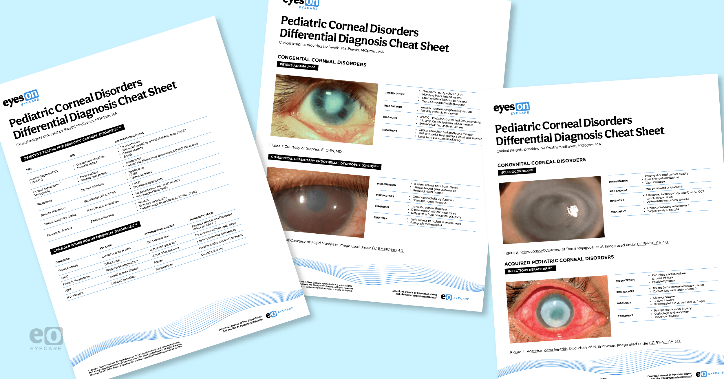
Pediatric Corneal Disorders Differential Diagnosis Cheat Sheet
This differential diagnosis cheat sheet features insights into objective testing, diagnostic pearls, common misdiagnoses, and slit lamp imaging of pediatric corneal disorders.

Acquired pediatric corneal disorders
Acquired pediatric corneal disease includes infectious, inflammatory, traumatic, neurotrophic, and ectatic conditions.
Infectious keratitis
Trauma remains the most common predisposing factor for pediatric microbial keratitis. Bacterial keratitis typically presents with rapid onset pain, redness, stromal infiltrates, and potential hypopyon formation.
Further, fungal keratitis often follows trauma involving organic material and may show satellite lesions. Early and aggressive treatment is critical to prevent permanent corneal scarring and amblyopia.6,8
In children with large, central, deep, atypical, or non-responsive ulcers, corneal scraping and microbiological culture should be considered. Obtaining cultures may require examination under anesthesia to ensure adequate sampling and patient comfort.
Treatment is experimental and depends on the suspected organism and severity of the ulcer:6,8
- Bacterial keratitis: Fourth-generation fluoroquinolones (moxifloxacin, gatifloxacin) are commonly used as first-line agents and are generally well tolerated in children.
- Severe ulcers: May require fortified antibiotics under corneal specialist supervision.
- Drug precautions: Avoid tetracyclines in young children; use aminoglycosides cautiously due to epithelial toxicity.
- Herpex simplex virus (HSV) keratitis: Treated with topical antivirals with or without oral acyclovir depending on age and severity.
Early treatment is essential to prevent corneal scarring, amblyopia, and permanent vision loss.5,8,9
Pediatric blepharokeratoconjunctivitis (PBKC)
Pediatric blepharokeratoconjunctivitis (PBKC) is a chronic inflammatory disorder involving the eyelid margins, conjunctiva, and cornea and is an important yet frequently underrecognized cause of pediatric corneal morbidity.9
Children may present with:9
- Recurrent red eye
- Photophobia
- Tearing
- Recurrent chalazia
- Peripheral corneal infiltrates or vascularization
Because it is often misdiagnosed as allergic conjunctivitis, delayed treatment can result in corneal scarring and amblyopia. Management includes meticulous lid hygiene, warm compresses, topical antibiotics, and short courses of topical corticosteroids when significant inflammation is present.9 Long-term maintenance therapy is often required to prevent recurrence.
Pediatric keratoconus
Pediatric keratoconus is increasingly recognized as a major cause of visual impairment in adolescents. Compared with adult disease, pediatric keratoconus demonstrates faster progression, greater severity at diagnosis, and stronger associations with ocular allergy and eye rubbing.1-3
Some studies report progression in up to 88% of pediatric cases within one year of diagnosis, supporting early corneal collagen cross-linking.1-3 The pathophysiology is multifactorial and includes genetic predisposition, inflammatory cytokine imbalance, oxidative stress, and enzymatic collagen degradation.
Associated systemic conditions include:1-3
- Down syndrome
- Marfan syndrome
- Ehlers-Danlos syndrome
Short on time? Download the Pediatric Corneal Disorders Differential Diagnosis Cheat Sheet!
Trauma-related corneal disease
Trauma remains a leading cause of pediatric corneal morbidity worldwide.6,10 Central corneal scars may cause irregular astigmatism and amblyopia if not treated appropriately.
Based on the extent and location of the injury, treatment options may include lubrication, topical antibiotics, bandage contact lenses, cyanoacrylate tissue adhesive for small perforations.6,10
Surgical intervention such as corneal laceration repair or keratoplasty are considered in severe cases. Subsequent visual rehabilitation often requires refractive correction, rigid gas permeable or scleral contact lenses for irregular astigmatism, and amblyopia therapy.6,10 Preventive strategies including eye protection education remain critical.8
Neurotrophic and exposure keratopathy
These conditions occur in children with cranial nerve dysfunction, eyelid abnormalities, or neurological disease.6 Reduced corneal sensation or inadequate eyelid closure can result in persistent epithelial defects, stromal ulceration, and scarring.
Management should be focused on restoring and maintaining ocular surface integrity with options like:6
- Preservative-free lubricants
- Lubricating ointments
- Moisture chambers
- Punctal occlusion
- Prophylactic topical antibiotics when epithelial defects are present
Bandage contact lenses or amniotic membrane transplantation may be considered for persistent defects, while temporary or permanent tarsorrhaphy may be necessary in severe cases.6
Treating the underlying neurologic or eyelid disorder is equally important. Long-term follow-up is essential to monitor healing, prevent amblyopia, and detect recurrent epithelial breakdown.6
Diagnostic approaches in pediatric patients
Examination techniques vary significantly based on age. Infants often require examination under anesthesia for accurate anterior segment evaluation and intraocular pressure measurement. Older children can typically undergo slit lamp examination and subjective refraction.10,11
Essential diagnostic tools
Key diagnostic modalities include:1-3,11
- Corneal topography and tomography for ectatic disorders
- Anterior segment OCT for structural assessment
- Pachymetry for corneal thickness evaluation
- Specular microscopy in selected endothelial disorders
These tools are critical for early detection of subtle corneal abnormalities, particularly in pediatric keratoconus.1-3,11
Red flags requiring urgent referral
Urgent referral should be considered in:1-3,11
- Central corneal opacity in infants
- Rapidly progressive ectasia
- Corneal ulcer with stromal thinning or melt
- Corneal opacity associated with systemic developmental delay
As an eyecare provider it is important to keep in mind that early specialist involvement significantly improves visual outcomes.4,5
Surgical considerations and long-term monitoring
Pediatric keratoplasty remains technically challenging due to smaller ocular anatomy, increased vitreous pressure, and the need for repeated examinations under anesthesia.10,12 Surgical outcomes depend on primary diagnosis, timing of surgery, and post-operative compliance.
Lamellar keratoplasty techniques are increasingly preferred when feasible due to lower rejection risk and improved structural stability. However, penetrating keratoplasty remains necessary in many congenital corneal opacity cases.4,10
Post-operative care must include aggressive amblyopia therapy, as graft clarity alone does not guarantee functional visual recovery.10,12
5 key takeaways1-4
- Pediatric corneal disease is a visual development emergency.
- Congenital corneal opacity requires systemic and genetic evaluation.
- Pediatric keratoconus progresses faster and requires early screening and intervention.
- Trauma and infection remain major preventable causes of pediatric corneal morbidity globally.
- Long-term visual outcomes depend heavily on amblyopia therapy and follow-up compliance.
Conclusion
Pediatric corneal disorders encompass a wide spectrum of congenital, infectious, inflammatory, and ectatic diseases. The major clinical challenge lies in balancing early intervention with long-term visual rehabilitation.
In children, corneal disease must be approached not only as an ocular condition but as a neurodevelopmental visual risk. Early diagnosis, appropriate imaging, and timely management are essential to prevent amblyopia and lifelong visual disability.
Multidisciplinary care involving optometrists, pediatric ophthalmologists, cornea specialists, and low vision rehabilitation teams is often required. Ultimately, early recognition and proactive management remain the most important determinants of visual outcomes in pediatric corneal disease.1,4,6

