


As a pediatric optometrist, one of the most common concerns that I hear from parents is the question of whether or not their child is color blind.
While it may seem like a straightforward yes or no question, the nuances of color vision deficiencies and how they relate to the health of the eyes can snowball into a much more in-depth discussion.
Some color deficiencies are associated with ocular pathology, while others are completely benign. It is important to know the differences and when additional testing is needed.
Overview of color vision deficiency
The term “color blindness” is a bit misleading, as color vision deficiency (CVD) exists in varying degrees, and most “color blind” patients do perceive color, only in a different manner than patients with normal color vision. There is a difference in the configuration of their cones (i.e., the color-sensing cells of the retina).
There are three main types of CVD depending on which cones are affected:
- Red-green
- Blue-yellow
- Achromatopsia
Patients with a red-green or blue-yellow color deficiency may have a partial deficiency (an -anomaly or anomalous trichromacy) or a complete lack of a certain cone type (an -anopia, or dichromacy). Achromatopsia, or rod monochromacy, is a complete lack of cones and color vision.
Worldwide, around 300 million people have some form of CVD. Red-green color blindness, which is usually inherited, affects around 1 in 12 males and 1 in 200 females. Blue-yellow color deficiency affects around 1 in 10,000 people, and achromatopsia affects around 1 in 30,000; neither of these has a gender predilection.1
Patients with CVD may be unaware of their condition because they have no comparison—this is how they have always seen the world around them. Family members or others in their social circle may notice that they have trouble differentiating between certain shades or colors.
Children starting school may have difficulty with certain learning materials relying on correct color differentiation. These symptoms will be more pronounced in dichromats than in anomalous trichromats.
Red-green color deficiency
Red-green color deficiency, the most common CVD, can be further subdivided into a deutan deficiency or a protan deficiency. A deutan deficiency affects the patient’s M cones, which are responsible for the perception of green light, while protan defects impact the L cones responsible for red light perception.
Red-green color deficiency is most frequently inherited with an X-linked recessive inheritance pattern, which is why it impacts men more frequently than women.
Blue-yellow color deficiency and achromatopsia
Blue-yellow color deficiencies can be inherited or the result of trauma or disease and affect the S cones, which perceive blue light in normal patients. They can be acquired through trauma (chemical exposure, welding lights, etc.), certain medications, ocular pathology (glaucoma, cataracts, age-related macular degeneration, etc.), and nervous system disorders.2
Achromatopsia occurs when all of a patient’s cones are either missing or working improperly. This is frequently associated with amblyopia, nystagmus, light sensitivity, and poor vision.1 Inherited blue-yellow CVD and achromatopsia do not involve X chromosomes.
Table 1 lists a broad overview of color vision deficiencies and their prevalence.3
| Color Vision Deficiency | Type | Cones Affected | Prevalence (Males/Females) |
|---|---|---|---|
| Protanopia | Dichromacy | L cones | 1%/0.01% |
| Deuteranopia | Dichromacy | M cones | 1.5%/0.01% |
| Tritanopia | Dichromacy | S cones | 0.008%/0.008% |
| Protanomaly | Anomalous Trichromacy | L cones | 1%/0.01% |
| Deuteranomaly | Anomalous Trichromacy | M cones | 5%/0.4% |
| Tritanomaly | Anomalous Trichromacy | S cones | Rare/Rare |
| Achromatopisa | Monochromacy | Most or all cones | Rare/Rare |
Table 1: Courtesy of Marni Robbins, OD.
Available testing for color vision deficiencies
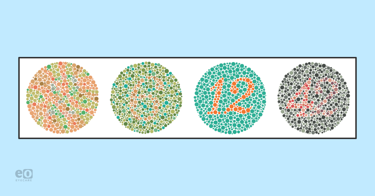
There are several different tests for color vision deficiencies. The most common test is a color plate test, during which the test administrator will present a series of plates featuring colored symbols against a contrasting background.
This symbol is most frequently a number, but color plates for preschool-age children may utilize shapes; for example, Color Vision Testing Made Easy (CVTME) is a shape-based test appropriate for patients as young as 3 years old.4
Patients with normal color vision will be able to identify most, if not all, of the symbols, while those with color deficiency will only identify symbols on plates designed to test for their deficiency. These tests are easy to incorporate as part of the workup during a comprehensive eye exam and can be performed by the practitioner or a technician.5
Ishihara and HRR
The two main color plate tests are the Ishihara and HRR tests. The Ishihara is geared towards red-green deficiencies and features both general screening plates and more sensitive plates differentiating between protan and deutan deficiencies.
The HRR is unique in that it can also test for blue-yellow defects, making it valuable in evaluating acquired color vision deficiency.5
Figure 1 demonstrates an example of an Ishihara color plate.

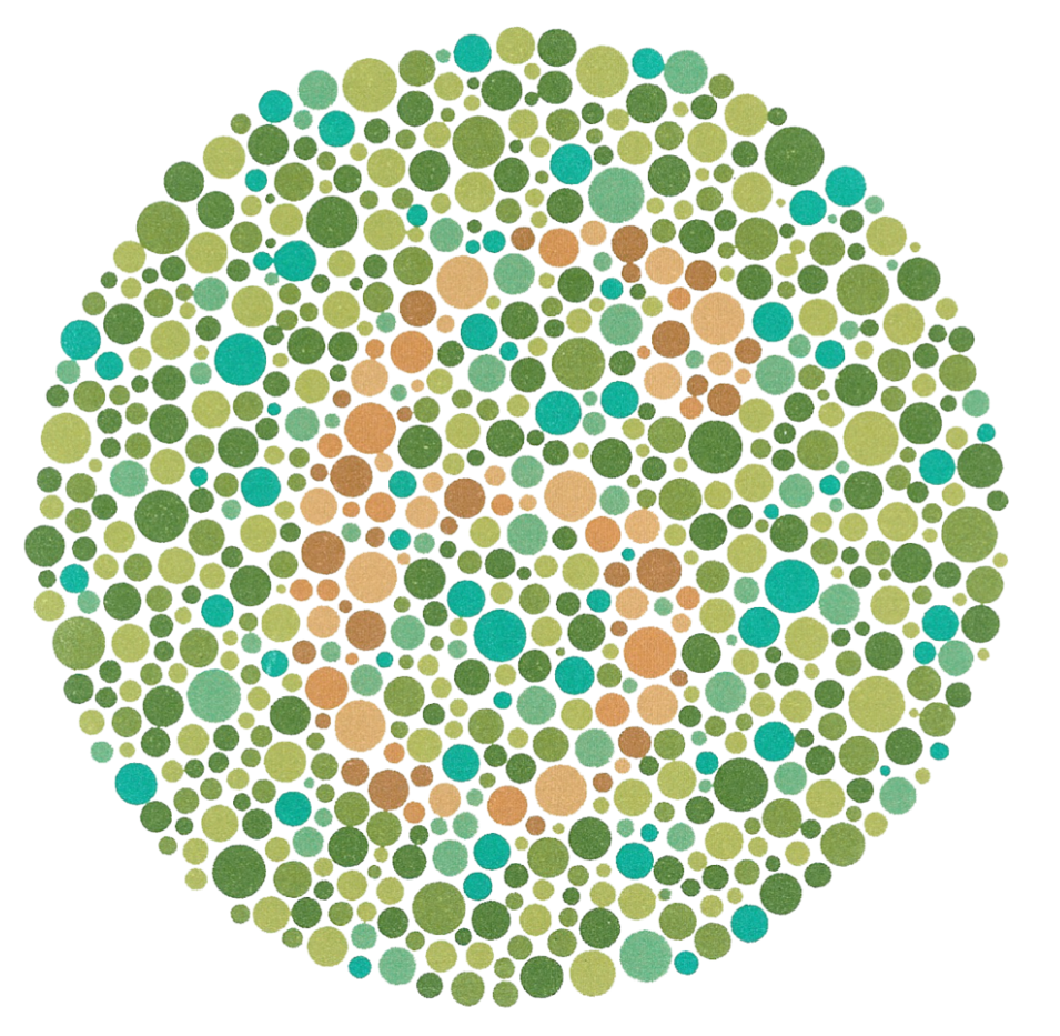
Figure 1: Courtesy of Marni Robbins, OD.
Anomaloscope test
The anomaloscope test, considered the gold standard in color vision testing for protan and deutan defects, requires patients to look through an eyepiece at two different lights and manipulate knobs to make the lights match in color.
Anomaloscope testing can distinguish between dichromats and anomalous trichromats, which is an important distinction for some career-dependent color vision testing, but it is specific to inherited color vision deficiency and has limited application for acquired color vision defects.6
Hue testing technique
In hue testing, the examiner presents the patient with a set of blocks or tiles of different colors and has them arrange the blocks in order based on two fixed colors. The Farnsworth-Munsell D15 tests patients on 15 different color hues and provides information on color vision defects.
Similarly, the Farnsworth-Munsell 100 Hue Test, the most well-known hue test, requires patients to arrange 100 tiles by hue (four rows each, containing 25 variations of each hue). This more rigorous testing measures an individual’s color vision acuity and is intended for those whose careers require very accurate color acuity, such as graphic designers.5,6
Figure 2 illustrates the representative color hues of a hue test. The hues at either end are static; the hues in the middle would be manipulated and placed in order by the patient.

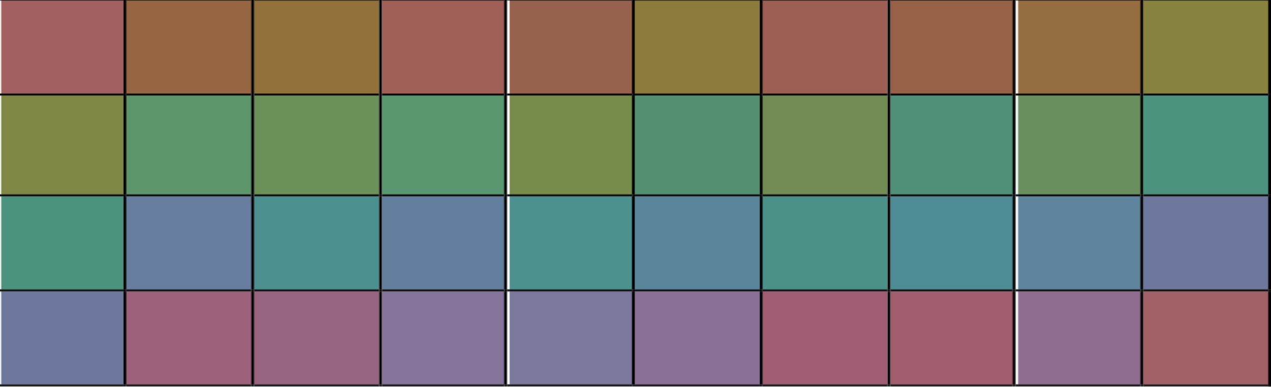
Figure 2: Courtesy of Marni Robbins, OD.
Management of color vision deficiencies
Inherited color vision deficiencies cannot be cured, but most people affected are able to manage the condition well. Certain accommodations may need to be made in the classroom for children, and as they get older, they may need career counseling to ensure that they are aware of how their color vision may impact job performance.2
Some acquired color vision deficiencies, such as those resulting from cataracts or medication, can improve or resolve with treatment of the condition or cessation of the medication. Those resulting from other pathologies, however, may be permanent and require adaptation and accommodations for the patient.5
Specialty glasses and contact lenses
There has been a recent surge in interest in special glasses and contact lenses for people with color vision deficiencies. Patient testimonials on social media are often very emotional and appreciative of the changes they have experienced with the filters.
Research has found, however, that while these filters may alter the appearance of colors, they do not replicate normal color vision and do not improve results on color plate or hue testing.7
Gene therapies
Gene therapy has been proposed as a possible treatment for color vision deficiencies. Most research has been conducted in animal models, but there are currently clinical trials evaluating whether human children born with achromatopsia treated with gene therapy can develop color vision.
Early results have been promising, but more research is needed.8
CVD patient education/resources
When a patient is diagnosed with a color vision deficiency, it is important to reassure them that the condition is common and manageable. Providers who see a high volume of pediatric patients may want to have handouts available to provide parents with more information.
The Cleveland Clinic and National Eye Institute both have excellent resources for the public explaining color vision deficiencies.
For patients interested in pursuing colored filters to alter their color perception, practitioners can contact their preferred optical lab to determine what they offer, or they can partner directly with companies specifically geared towards colored filters for color vision deficient patients.
Conclusion
Color vision deficiencies are a common visual impairment that, in most cases, are easily manageable once identified. Their diagnosis can provide patients and their families with a solid foundation on how to proceed and ensure that their varied perception of the world around them does not hinder their ability to function.
Color plate testing is an easy and quick way to determine whether or not a patient has a color vision deficiency and if additional testing is warranted.
For primary eyecare providers, it is important that color vision testing is performed on every new patient and on any patient who reports changes to their color perception.
