


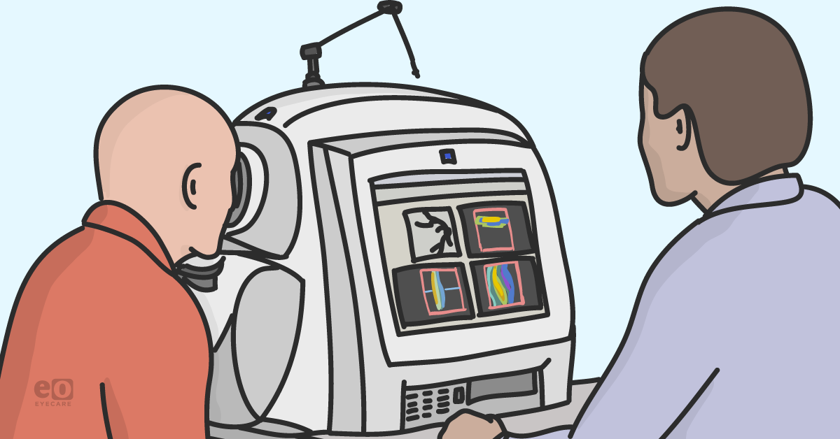
Eyecare has quickly become a high-tech medical specialty, relying on advanced diagnostics to identify and guide treatment for a myriad of ocular conditions. The OCT has become the workhorse of every medical eyecare practice, with utility in retinal, optic nerve, and corneal disease (and contact lens fits!). Though the OCT generates extremely useful reports with advanced quantitative metrics, we cannot forget to qualitatively analyze the individual b-scans, as these are the foundations for our analytics.
Welcome to Diagnostics & Imaging Monthly, a new column from Daniel Epshtein, OD, FAAO! This column will cover diagnostics and imaging of the anterior and posterior segment and will share both basic and advanced educational tips to help optometrists and ophthalmologists.
While you're waiting for the next column, check out these articles:
- OCT Diagnostics in 2021: When 45 Degrees Isn’t Enough
- The Ultimate Guide to SMILE Refractive Surgery for Optometrists
- Pathogenesis, Classification, and Treatment of Diabetic Retinopathy
- A Complete List of Ocular Diseases with Optical Coherence Tomography (OCT)
- How to Use Ultra-widefield HD 2.0 True Color Imaging in Full-Scope Optometry
- A Complete Guide to the Clarus 500 Ultra-widefield Retinal Camera
Eyecare has quickly become a high-tech medical specialty, relying on advanced diagnostics to identify and guide treatment for a myriad of ocular conditions. The OCT has become the workhorse of every medical eyecare practice, with utility in retinal, optic nerve, and corneal disease (and contact lens fits!). Though the OCT generates extremely useful reports with advanced quantitative metrics, we cannot forget to qualitatively analyze the individual b-scans, as these are the foundations for our analytics.
I’ve always been a firm believer that to understand pathology, one must first truly understand normal anatomy and its variations. Early on in my career as an optometry student, when analyzing an OCT image, I would often refer to a normal OCT image that I had saved on my phone. Having the side-by-side comparison made it much easier to detect changes in the retinal anatomy, considering I had not yet committed all the pathological patterns to memory.
So with my young student self in mind, I would like to start the inaugural voyage of this column by going over the basics of normal OCT anatomy. Hopefully, it will be as useful to you, as it was to me years ago.
Let's begin by taking a look at an image of a normal OCT with the appropriate labels.

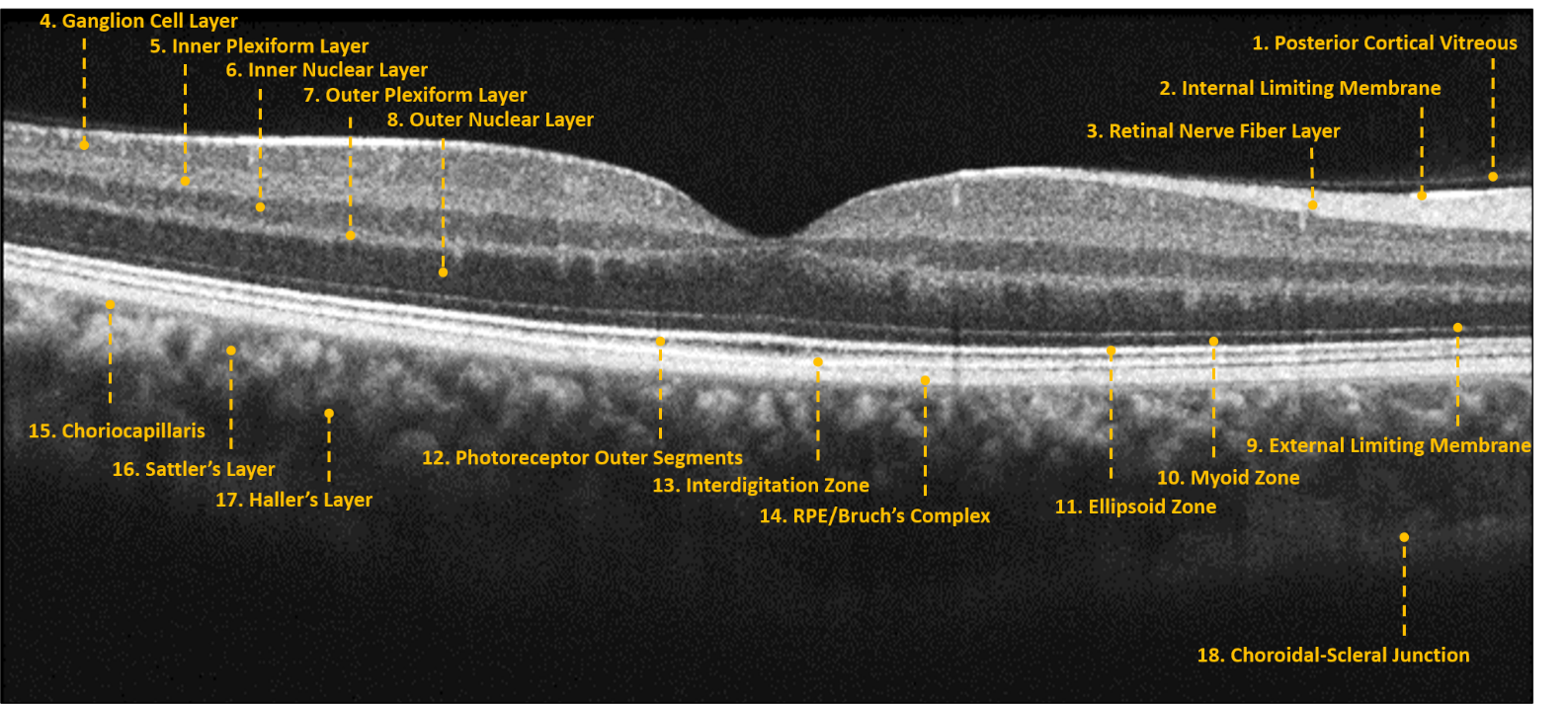
OCT technology takes advantage of the varied light reflectance properties of ocular tissues. Homogenous structures, such as the vitreous and outer nuclear layer, allow light to pass relatively undisturbed and appear as black/dark grey (or cooler colors if using the color scale). Heterogenous structures, such as the retinal nerve fiber layer and RPE, tend to reflect/scatter more light and will appear as white/bright grey (warmer colors on color scale) areas on the OCT image.
Though OCT imaging provides near histological resolution of ocular tissues, we must always be aware that we are viewing the retina as the OCT views it and not as we would see it with a microscope. Due to the complex intracellular and intercellular retinal structure, OCT structures may not always correlate to retinal anatomy.
The bright outer retinal bands are a perfect example of this, with the latest nomenclature preferring to use “zone” for the anatomically ambiguous OCT structures myoid zone, ellipsoid zone, and interdigitation zone.1 Expert researchers have a good idea of what these OCT structures correlate to anatomically, but wished to infuse their uncertainty into the names of the OCT layers themselves, leaving wiggle room to change the names in the future.
Though the nomenclature debates continue to rage on in the halls of academia, the clinical importance of these outer retinal structures has been cemented in countless articles and hours of clinical use. The bright outer retinal bands, external limiting membrane, ellipsoid zone, and RPE/Bruch’s complex correlate very well with visual acuity and are one of the most important areas to scrutinize when viewing a retinal OCT.
Moving anteriorly, the inner retinal contour should be relatively flat with a smooth curve representing the foveal depression. Macular edema or vitreomacular interface disease can alter or even obliterate the foveal depression, correlating to visual changes such as blur and metamorphopsia. In rare cases of foveal hypoplasia, the foveal depression is under-developed and represents normal retinal anatomy for that particular patient. Overall retinal thickness and contour can vary significantly based on axial length, this is most evident in highly myopic patients where the retina tends to be thin and curved.
Though pattern recognition is extremely important in OCT analysis, having a deep understanding of the normal retinal architecture and its inter-patient variability provides an important foundation for deciphering pathological OCT images. In future editions of this column, we will begin our deep dive into the various OCT pathological patterns, reviewing various common and uncommon posterior segment conditions.
References
- Staurenghi, Giovanni, et al. "Proposed lexicon for anatomic landmarks in normal posterior segment spectral-domain optical coherence tomography: the IN• OCT consensus." Ophthalmology 121.8 (2014): 1572-1578.
We have a direct partnership with ZEISS!

