


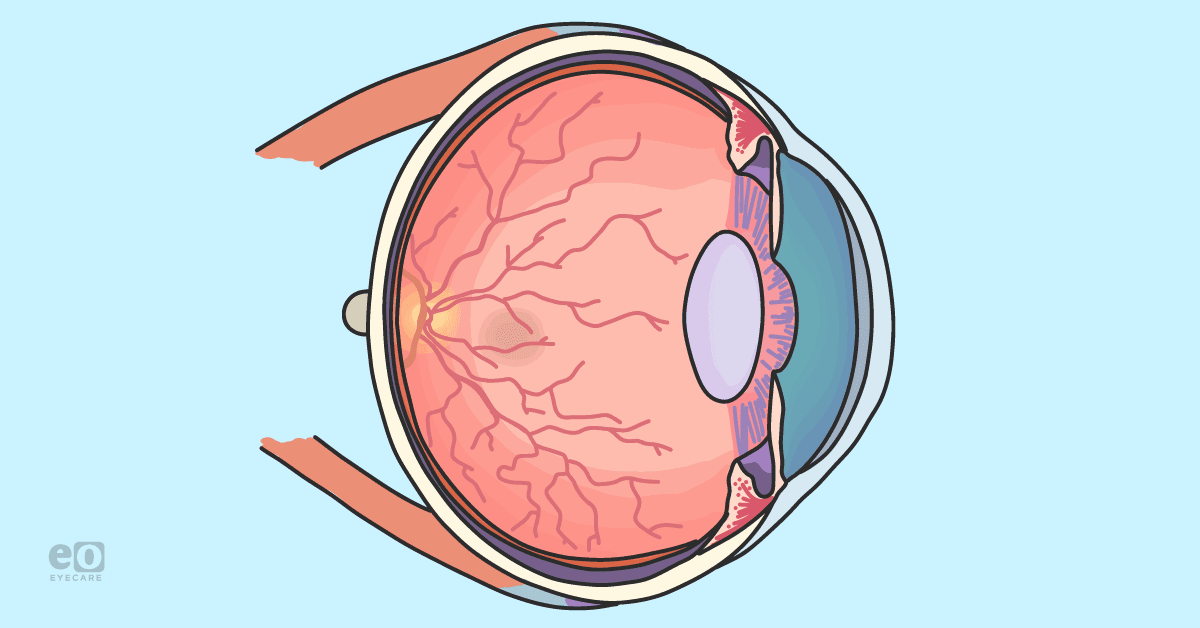
The macula is a cone-rich area of the retina that provides the highest visual acuity, is responsible for optimal vision, and has the highest exposure to light. Macular pigmentation (MP) includes a structural isomer zeaxanthin (Z), carotenoids lutein (L) and a zeaxanthin stereoisomer, meso-zeaxanthin (meso-Z). In the eye, L and Z are distributed in the lens and ciliary body, but have the highest concentration in the fovea, specifically within the outer plexiform layer (OPL) in the retina, ideally protecting the outer retina from damage.1,2
In this article we’ll review the importance of macular pigment for optimal ocular health as well as its role in various ocular diseases.
Biological functions of macular carotenoids
- Light absorption: the macula is protected from light related damage by the macular carotenoids absorbing 40-90% of blue light.1,2,3
- Protection against oxidative stress: L and Z are strong antioxidants that scavenge for free radicals to reduce damage from oxidative stress. L and Z indirectly reduce oxidative damage by absorbing blue light, and directly promote oxidative metabolism in the retinal pigment epithelium (RPE) because they are the two predominant carotenoids contained in RPE membranes.1,2,3
- Protection against inflammation: L can protect against inflammation by indirectly influencing ocular inflammation and reducing the expression of specific systemic inflammatory pathways. This includes preventing the upregulation in expression of inflammation-related genes and more specifically reducing the increase in oxidation-induced cytokines.1,2,3
Changes to macular pigmentation
The methods that are available to measure MP can be separated into physical tests and psychophysical tests, which are known to generate objective and subjective measurements, respectively.4 Color matching, motion photometry, heterochromatic flicker photometry (HFP), and customized heterochromatic flicker photometry (cHFP) are among the psychophysical methods that are accessible.4 Resonance Raman spectroscopy, fundus autofluorescence, and fundus reflectance are of the physical methods used to measure MP.4
Individual macular levels of L and Z differ significantly, according to various studies.5 Carotenoids that are present in the body are either directly derived from food or partially altered by metabolic processes from consumed meals.5 The last trimester of pregnancy and lactation are two crucial times for the development of the retina and the brain. During these times, L and Z concentrations rise and are preferentially deposited in the retina and the brain.5,6
The developing baby normally receives L from cord blood, breast milk, and infant nutrition. The overall presence of this carotenoid is dependent on the mother’s consumption of fruits and vegetables and is typically more bioavailable in breastmilk. Carotenoids give colostrum (“first milk”) its distinctive yellow hue because of the high concentration. Throughout the entire nursing period, L and Z have been found in breast milk. Concentrations are typically lower in mature milk when compared to colostrum.5,6
Fruits and vegetables are often the primary dietary sources of carotenoids.7 The most prevalent carotenoid in the human diet, L, is frequently found in conjunction with Z. All dark green leafy vegetables, especially kale and spinach, are the richest food sources of L.7 Kale and spinach provide 40 mg and 12 mg of L per 100 g meal, respectively. Vegetables typically contain substantially less Z.7
According to a large body of evidence, numerous dietary, metabolic, and genetic factors can affect how retinal carotenoids are absorbed, transported, and accumulated 7,8 These numerous factors may affect the uptake and/or stabilization of L and Z in the retina, as well as the absorption and passage of these pigments in the blood.7,8 Individual reactions to dietary supplementation of L and Z vary considerably.7,8 The high interindividual heterogeneity in response to supplementation shows that the absorption, transport, and retinal capture of L and Z are subject to both external and endogenous effects.7,8,9
The role of macular pigment
Visual acuity
As determined by the capacity to recognize smaller letters at a specific distance, visual acuity shows the capacity to resolve items that are in sharp contrast to their background. Studies show that supplementing L and/or Z alone improves visual acuity. Individuals with AMD who took L and/or Z supplements for 6 months to 3 years showed a strong protective effect. There was a direct correlation between the improvement in visual acuity and the change in MPOD levels attained.10,11
Contrast sensitivity
The capacity to notice differences in degrees of brightness or darkness of an item, or of colors, relative to the object's backdrop, is referred to as contrast sensitivity. Contrast sensitivity typically degrades before visual acuity due to age or illness. The literature has shown, especially in young, healthy volunteers, that supplements containing L and Z, administered for 3 months to 3 years can increase contrast sensitivity. Long-term impacts of L and Z status on contrast sensitivity in later life would include not just these short-term benefits, but also protection against age and/or disease-related ocular changes.12
Glare reduction
Observational research shows that increased MP levels can decrease the time required to recover from bright light exposure as well as improve vision in the presence of significant glare.13
Visual processing speed
Adults aged 18 to 32 who took L and Z supplements for four months dramatically increased their visual processing speed. These findings imply that high MP levels may improve eye function for patients with occupations that need quick reaction times, such as athletes, military, or computer-related employment.14
Dark adaptation
Dark adaptation is essential to recover visual sensitivity when transitioning from bright to low illumination. Deficiencies in dark adaptation are thought to be caused by the buildup of hydrophobic lipids in the retinal pigmented epithelium (RPE) causing a delay in the supply of nutrients (e.g., Vitamin A) to rod cells. Impaired delivery would delay the rate of rhodopsin regeneration inside rod photoreceptors, making dark adaptation take longer.15,16
MP may protect against deficiencies in rod-mediated dark adaptation, though, the exact biological mechanism is not known at present. Two short observational studies found those with low levels of MP take longer to dark adapt.15,16
Macular pigment in ocular disease
AMD
L and Z, which have been shown to reduce oxidative stress and inflammation, can help protect against AMD. These carotenoids are particularly prevalent in the fovea, and absorb 40% to 90% of the short-wavelength light that would otherwise damage the retina. L supplementation has also been shown to reduce inflammatory responses and the development of AMD by reducing circulating levels of proteins that participate in the complement activation pathway.17,18
L and Z may also have a more pronounced protective impact in persons who have a high hereditary risk for AMD.17,18 This is supported by prospective cohort studies (eg. Rotterdam and Blue Mountain Eye studies) that found substantial interactions between L and Z consumption and genotype in AMD risk.19,20
In the CAREDS study cohort, a similar trend of reduced genetic risk for AMD was identified among persons with high L and Z consumption.21 Individuals ingesting 5 to 6 mg/day had the lowest risk of AMD.21 The amounts required to reduce AMD risk may be higher in patients with phenotypes and genotypes linked with inadequate absorption, uptake, or retention in the retina.19,20,21
Cataracts
Oxidative stress can lead to cataract formation. L and Z are carotenoids that protect against the development of lens opacities and advanced cataracts. Experimental results show that these carotenoids prevent oxidative stress and lens damage, which lead to cataract formation.22,23
Adding L and Z to other antioxidant supplements did not significantly delay the need for cataract surgery in one major multisite clinical trial over 5 years, with the exception of people with the lowest dietary amounts. This finding suggests that L and Z are likely to be two dietary components that protect against nuclear cataract development, but the amount needed to protect against cataract formation is likely to be 0.5 to 1 mg/day - significantly lower than the average intake in American adults today.22,23
Glaucoma and diabetic retinopathy
According to current research in animals and humans, pathogenic processes underlying glaucoma include damage to retinal ganglion cells caused by inflammation, hypoxia, and oxidative stress. L has been shown to protect retinal ganglion cells in rats. Furthermore, L and Z found in the ciliary body, may protect the trabecular meshwork from injury and minimize the risk of high intraocular pressure.24
A subset of data from animal and human observational research shows that L and Z may have a role in preventing diabetic retinopathy as well. In diabetic mice, consuming L and Z-rich diets reduced retinal vascular abnormalities and increased the expression of genes involved in the absorption of L and Z in the retina, thus protecting against oxidative stress.25
Tips to maintain macular pigment
Diet
Leafy green vegetables, corn tortillas, eggs, orange juice, and broccoli were substantial contributors of L, whereas corn tortillas, eggs, orange juice, and broccoli were major contributors of Z.7
Supplements
The American Academy of Ophthalmology has suggested that those with intermediate or advanced age-related macular degeneration take a high-dose antioxidant supplement including L and Z, based on the previously stated AREDS2 results. Furthermore, those with a family history of AMD may prefer to supplement with comparable formulas, even though there is no evidence that L and Z supplementation prevents AMD in these particular individuals.26
Techniques for assessing macular pigment optical density (MPOD) are being marketed for use in optometric and ophthalmological clinics. These new technologies will likely lead to the identification of patients with low MP levels and an increased predisposition for those with low levels to take supplements.27 L and Z have been included in the majority of eye supplements since 2014.27
Below is a list of supplements in USA and Canadian markets that contain lutein (L), zeaxanthin (Z), and meso-zeaxanthin (Meso-Z).
Supplements with L, Z, and Meso-Z
Supplements with L, Z, and Meso-Z
L + Z
- Vatalux Advanced (capsule or chewable): Made in Canada
- EyePromise Z + L (soft gel): Made in USA
- PreserVision AREDS2 (soft gel): Made in USA
- Ocuvite Gummies: Made in USA
- Doctor's Best L+Z (gummies): Made in USA
L+ Z + Omega 3
- Vitalux Advanced Plus Omega 3 (soft gel): Made in Canada
- Ocuvite Eye Vitamins(soft gel): Made in USA
L + Z + Daily Vitamins
- Vitalux Healthy Eyes (soft gel): Made in Canada
- Vitalux Advanced Plus Multi-Vitamins (chewable): Made in USA
L + Z + Meso-Z
- Vision Essence Early Defense (capsule): Made in Canada
- Life Extension MacuGuard (soft gel): Made is USA
L + Z + Meso-Z (beef gelatin tablets)
- MacuHealth with LMZ3 (soft gel): Made is USA
- MacuHealth Plus with LMZ3 + AREDS2 + zinc (soft gel): Made is USA
Table 1
Conclusion
Macular pigment plays a crucial role for optimal ocular health and visual function. It is important for today’s eyecare providers to educate their patients on the importance of MP, especially those at risk for retinal disease like AMD. Given the growing body of evidence linking carotenoid supplementation and lower risk of ocular disease progression, clinicians must also be ready to give specific dietary recommendations to patients when appropriate.
As diagnostic technologies continue to develop, measuring MPOD will likely become standard-of-care for our patients, improving early detection and treatment outcomes for macular disease.
