


As a cataract surgeon, I’ve found that most patients deciding to have cataract surgery are curious regarding the recurrence of their cloudy lens. Will they need to anticipate another surgery in a few years? How long will the surgery "last"? Is cataract surgery a “curative” procedure, or can cataracts grow back—and if so, how?
In this article, I will cover the basics of cataract formation, cataract surgery, and how we can address our patients’ fears of cataract recurrence.
Anatomy of a cataract
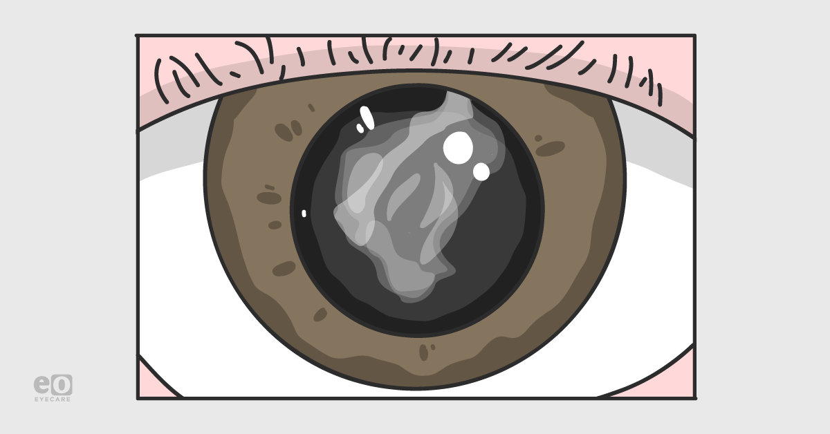
A cataract is a clouding of the eye’s natural crystalline lens, often related to aging.1 In fact, more than 50% of Americans 80 years and older have cataracts or have had cataract extraction surgery.1 Cataracts typically evolve gradually over time; therefore, early cataracts can be overlooked if they are not causing symptoms of blurry vision, or if they are not identified on a routine eye exam.
Cataracts are formed from the natural crystalline lens, which is typically clear at birth.1-3 The crystalline lens is an avascular, oval-shaped, transparent structure that aids in focusing light rays on the retina.1-3 It is located in the posterior chamber of the eye, posterior to the iris, and anterior to the vitreous.1-3 It is suspended within the eye by 360 degrees of zonular fibers from the ciliary body.1-3
Early in life, the lens is relatively malleable; ciliary muscle contraction changes zonular fiber tension, causing a change in lens shape. This change in lens shape can increase the dioptric power of the eye (accommodation), thereby allowing near images to come into focus.1-3
Crystalline lens transparency is a function of the absence of blood vessels, few cellular organelles, and an orderly arrangement of lens fibers.1-3 The crystalline lens absorbs most of the ultraviolet (UV) light to which it is exposed, causing lens oxidation by free radicals.1-3 This free radical damage degrades lens proteins and alters the normal anatomical orientation.1-3
UV light exposure also increases chromophore concentration, leading to the accumulation of yellow pigments in the center of the lens, which may progress to a dark brown hue (“lens brunescence”) with time.1-3 For most individuals, lens protein denaturation starts in the fifth decade.1-3 As the cataract increases in density and opacity, vision also declines.
Risk factors for cataracts include diabetes, smoking, alcohol consumption, history of eye trauma, family history, previous head or neck radiation, sun (UV) exposure, and long-term corticosteroid use.1-5 There is no proven prevention strategy for cataract formation or risk factor mitigation; however, reducing sun exposure, abstaining from smoking, eating a well-balanced diet, and having a dilated eye exam every year starting at age 60 are common recommendations.1-3
Cataracts can be categorized as age-related, traumatic, radiation-induced, and pediatric.1-5 Age-related cataracts are most common and occur due to natural changes in the lens with time. Serious eye injuries can damage the lens and cause a cataract to form immediately; alternatively, the cataract can form many years after the initial injury if non-penetrating in nature (e.g., a blunt ocular trauma).1-5 Certain types of radiation can cause cataracts; including UV rays from the sun as well as radiation from certain cancer treatments.1-5
Pediatric cataracts can be congenital (due to infections or complications in-utero), or acquired (they may develop later due to illness, inflammation (i.e., uveitis), and tumors in the eye).1-5 If pediatric cataracts are large enough to cause visual decline, they require urgent treatment to prevent amblyopia.1-5
The only definitive treatment for cataracts is surgery, though early cataracts may be corrected with glasses or contact lenses to help improve vision.1-5
Laser vision correction surgery such as LASIK, PRK, or SMILE will not eliminate blurry vision due to cataracts as these procedures treat the cornea, not the crystalline lens.
Can cataracts grow back after surgery?
During cataract surgery, a tiny, self-sealing incision is made in the eye and an opening (capsulorhexis) is created in the lens capsule that holds the cloudy lens in place.1-3 An ultrasound probe is then used to break up the cataract, making it easier to aspirate the lens pieces from the eye. The lens capsule is carefully preserved during the surgery so that it can house the new artificial intraocular lens implant (IOL) that replaces the cataractous lens.1-3
“Cataracts cannot grow back after surgery; the artificial lens that has been implanted cannot develop cloudy or opaque areas in the same way the natural crystalline lens can.”
What can happen, however, is that the lens capsule can become opacified due to the growth of residual microscopic lens cells inside of it. This is called posterior capsular opacification (PCO) and occurs in 50% or more of patients.1-7
Posterior capsule opacification
PCO can cause symptoms that are consistent with cataracts, which is why it is essential to inform your patients of this before surgery. Some patients may complain that their vision is blurrier than just after their surgery; some may experience visual disturbances such as glare, starbursts, and haloes around lights.6,7 Even more confusing, PCO is sometimes referred to as a “secondary cataract,” and this misnomer may lead patients to think that their cataract did in fact ‘grow back.’ However, the mechanism of PCO formation is different from cataract formation.6,7
PCO results from the proliferation of lens epithelial cells on the posterior capsule; it is reported in 8-50% of cases following cataract surgery within 5 years postoperatively.1,4,6,7 Some reports have noted that PCO incidence is increased in patients who have a large capsulorhexis (6-7mm) and those with a history of uveitis.1,4,6,7
In addition, diabetic patients can develop significantly more frequent and denser PCO than non-diabetics.1,4,6,7 On a microscopic level, aberrant lens cells attached to the capsule try to form new lens fibers.1,4,6,7 PCO causes visual decline due to the formation of multiple microscopic layers of lens epithelium; this creates frank opacity.1,4,6,7 Also, fibroblastic differentiation of the lens epithelium occurs, which produces contraction and wrinkles in the posterior capsule, as well as possible visual distortion.1,4,6,7
Fortunately, ophthalmologists can efficiently and painlessly treat PCO to help restore clearer vision. PCO treatment is done via a neodymium-yttrium aluminum garnet (Nd:YAG) laser in the office or outpatient surgical setting.6-9 Treatment involves pupillary dilation, and the laser is specifically focused on the area of PCO, allowing for the dissolution of the fibrous tissue.6-9 The laser does not touch the eye, no physical incisions are made, and no wounds are involved.
Following laser treatment, the vision may be blurry for a few hours; however, most patients can resume normal activities the same day without restrictions.6-9
While there has been research into pharmacologic treatments for PCO (including intracameral chemotherapeutic agents in some studies), the most effective treatment for PCO remains Nd:YAG laser capsulotomy.6-9 The procedure is relatively low risk, and adverse events are not common.6-9 In addition, Nd:YAG laser capsulotomy for PCO is generally covered by insurance.
Common codes related to Nd:YAG laser capsulotomy
- CPT code: 66821
- -50 modifier if done bilaterally
- -LT or -RT modifier if performed on one eye only
- -78 modifier if performed within 90 days of cataract surgery
- ICD-10 codes
- H26.40 - unspecified secondary cataract
- H26.411 - Soemmering's ring, right eye
- H26.412 - Soemmering's ring, left eye
- H26.413 - Soemmering's ring, bilateral
- H26.491 - Other secondary cataract, right eye
- H26.492 - Other secondary cataract, left eye
- H26.493 - Other secondary cataract, bilateral
In conclusion
Cataracts are one of the most common causes of preventable blindness in the world.1,2 In the United States, approximately 4 million cataract surgeries are performed per year. Most patients have very favorable outcomes, and patients should be educated that their cataracts will not return or regrow. Should PCO develop, this can usually be managed by the treating surgeon in a safe and straightforward manner.
References
- Cataracts. National Eye Institute. https://www.nei.nih.gov/learn-about-eye-health/eye-conditions-and-diseases/cataracts. 2019
- Remington, Lee Ann, editor. “The Crystalline Lens.” Clinical Anatomy of the Visual System, Elsevier Health Sciences, 2014, pp. 92–101.
- S: The physical basis for transparency of the crystalline lens, Invest Ophthalmol 1:493, 1962
- Types of Cataract. National Eye Institute. https://www.nei.nih.gov/learn-about-eye-health/eye-conditions-and-diseases/cataracts/types-cataract 2019
- Yanoff, Myron, and Joseph W. Sassani, editors. “Surgical and Nonsurgical Trauma.” Ocular Pathology, 6th ed., Mosby, 2009, pp. 129–130.
- Hayashi K, Hayashi H, Nakao F, et al. Posterior capsule opacification after cataract surgery in patients with diabetes mellitus. Am J Ophthalmol. 2002; 134:10.
- Awasthi N, Guo S, Wagner J. Posterior Capsular Opacification: A Problem Reduced but Not Yet Eradicated. Arch Ophthalmol. 2009;127(4):555-562
- TM, Devlin HD, Hillon B. Use of Nd:YAG laser capsulotomy. Surv Ophthalmol. 2003;48(6): 594-612
- Karahan E, Er D, Kaynak S. An Overview of Nd:YAG Laser Capsulotomy. Med Hypothesis Discov Innov Ophthalmol. 2014;3(2)
