


Uveitis is defined as an inflammation of the uvea, the middle layer of the eye, which is divided into three parts, the iris, ciliary body, and choroid. Uveitis accounts for 10 to 20% of blindness in the United States and Europe, and as much as 25% in the developing world.1,2
Uveitis can be caused by autoimmune conditions, infectious processes, or even be idiopathic. Being able to accurately diagnose, manage, and treat uveitis can help prevent vision loss.
Anterior uveitis
Symptoms of anterior uveitis include photophobia, pain, and circumlimbal flush/injection are hallmark signs of anterior uveitis. Pain secondary from ciliary/iris spasm may also produce photophobia. Circumlimbal flush is due to enlargement of episcleral vessels around the limbus. Patients may also report blurry vision.
Causes of anterior uveitis:3
- Human leukocyte antigen B27 (HLA-B27)
- Patients with HLA-B27-associated uveitis may have systemic associations such as ulcerative colitis, Crohn’s disease, reactive arthritis, ankylosing spondylitis, and psoriatic arthritis
- Juvenile rheumatoid arthritis
- Herpes simplex and zoster
- Tuberculosis
- Syphilis
- Brimonidine
- A well-known glaucoma medication in our arsenal has been reported to cause granulomatous anterior uveitis and elevated intraocular pressures (IOPs) with chronic usage4
Causes of intermediate uveitis can include:3
- Behçet’s disease
- Lyme disease
- Multiple sclerosis
- Sarcoidosis
- Tubulointerstitial nephritis and uveitis (TINU)
- Vogt-Koyanagi-Harada (VKH) disease
- Masquerading syndromes
- Such as malignant melanoma and large cell lymphoma5
It is also estimated about 50% of anterior and intermediate causes are idiopathic—meaning no identifiable cause is found.6,7
Clinical signs of anterior uveitis
In cases of anterior uveitis, white blood cells (“cells”) are commonly noted. Fibrin (“flare”) is often noted due to a breakdown of the blood/aqueous barrier resulting in protein leakage.8 The Standardization of Uveitis Nomenclature (SUN) Working Group graded cell and flare from 0 to 4, as seen in Tables 1 and 2.9
Table 1: SUN grading of anterior chamber cells.9
| Grade | Cells in Field |
|---|---|
| 0 | <1 |
| 0.5+ | 1 to 5 |
| 1+ | 6 to 15 |
| 2+ | 16 to 25 |
| 3+ | 26 to 50 |
| 4+ | >50 |
Table 1: Courtesy of Jabs et al.
Table 2: SUN classification of anterior chamber flare.9
| Grade | Description |
|---|---|
| 0 | None |
| 1+ | Faint |
| 2+ | Moderate (iris and lens details clear) |
| 3+ | Marked (iris and lens details hazy) |
| 4+ | Intense (fibrin or plastic aqueous) |
Table 2: Courtesy of Jabs et al.
Download the cheat sheet to reference SUN Working Group classifications
Clinical features of anterior uveitis
With anterior uveitis, a hypopyon can form due to a large amount of white blood cells layering in the anterior chamber. Posterior synechiae may be noted; clumps of pigment on the surface of the crystalline lens suggest broken synechiae from a previous attack.
In addition, non-granulomatous or granulomatous deposits may be apparent on the corneal endothelium. Non-granulomatous is typically non-infectious in origin compared to granulomatous, which may suggest an underlying infectious or systemic etiology. Patients may also report floaters due to the spillover of cells into the posterior chamber.
Intraocular pressure (IOP) may initially be low due to hyposecretion of the ciliary body.
Intermediate uveitis
With intermediate uveitis, pain is less common; floaters and reduced visual acuity are noted more often. Snowbanking, or vitreous cells in the inferior ora serrata, and cellular aggregates in the inferior vitreous (snowballs) are key clinical indications in intermediate uveitis. In younger patients, a vitreous hemorrhage may also be noted.
Diagnosis of intermediate uveitis
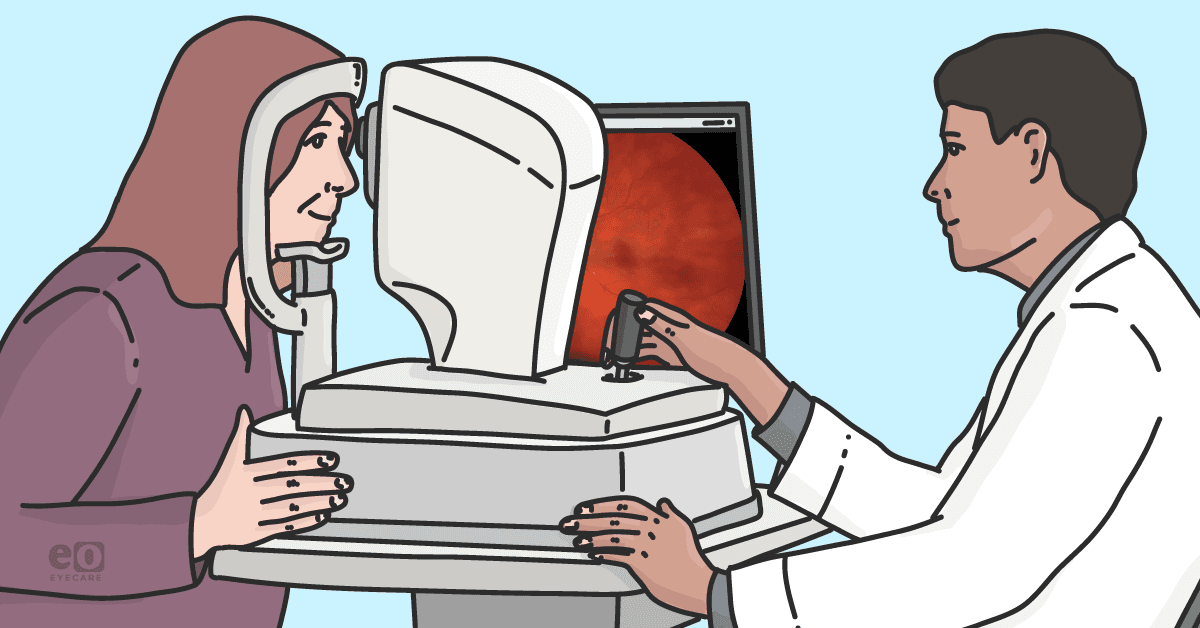
While slit lamp biomicroscopy remains the standard of care for assessing inflammation of the anterior segment and vitreous, imaging can help aid in the diagnosis. On macula optical coherence tomography (OCT), cystoid macular edema may be noted in some intermediate uveitis cases.10
B-scan ultrasound is beneficial when visualization of the fundus is obscured due to dense vitritis to ensure no other posterior segment complications are present, such as a retinal detachment.11 On fluorescein angiography, vascular leakage can be noted in some intermediate uveitis cases.12
Laboratory work-up is a critical component in uveitis, especially for recurrent, bilateral, intermediate, posterior, or panuveitis cases.
The following are some key labs to order for uveitis:13
- Erythrocyte sedimentation rate (ESR)
- C-reactive protein (CRP)
- Rheumatoid factor (RF)
- Antinuclear antibody (ANA)
- HLA-B27
- HLA-A29
- Angiotensin-converting enzyme (ACE)
- Rapid plasma reagin (RPR)
- Venereal disease research laboratory test (VDRL)
- Fluorescent treponemal antibody absorption test (FTA-ABS)
- Human immunodeficiency virus (HIV) screening
Table 3: Suggestions for which laboratory/ancillary testing should be ordered based on the patient’s clinical presentation.13
| Anatomic Type of Uveitis | Investigations |
|---|---|
| All types of uveitis | CBC, ESR, CRP, tuberculin skin test, syphilis serology, chest X-ray |
| Acute anterior uveitis (non-granulomatous) | HLA-B27, sacroiliac imaging if back pain |
| Acute anterior uveitis (granulomatous), unexplained | Anterior chamber tap and polymerase chain reaction (PCR) for herpes virus after herpes simplex virus (HSV), varicella zoster virus (VZV), and cytomegalovirus (CMV) serology |
| Chronic uveitis | ACE, interferon-gamma release assay (IGRA), chest computed tomography (CT) scan |
| Chronic intermediate or posterior uveitis (>40 years of age) | Brain magnetic resonance imaging (MRI), toxoplasma serology if focal retinitis is present |
| Bilateral papillary edema | Lumbar puncture |
| Intermediate uveitis (>40 years of age) | Anterior chamber tap evaluating for interleukin-10 and interleukin-6 |
| Steroid treatment-resistant uveitis | Anterior chamber tap: cytology, interleukins, herpes PCR, M. tuberculosis PCR, universal PCR, vitrectomy: lymphoma evaluation |
Table 3: Courtesy of Sève et al.
Treatment/Management of uveitis
Treatments for uveitis range from topical therapies to injections to surgical intervention, depending on the severity of the condition.
Topical therapies for uveitis
In both anterior and intermediate uveitis, cycloplegics such as cyclopentolate 1% eye drops BID are used for mild to moderate inflammation and atropine sulfate 1% BID for severe inflammation.14 They reduce pain by preventing spasm of the ciliary body and help prevent posterior synechiae. These typically are tapered as the inflammation diminishes and are stopped once there are no more cells in the anterior chamber.
Steroids are the mainstay of uveitis treatment to reduce inflammation. Prednisolone acetate 1% q1h to 6h is often used, depending on the severity. This is also tapered as the inflammation starts to resolve, and discontinued once the anterior chamber is quiet.15
If the presence of cells is not decreasing despite prescribed steroid usage, the practitioner should question if the patient is using the drops appropriately, including shaking the bottle, or if there is a herpetic component.
Difluprednate may also be used, especially if there is no to minimal response to prednisolone acetate; however, as it is a stronger steroid, the IOP needs to be carefully monitored.
Non-topical therapies for uveitis
Intermediate uveitis may require an aggressive approach in more severe cases. Periocular injections of corticosteroids such as triamcinolone acetonide with or without accompanying systemic corticosteroids is the first-line treatment to treat cystoid macular edema.16 In recurrent cases of uveitis, a subtenon injection of triamcinolone may be considered.17
Additionally, some retina specialists may give intravitreal injections of steroids to manage noninfectious uveitis and associated cystoid macular edema.18 Transscleral cryotherapy can be considered in cases of steroid-resistant uveitis and be applied to areas of snowballing and snowbanking to stabilize inflammation; however, this treatment approach is becoming less common due to potential complications such as retinal detachment.19
Pars plana vitrectomy is another surgical option in patients in unmanageable cases and addresses posterior segment complications associated with uveitis, such as vitreous hemorrhage, epiretinal membrane, and retinal detachment.20
Medical management of uveitis
Within the past 10 years, the FDA approved monoclonal antibodies that inhibit tumor necrosis factor (TNF) to reduce the likelihood of developing anterior uveitis in the future, such as certolizumab and golimumab.21
Other immunosuppressive agents that can be used for intermediate, posterior, and panuveitis include azathioprine, methotrexate, and adalimumab.22 Acthar gel, a repository corticotropin injection, has been gaining traction for the treatment of non-infectious anterior, intermediate, posterior, and panuveitis.23
Follow-up care for uveitis
In anterior uveitis, a follow-up is recommended after 1 to 7 days, depending on the severity of the case. At each visit, the IOP should be checked along with a posterior/fundus examination. Once the inflammation is resolved, the patient should be followed up at 1- to 6-month intervals, depending on the severity.
In the acute phase of intermediate uveitis, patients are examined after 1 to 4 weeks, depending on the severity. In the chronic phase, patients are examined every 3 to 6 months.24
A referral to a uveitis specialist should be considered if the uveitis is bilateral and severe, or if there is no improvement on maximum steroid therapy. Interdisciplinary care may be required for patients, such as rheumatology and infectious disease specialists.
Case Report 1: Moderate recurrent anterior unilateral non-granulomatous uveitis
A 27-year-old male presented to the eye clinic due to ocular redness and pain in his right eye for 5 days. He also reported photophobia and presented to the exam room wearing sunglasses.
The clinical examination revealed:
- Medical history: Pertinent for past tobacco use and history of left Achilles tendonitis
- Ocular history: Positive for anterior uveitis in the left eye (OS) 2 years prior and myopia in both eyes (OU)
- Family history: No ocular diseases reported nor history of ocular surgeries
- Medications: None
- Allergies: None
- Visual acuity: Best corrected visual acuities (BCVAs) measured 20/30 in the right eye (OD) with pinhole improvement to 20/20; visual acuity OS was stable at 20/20
- Pupils: Equal, round, and reactive to light without an afferent pupillary defect, motilities were full OU, and confrontational facial fields were full OD and OS
- Anterior segment: OS was unremarkable.
- Posterior segment: Unremarkable for 360° OU
His conjunctiva OD had 2+ injection and the anterior chamber was noted to have 2+ cells, no flare, no hyphema, no hypopyon, and no keratic precipitates. Posterior synechiae was also noted in the right eye. Intraocular pressure was measured to be 14mmHg OU by Goldmann applanation tonometry.
Diagnosis
The patient was initially diagnosed with moderate recurrent anterior unilateral non-granulomatous uveitis OD. He was prescribed prednisolone acetate 1% QID OD and atropine sulfate 1% BID OD. He was asked to return 2 to 3 days later for a follow-up.
As this was his second occurrence in 2 years, he was asked about any systemic symptoms, such as gastrointestinal discomfort and joint pain. The patient denied any symptoms, stating he only had the Achilles tendonitis a few years ago, right before his first incidence of uveitis.
Co-management with PCP
The patient’s primary care physician (PCP) was consulted, and HLA-B27 labs were ordered in addition to:
- CBC
- CRP
- ESR
- RF
- ANA
- ACE
- Lyme antibodies
- RPR
- VRDL
- FTA
- QuantiFERON
HLA-B27 was ordered due to this being a unilateral, recurrent anterior uveitis in a young, adult male. Despite the patient lacking constitutional symptoms or any infectious signs such as granulomatous keratic precipitates noted, CRP, ESR, RF, ANA, ACE, Lyme, syphilis, and QuantiFERON were ordered for him due to the patient having recurrent, unilateral, anterior uveitis.
Follow-up
The patient was followed over the next 2 weeks and had both drops tapered as improvement was noted. The patient’s uveitis resolved without consequences.
Additionally, his labs came back as positive for HLA-B27. His primary care physician placed a consult to rheumatology along with X-rays of lumbosacral spine, sacroiliac joint, ankles, feet, and an arthritis panel. However, the patient chose to decline any further follow up care as he was not currently in any discomfort, and as a result, a final diagnosis could not be established.
Figure 1: X-ray of the patient’s spine showing narrowing between L1 and L2 and L5 and S1. This X-ray revealed subchondral sclerosis of the left sacroiliac joint with erosive changes consistent with sacroiliitis.


Figure 1: Courtesy of Jill Gottehrer, OD, FAAO.
Case 2: Bilateral intermediate uveitis
A 61-year-old female came into the office reporting blurriness to the left eye for 4 days. She had gone to the emergency room and was diagnosed with conjunctivitis, where she was given antibiotic eye drops that did not provide any relief.
Over the last few days, she had noticed a grey haze over half the eye and an increased number of floaters.
The clinical examination revealed:
- Medical history: Positive for hypothyroidism, hyperlipidemia, and cardiomyopathy
- Ocular history: Positive for compound myopic astigmatism OU with presbyopia
- Family history: Maternal history of glaucoma
- Medications: Levothyroxine, metoprolol, and valsartan
- Allergies: None
- Surgeries: None
- Visual acuity: BCVA OD remained stable at 20/20; Visual acuity OS was reduced to 20/200 compared to 20/20 6 months prior without improvement upon pinhole or refraction
- Pupils: Equal, round, and reactive to light without an afferent pupillary defect, motilities were full OU, and confrontational facial fields were full OD and OS
- Anterior segment: OD was unremarkable. OS was noted to have 3+ conjunctival injection along with posterior synechiae and 1+ anterior chamber cells
- Posterior segment: Due to the dense vitritis, it was difficult to obtain a distinct view of the posterior pole
No flare was noted, nor keratic precipitates, hyphema, or hypopyon. Her IOP by Goldmann applanation tonometry was 14mmHg OD and 10mmHg OS. On the dilated fundus exam, OD was within normal limits. Diffuse vitreous cells and snowbanking, or an aggregation of vitreal exudates, inferiorly were noted OS. Both macula OCT (Figures 2 and 3) and Optos photos (Figures 4 and 5) were obtained.
Figure 2: Macula OCT OD showing no indication of vitritis or cystoid macular edema.


Figure 2: Courtesy of Jill Gottehrer, OD, FAAO.
Figure 3: Macula OCT OS showing dense vitritis and no frank cystoid macular edema, albeit a poor image due to obscured view.


Figure 3: Courtesy of Jill Gottehrer, OD, FAAO.
Figure 4: Optos photos OD showing no evidence of inflammation.


Figure 4: Courtesy of Jill Gottehrer, OD, FAAO.
Figure 5: Optos photos OS showing heavy vitritis causing reduced visibility of the optic nerve head and macula.


Figure 5: Courtesy of Jill Gottehrer, OD, FAAO.
Co-managing uveitis with a specialist
The patient was referred the same day to a uveitis specialist for further evaluation and treatment. At this appointment, she was started on difluprednate six times a day OS and atropine sulfate 1% QHS OS.
The following labs were ordered:
- HSV1 and 2
- Immunoglobulin G (IgG)
- Lyme antibodies
- RPR
- VDRL
- QuantiFERON
- HLA-B27
- ACE
Shortly after her initial visit, her right eye began to also show signs of intermediate uveitis, and she was co-managed over the next several months by a uveitis specialist. Unfortunately, while no cause of her uveitis was identified, due to prompt diagnosis and treatment, she is now stable with a BCVA of 20/20 OD and OS.
Conclusion
Uveitis is commonly encountered by eyecare providers. It is important to be able to recognize the symptoms and be able to make an accurate diagnosis. This is to not only prevent blindness in patients but also prevent any systemic complications.
