


The Topcon Maestro 3D OCT combines anterior and posterior segment optical coherence tomography (OCT) with a fundus camera. This novel instrument not only saves a lot of space in your office with its multi-functional capabilities but also a lot of time. The Topcon Maestro is able to take all of its measurements in a completely automated way in just a few seconds. The Topcon Maestro has been a crucial instrument in our office and here we’ll share our top 5 uses of it.
1) Fundus photography
In addition to the plethora of other imaging modalities on the Topcon Maestro, it is able to capture single frame fundus photos simultaneously while scanning other images. Despite being one of the oldest forms of ophthalmic imaging, fundus photography is still an essential part of today’s clinical practice, from documenting retinal lesions to monitoring glaucomatous optic nerves. The Topcon Maestro can also compile montages of several photos taken in different retinal quadrants.
Figure 1 shows fundus photos of severe nonproliferative diabetic retinopathy.

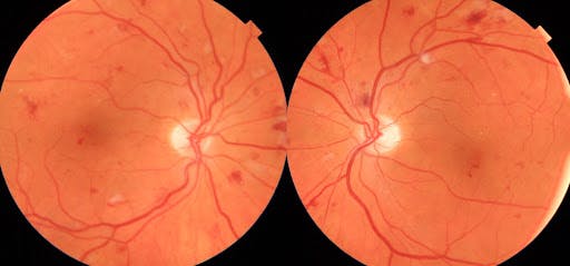
Figure 1
In Figure 2, we see a fundus photo montage of posterior poles with moderate nonproliferative diabetic retinopathy.

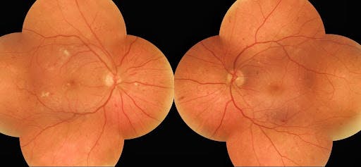
Figure 2
2) Retina/macula OCT
The ability to assess disorders of the retina and macula via OCT is essential for clinical practice today. The Topcon Maestro’s ability to take a single scan OCT as well as cube raster scans of the macula enhances diagnosis and treatment for retinal pathology. All OCT images are taken simultaneously with a corresponding photograph.
Figure 3 represents macula OCT of moderate nonproliferative diabetic retinopathy with macular edema.

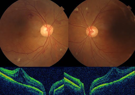
Figure 3
Figure 4 demonstrates moderate NDPR with macular edema as seen in an OCT scan with accompanying fundus photos.

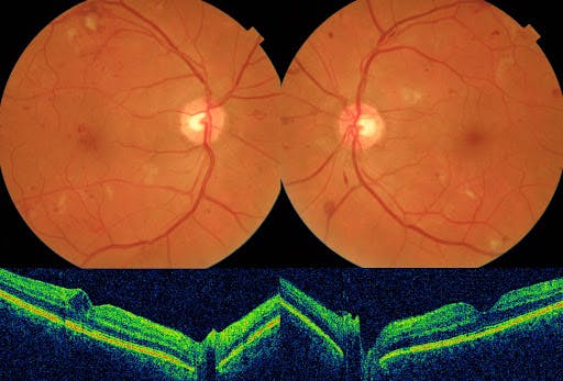
Figure 4
In Figure 5, we see a macula scan of resolving central serous chorioretinopathy OD.

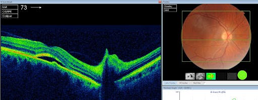
Figure 5
3) Optic nerve OCT
Having a technique to image and assess the optic nerve is crucial for today’s eye care practitioners. OCT has revolutionized the way we diagnose and treat optic nerve pathology. This provides the clinician a simple way to assess optic nerve structure, as well as the integrity of the retinal nerve fiber (RNFL) and ganglion cell layers.
As seen in Figure 6 of the optic nerve OCT scan for primary open angle glaucoma of the right eye, the red-free filter is also a nice feature, allowing for a closer look at various disorders of the retina and optic nerve. Here, the red-free image shows a superior wedge defect which corresponds with RNFL thinning.

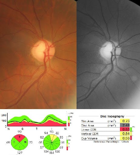
Figure 6
Figure 7 of an optic nerve OCT scan for traumatic optic neuropathy OS.

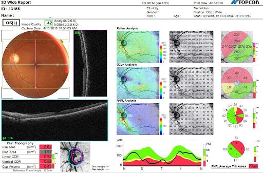
Figure 7
4) Glaucoma: anterior segment (pachymetry and anterior chamber depth)
Gone are the days of invasive techniques to assess corneal thickness. Anterior segment OCT makes pachymetry measurements simple and easy (and are billable procedures to boot!).
In Figure 8, we see pachymetry taken via the Topcon Maestro’s anterior segment OCT.

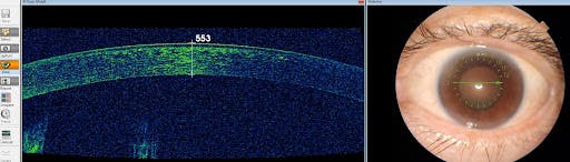
Figure 8
Assessing anterior chamber anatomy is a useful tool for both glaucoma patients and those with narrow angles. This feature is a nice adjunct to gonioscopic assessment of the angle.
Figure 9 demonstrates wide open nasal anterior chamber angle assessment OD.
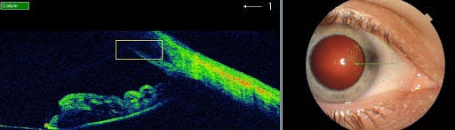
Figure 9
5) Assessing scleral contact lenses
Last but not least, we use the anterior segment capabilities of this instrument to assess the fit of scleral contact lenses. Determining adequate central clearance, as well as satisfactory scleral landing zones, are two ways to implement anterior segment OCT when fitting scleral lenses.
While many other factors and considerations go into the final scleral lens fitting, implementing anterior segment OCT can facilitate and optimize the fitting process for clinicians.
Figure 10 shows the use of anterior seg OCT to assess central clearance on scleral lens fitting.

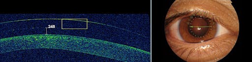
Figure 10
Figure 11 represents anterior seg OCT to assess the temporal scleral landing zone.

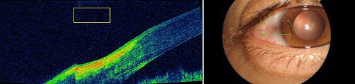
Figure 11
Conclusion
Combining OCT and fundus photography makes for an invaluable tool for a medical-based eyecare practice. We use the Topcon Maestro as a screening tool on all our patients to obtain a baseline OCT and fundus photograph. We can then fine-tune our scans with specific macula, retina, or optic nerve imaging if there is a particular pathology we want to investigate more deeply. Anterior segment use provides further insight for glaucoma and scleral contact lens fittings.
