


If you were to ask someone what organ they associate with the term “melanoma,” their answer would most likely be the skin. While this is the most commonly occurring cancer type globally, there is a subtype of melanoma that most people aren’t aware of, and is sight- and globe-threatening: uveal melanoma. Uveal melanoma is associated with significant morbidity and mortality, making both diagnosis and treatment crucial. Below outlines an overview as well as risk factors for uveal melanoma.
Uveal melanoma: An overview
Uveal melanoma is the most common primary malignant tumor of the eye, representing 3-5% of all melanomas. It is derived from choroidal melanocytes and is therefore usually pigmented. The uvea consists of the choroid, iris, and ciliary body; uveal melanoma can arise from any of these three different regions (and may be present in more than one location at the time of diagnosis). Symptoms, which depend on the location of the tumor, usually begin when the tumor extends to the optical axis or if there is concomitant retinal detachment or interference with the function of the ciliary body or pupil.
Symptoms to watch out for can include the following:
- Blurred or changed vision
- Floaters
- Flashes of light
- A dark spot/lesion on the iris
- Changes in pupil size or shape
Due to the lack of early symptoms, except iris melanoma, uveal melanoma is often diagnosed late in the disease course. Although it may be detected during a regular fundoscopic exam and on B-scan ultrasonography, approximately 50% of patients with uveal melanoma go on to develop distant metastases, most commonly to the liver. Unfortunately, for those patients who develop metastases, 80% of those succumb to the disease within 1 year. With these statistics, it is clear that early screening methods are imperative to decreasing the mortality rate of this disease.
Epidemiology of uveal melanoma
The incidence of uveal melanoma is 6-10/1,000,000 per year in the United States. The median age of diagnosis is approximately 62 years; however, the peak range for diagnosis is between 70-79 years. It is most common in Northern European descent, particularly those with light eye color and fair skin. Males have a 30% greater incidence than females. Of the three possible locations within the eye, a choroidal tumor is the most common (85-90% of cases), followed by ciliary body (5-8% of cases), and finally iris (3-5% of cases).
A retrospective medical record review at the Ocular Oncology Service, Wills Eye Institute, Thomas Jefferson University in Philadelphia, Pennsylvania, elaborated on these findings. In this publication, of the 8,033 patients managed at this service diagnosed with uveal melanoma, the mean (median) age was 58 (59), and 98% were Caucasian. 7256 of these patients had a choroid tumor, 492 a ciliary body tumor, and 285 an iris tumor.
Etiology of uveal melanoma
There have been many risk factors identified with uveal melanoma. The presence of light eyes and fair skin has been associated with the disease. Other factors include an inability to tan, ocular melanocytosis, dysplastic nevus syndrome, and BAP1 mutations. Ocular melanocytosis is a congenital periocular pigmentary condition that leads to the development of uveal melanoma affecting an estimated 1 in 400 patients.
Interestingly, Shields et al found that patients with uveal melanoma associated with ocular melanocytosis had double the risk for metastasis compared to patients without ocular melanocytosis. Dysplastic nevus syndrome is an autosomal dominant disorder in which a patient develops benign acquired melanocytic neoplasms. A study by Weis et al. performed a meta-analysis of the literature on dysplastic nevus syndrome and uveal melanoma from 1966 to August 2007 and found an association between the two.
BAP1 is a tumor suppressor gene located on chromosome 3, which was found to be mutated in 47% of uveal melanoma cases. Patients with this loss of function mutation also had an 11% greater chance of developing second malignancies, such as renal cell carcinoma and cutaneous melanoma, than the general population.
Many studies have tried to find a correlation between sunlight exposure and uveal melanoma due to the risk factors of light eyes, fair skin, inability to tan, and Northern European descent. However, while there is ample evidence of this correlation with skin melanoma, the evidence in regard to uveal melanoma is insufficient and contradictory.
A recent study found that while some tumors with adenine-to-cytosine mutations were found to have increased incidence in illuminated areas of the eye, other tumors with adenine-to-thymine mutations were found to have increased incidence in unilluminated areas of the eye in patients with light eye color. This suggests that light eye color and sunlight exposure may be different risk factors associated with different anatomic and mutation profiles.
Prognosis of uveal melanoma
Age is a factor to consider regarding the prognosis of a uveal melanoma diagnosis. Shield et al found that out of 8,033 patients, 4,287 were 21-60 years old, 3,640 were older adults (60+ years old), and 106 were young (≤ 20 years old). While young patients had a higher incidence of iris melanoma, older patients averaged greater tumor thickness and tumor-related metastasis and death rates.
As in cutaneous melanoma, tumor size is an important clinical feature when assessing prognosis for uveal melanoma. It is often measured in chord or arc length of greatest basal diameter and greatest tumor thickness when measuring tumor size. Tumor thickness tends to be a more accurate measurement, with ultrasonographic calipers coming within 2 mm of histopathologic measurement in 90% of eyes.
A meta-analysis concluded that with increasing tumor thickness in uveal melanoma, the higher the risk of metastasis and thus the higher risk for mortality. Iris melanoma had a significantly smaller average tumor size than ciliary body melanoma, while the average ciliary body and choroidal melanoma tumor sizes did not have significant differences. Iris melanoma also had a significantly smaller average tumor thickness than choroidal melanoma, with a smaller average tumor thickness than ciliary body melanoma.
Over 3-, 5-, 10-, and 20-year follow-ups, metastasis rates continuously increased as the original tumor size increased. Once a patient develops metastatic disease from uveal melanoma, survival rates are poor, with median overall survival of 13.4 months and only 8% of patients surviving 2 years.
Treatment of uveal melanoma
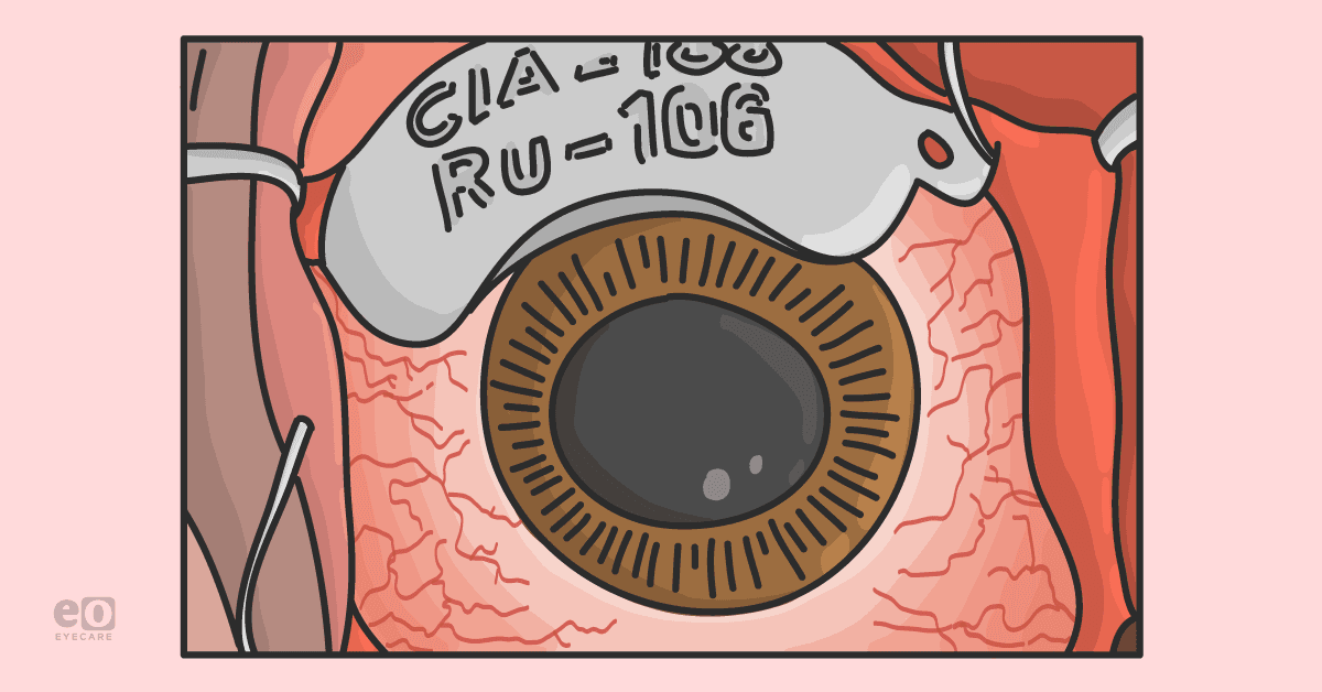
The management of localized uveal melanoma can be broken down into globe-preserving therapy or enucleation surgery to remove the eyeball. Globe-preserving treatments can be further categorized into radiation, surgical, or laser therapy. Most uveal melanoma cases in the United States are treated with plaque brachytherapy, a type of radiation therapy. While this provides excellent local control and globe preservation, long-term vision loss is common.
In some cases, enucleation may be performed if a patient is already experiencing vision loss, or the tumor is beginning to invade the optic nerve or metastasize. No difference in mortality was observed between the two treatment modalities at up to 15 years of follow-up.
Exciting new pharmaceutical therapies are starting to be explored to treat uveal melanoma. One example is sunitinib malate, an oral multi-targeted tyrosine kinase inhibitor with anti-tumor and immune-modulating effects. It is normally used on gastrointestinal stromal tumors that fail on other therapies, and it is used to prevent renal cell carcinoma in high-risk patients. Carol L. Shields MD conducted a study out of the Wills Eye and Thomas Jefferson University on 20 patients with uveal melanoma metastasis who failed other treatments. In 30% of cases, there was progression-free survival at 6 months, which Dr. Shields described as a modest effect.
However, when using low-dose sunitinib malate in high-risk uveal melanoma patients with no metastasis but high-risk cytogenetics, there was improved survival of 85% at 6 years compared with only 40% in those not using additional medications. These benefits were found in patients under 60 years of age; therefore it would be a logical next step to see how older patients would fare with this treatment.
Other novel therapeutic strategies are being explored for the treatment of primary uveal melanoma, some of which include the targeting of tissue factor and heparan sulfate proteoglycans. A 2017 study supported further clinical development of the use of heparan sulfate proteoglycans as targeted therapy for early-stage uveal melanoma patients.
For metastatic disease, understanding the pathophysiology behind uveal melanoma will hopefully begin to yield targeted therapies for dysregulated cellular pathways, such as the BAP1 mutation. Agents that modify gene expression and epigenetics should be further studied and next-generation immunotherapies will then have the opportunity to move through development. However, for this to occur, there is much more research to be done, and there must be continued support and funding of clinical trials for these novel agents.
Final thoughts
At this time, the prognosis of a patient diagnosed with uveal melanoma varies greatly depending on when the disease is caught. If the patient is lucky enough to be diagnosed before metastasis occurs, mortality rates are concerning but overall; the odds are in the patient’s favor. However, if the disease can metastasize, the mortality rates become alarmingly high.
“The key to reducing mortality rates would be early screening methods and more targeted immunotherapy options. A yearly fundoscopic exam is a great stepping-off point for early detection.”
Still, it appears more methods need to be implemented as half of the patients diagnosed go on to develop metastatic disease. Once a patient is diagnosed with uveal melanoma, a cytogenetic profile should be obtained so physicians can target any specific genetic abnormalities that may be found. With this information, next-generation immunotherapies can potentially make a great difference in patients’ prognoses. Unfortunately, there is no way to guarantee this ideal outcome as these immunotherapies are not available as of now. Clinical trials are imperative to start the process of lessening the disease burden of uveal melanoma.
Works cited
- Charters, Lynda. “Pearls for Managing Care of High-Risk Uveal Melanoma Patients.” Ophthalmology Times, Ophthalmology Times, 10 Oct. 2019, https://www.ophthalmologytimes.com/view/pearls-managing-care-high-risk-uveal-melanoma-patients.
- Collaborative Ocular Melanoma Study Group. “The COMS Randomized Trial of Iodine 125 Brachytherapy for Choroidal Melanoma: V. Twelve-Year Mortality Rates and Prognostic Factors: COMS Report No. 28.” Archives of Ophthalmology (Chicago, Ill. : 1960), U.S. National Library of Medicine, Dec. 2006, https://pubmed.ncbi.nlm.nih.gov/17159027/.
- de Lange, MJ, et al. “Distribution of GNAQ and GNA11 Mutation Signatures in Uveal Melanoma Points to a Light Dependent Mutation Mechanism.” PloS One, U.S. National Library of Medicine, Sept. 2015, https://pubmed.ncbi.nlm.nih.gov/26368812/.
- Harbour, J William, et al. “Frequent Mutation of BAP1 in Metastasizing Uveal Melanomas.” Science (New York, N.Y.), U.S. National Library of Medicine, Dec. 2010, https://pubmed.ncbi.nlm.nih.gov/21051595/.
- Kines, Rhonda C, et al. “An Infrared Dye-Conjugated Virus-like Particle for the Treatment of Primary Uveal Melanoma.” Molecular Cancer Therapeutics, U.S. National Library of Medicine, Feb. 2018, https://www.ncbi.nlm.nih.gov/pmc/articles/PMC8063570/.
- Jovanovic P, Mihajlovic M, Djordjevic-Jocic J, Vlajkovic S, Cekic S, Stefanovic V. Ocular melanoma: an overview of the current status. Int J Clin Exp Pathol .2013; 6(7): p.1230-44. pmid: 23826405.
- Krantz, Benjamin A, et al. “Uveal Melanoma: Epidemiology, Etiology, and Treatment of Primary Disease.” Clinical Ophthalmology (Auckland, N.Z.), Dove Medical Press, 31 Jan. 2017, https://www.ncbi.nlm.nih.gov/pmc/articles/PMC5298817/.
- Kujala, Emma, et al. “Very Long-Term Prognosis of Patients with Malignant Uveal Melanoma.” Investigative Ophthalmology & Visual Science, U.S. National Library of Medicine, Nov. 2003, https://pubmed.ncbi.nlm.nih.gov/14578381/.
- McLaughlin, Colleen C, et al. “Incidence of Noncutaneous Melanomas in the U.S.” Cancer, U.S. National Library of Medicine, Mar. 2005, https://pubmed.ncbi.nlm.nih.gov/15651058/.
- Shields , CL, et al. “Association of Ocular and Oculodermal Melanocytosis with the Rate of Uveal Melanoma Metastasis: Analysis of 7872 Consecutive Eyes.” JAMA Ophthalmology, U.S. National Library of Medicine, Aug. 2013, https://pubmed.ncbi.nlm.nih.gov/23681424/.
- Shields, Carol, et al. “Clinical Spectrum and Prognosis of Uveal Melanoma Based on Age at Presentation in 8,033 Cases.” Retina (Philadelphia, Pa.), U.S. National Library of Medicine, July 2012, https://pubmed.ncbi.nlm.nih.gov/22466491/.
- Shields, Carol L, et al. “Metastasis of Uveal Melanoma Millimeter-by-Millimeter in 8033 Consecutive Eyes.” Archives of Ophthalmology, JAMA Network, 1 Aug. 2009, https://jamanetwork.com/journals/jamaophthalmology/fullarticle/423790.
- Singh P, Singh A. Choroidal melanoma. Oman J Ophthalmol .2012; 5(1): p.3-9. doi: 10.4103/0974-620X.94718
- “Skin Cancer Facts & Statistics.” The Skin Cancer Foundation, 10 Aug. 2021, https://www.skincancer.org/skin-cancer-information/skin-cancer-facts/.
- “Uveal Melanoma: Skin Cancer.” UPMC HIllman Cancer Center, https://hillman.upmc.com/cancer-care/melanoma-skin/types/uveal.
- Weis, Ezekiel, et al. “The Association of Cutaneous and Iris Nevi with Uveal Melanoma: A Meta-Analysis.” Ophthalmology, U.S. National Library of Medicine, Mar. 2009, https://pubmed.ncbi.nlm.nih.gov/19167086/.

