


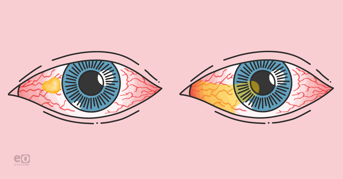
Pterygium and pinguecula are both conditions that affect the ocular surface. They are both classified as growths of the conjunctiva and can be caused by various risk factors including hereditary influence and environmental exposures, such as ultraviolet (UV) light.1
Individuals located closer to the equator have a higher predilection for pterygium and pinguecula development due to increased UV exposure.2 With a variable prevalence of 22.5% to 97%, pinguecula is more commonly found than pterygium, which has a 1.3% to 5.9% prevalence.3
Pterygium
Overview
Exposure to environmental factors such as wind and UV light can form pterygia, which are benign collections of conjunctival subepithelial degeneration, referred to as “surfer’s eye.”
There is a higher occurrence of pterygium development associated with people living in arid regions, individuals of African, Asian, American and Hispanic descent, history of dry eye disease, age under 40, and male gender.4 These risk factors ultimately cause excess collagen proliferation and blood vessel growth that can eventually involve the cornea.1 The result is a conjunctival growth that expands in a wing-like orientation from the limbus onto the cornea.
Pterygium development is stimulated by inflammation with increased growth factors, matrix metalloproteinases, angiogenesis, and fibrovascular proliferation.1
Few studies have been done regarding the hereditary influence of pterygium development, but research shows that pterygia present with multifactorial, familial, and autosomal dominant patterns of inheritance.1
Figure 1 illustrates pterygia with a bilateral hairlike extension growth.10

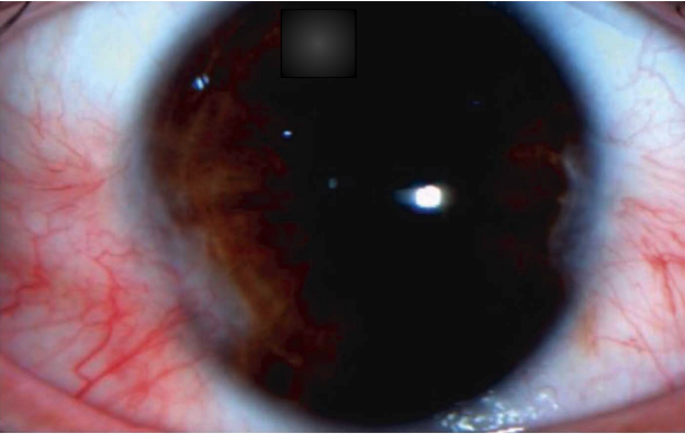
Figure 1: Elhamaky, RT and Elbarky, AM (Journal of Ophthalmology)
Symptoms of pterygium
Common symptoms include generalized ocular irritation, irritation with blinking, and blurred vision. Other complications include aesthetic changes, such as conjunctival redness. A slit lamp exam can be used to better visualize this conjunctival growth.
If the pterygium is extensive, there can be restrictions in eye movement since the pterygium can incorporate itself into the extraocular muscle and tendon tissue, or even act as a tether to the adjacent normal conjunctiva.
Pterygium expansion into the visual axis has been associated with corneal distortion that adversely affects visual acuity as the pterygium growth alters the cornea's curvature.5 This corneal curvature change results in irregular astigmatism, specifically referred to as “with the rule astigmatism,” where the eye is steeper at 90 degrees.5 The depth of penetration of the pterygium into the cornea affects the severity of astigmatism.5
Figure 2 shows histological imaging of overlying pterygium in conjunctiva.11

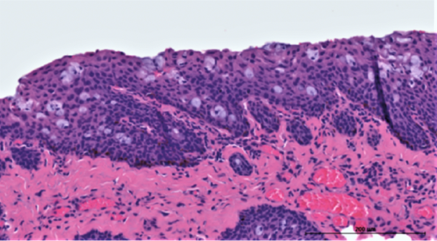
Figure 2 : Bergeron S, Ito H, Dosseus YE (Journal of Ophthalmology)
Treatment for pterygium
If the pterygium does not extensively involve the cornea, artificial tears and sunglasses can be helpful to diminish further growth.4 Surgical removal is indicated for more advanced pterygia, although resection carries the risk of postoperative recurrence.
When visual disruption becomes significant, surgical resection utilizing a conjunctival autograft, with or without antimetabolite therapy, is preferred.6 If it is not possible to use a conjunctival autograft after resection, placement of an amniotic membrane is a well-established alternative.4
The use of the Pterygium Extended Removal Followed by Extended Conjunctival Transplantation (P.E.R.F.E.C.T.) technique often results in a decreased likelihood of primary lesion recurrence at 0.1%, with no secondary recurrence.7
The table below lays out the P.E.R.F.E.C.T technique for reduced to no pterygium recurrence.6
| P.E.R.F.E.C.T. technique | Procedure |
|---|---|
| Stage 1 | Pterygium is removed from limbus including Tenon and conjunctival-involved areas, while medial and lateral rectus muscles are sutured. |
| Stage 2 | Downward rotation of the eye facilitates the marking of graft placement. |
| Stage 3 | The graft is sutured to the sclera while conjunctival edges are sutured to the cornea, leaving a distance from the limbus. |
P.E.R.F.E.C.T involves excising both the affected conjunctiva and Tenon’s layer and the fascial membrane that envelopes the eye from the orbit, leaving the sclera bare for autograft placement. The P.E.R.F.E.C.T technique can also be used with amniotic membrane transplantation and adjuvant therapy, like mitomycin C.
Pinguecula
Overview
Pinguecula is a benign conjunctival degradation that is unlikely to affect vision.8 It is classified as a nodular conjunctival growth and may be fatty or calcific. Pinguecula does not display angiogenic growth or progress onto the cornea the way a pterygium does.
While there is no predilection based on race or sex, contact lens wearers and individuals with lysosomal storage disorders have an increased predisposition to pinguecula development.8
Figure 3 features pinguecula and dilated vessels without invasion.12

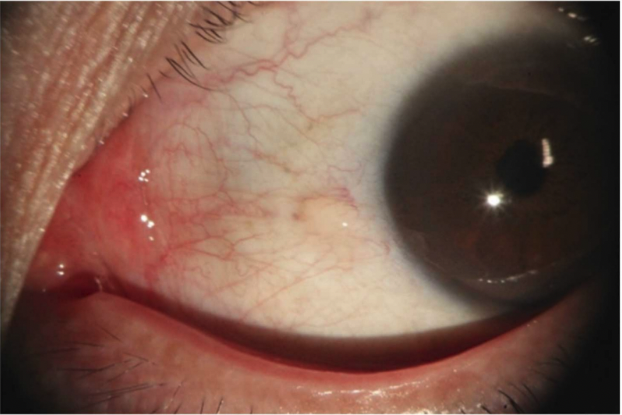
Figure 3: Jeong J, Rand GM, Kwon K (Journal of Ophthalmology)
Symptoms of pinguecula
Environmental factors like wind and dust, along with UVB light damage, can be related to the development of pinguecula due to increased oxidative stress and advanced glycation end products.8,9
Although pinguecula does not affect visual acuity; the conjunctival surface results in generalized irritation, foreign body sensation, tearing upon blinking, itching, and a burning dry eye.8
While pinguecula does not affect visual acuity, the conjunctival surface results in:8
- Generalized irritation
- Foreign body sensation
- Tearing upon blinking
- Itching
- Burning dry eye
Pingueculae are diagnosed clinically, based on appearance during a slit lamp examination.8
Treatment for pinguecula
For pingueculitis (inflammation of the pinguecula), treatment can range from artificial tears to topical steroids based on the degree of inflammation present.
While pingueculae are benign, if a patient wishes to have the lesion removed for cosmetic purposes, available options include surgical excision and argon laser photocoagulation.8 The use of laser photocoagulation allows for more precise lesion removal and decreases complications.
Conclusions
Prevention remains the gold standard in reducing the likelihood of conjunctival growth. For both pterygium and pinguecula prevention, sunglasses for eye protection from UV exposure during daylight hours are key.9 Differential diagnoses between pterygium and pinguecula can be readily established through slit lamp examination.
Risk factors should be evaluated to decrease the risk of growth and potential complications of inflammation for both types of lesions. Conjunctival involvement with ocular lesions can be managed conservatively through eye protection, but medical management and surgical resection remain viable options for both pterygium and pinguecula.
References
- Anguria P, Kitinya J, Ntuli S, Carmichael T. The role of heredity in pterygium development. Int J Ophthalmol. 2014;7(3):563-73.
- Planomaster. Pinguecula and Pterygium. Plano. 6 July 2022, https://plano.co/eye-conditions/other-eye-conditions/pinguecula-and-pterygium/.
- Viso, E., Gude, F. & Rodríguez-Ares, M. Prevalence of pinguecula and pterygium in a general population in Spain. Eye. 2015; Vol. 25; 350–357.
- Sarkar P, Tripathy K. Pterygium. [Updated 2022 Feb 21]. In: StatPearls [Internet]. National Library of Medicine: StatPearls Publishing. 2022 Jan-.
- Lin A, Stern G. Correlation between pterygium size and induced corneal astigmatism. Cornea. 1998; Volume 17; 28-30.
- Jaros, PA. DeLuise,VP. Pingueculae and pterygi. Surv Ophthalmol. 1988;(33)1: 41-49.
- Hirst, LW. Recurrence and complications after 1000 surgeries using pterygium extended removal followed by extended conjunctival transplant. Ophthalmol. 2012; 2205-2210.
- Somnath A, Tripathy K. Pinguecula. [Updated 2022 Feb 21]. In: StatPearls [Internet]. National Library of Medicine: StatPearls Publishing. 2022 Jan-.
- Notara, M., Behboudifard, S., Kluth, M.A. et al. UV light-blocking contact lenses protect against short-term uvb-induced limbal stem cell niche damage and inflammation. Sci Rep. 2018; Vol 8.
- Elhamaky, RT and Elbarky, AM. Outcomes of vertical split conjunctival autograft using fibrin glue in treatment of primary double-headed pterygia. J Ophthalmol. 2018; Vol. 2018;1–6.
- Bergeron S, Ito H, Dosseus YE, et al. Histopathological variability and concomitant lesions in pterygium in a large case series. J Ophthalmol. 2021; Vol. 2021;1–7.
- Jeong J, Rand GM, Kwon K, et al. The improvement of dry eye symptoms after pinguecula excision and conjunctival autograft with fibrin glue. J Ophthalmol. 2019; Vol. 2019; 1–6.
