


Gone are the days when you had to guess a c/d of 0.3 because you missed writing it during the exam and now you do not remember while doing charts. These are the benefits of retinal screenings!
Retinal screening/imaging is a non-invasive tool used in the diagnosis and management of retinal conditions. These screenings use multiple technologies to gather different images and scans of the retina, optic nerve, blood vessels and other associated structures of the back of the eye.
Advancement in technology has not only provided more accurate images of the retina but also provides detailed analysis of the layers of the retina which can further enhance monitoring for disease progression and management strategies.
Types of retinal screenings
There are multiple types of retinal screenings that can be done in an eyecare setting from fundus photography to Heidelberg Retinal Tomography. The three major retinal screenings we will be discussing today include fundus photography, optos, optical coherence tomography (OCT), and OCT-Angiography (OCT-A). Although dilation is the standard of care, retinal screening offers additional benefits in providing enhanced care to our patients including detailed diagnosis of their condition to monitoring changes over time.
Fundus photography
Fundus photography or commonly known as a retinal image is a simple yet effective tool to screen for fairly common conditions that affect the retina. This imaging technology provides high-resolution images of the fundus and typically involves a minute or two to do this test. This test is generally painless and often involves a bright flash of light for the patient. The only issue of fundus photography is that it only captures the central 30 to 45 degrees of the retina—the posterior pole.
Figure 1 pictures a Nidek fundus photography camera.

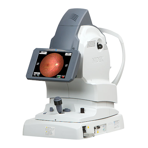
Figure 1
Optomap/Optos ultra-widefield imaging
Optomap is another imaging system which captures the digital ultra-widefield image of the retina. It produces a high-resolution image of 82% or 200 degrees of field of view of the retina in a single capture whereas fundus photography only captures up to 45 degrees of the retina. This wide angle provides a great advantage for your eye care professional to detect, diagnose, and manage retinal disease, especially the ones that affect the peripheral retina.
In Figure 2, a Nikon Optos imaging system is represented.

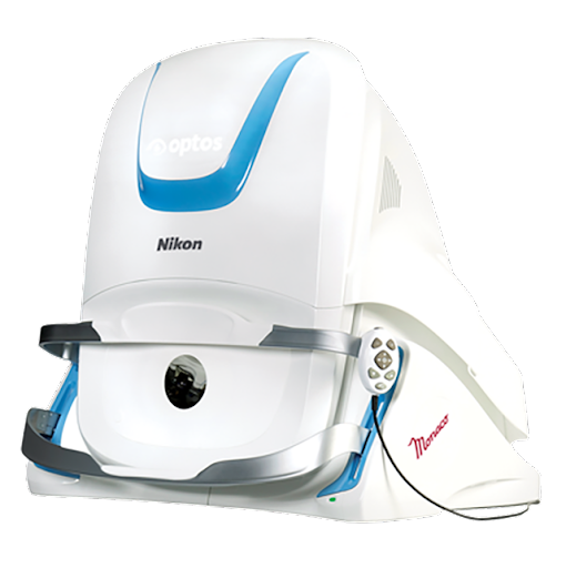
Figure 2
Optical Coherence Tomography (OCT)
OCT provides high resolution images of the structures behind the eye . These structures include the optic nerve, retina, choroid, and vitreous humor. This imaging modality provides a cross-sectional view of the layers of structures at the back of the eye along with quantitative measures to detect thickness and anatomical changes at various structures. OCT can also be used to view anterior structures of the eye including cornea, iridocorneal angle, iris abnormalities, and the anterior lens. OCT is like an ultrasound of the eye but instead of sound, it uses light.
This procedure is generally comfortable, painless, and takes less then a few minutes to be completed.
Refer to Figure 3 for a Zeiss OCT machine.

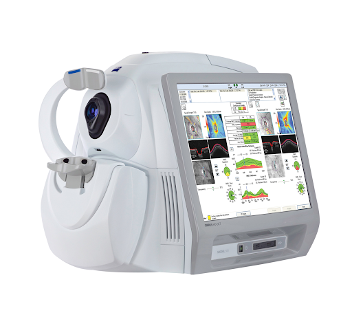
Figure 3
Conditions identified by retinal screenings
There are multiple conditions that can be identified, managed, and treated using retinal screenings. Some major ones are discussed below:
Diabetic retinopathy
Diabetic retinopathy is the leading cause of blindness among adults between the ages of 20-74 years in the United States, although with early detection, this condition can be treated, reducing the risk of severe vision loss by over 90 %. With recent advances in digital imaging, there have been new avenues for assessing retinopathy and providing better management, treatment, and diagnosis of this sight-threatening condition.
Retinal imaging tests such as OCT can help determine the thickness of retinal layers especially in patients with clinically significant macular edema. Fundus photography or Optos can determine changes in retinal structure caused by diabetic complications such as hemorrhages, exudates, neovascularization, and intraretinal microvascular abnormalities (IRMA).
More recent advancements in OCTA imaging can be used to determine the extent of neovascularization in patients with proliferative diabetic retinopathy (PDR).
See Figure 4 for fundus photography with diabetic retinopathy.

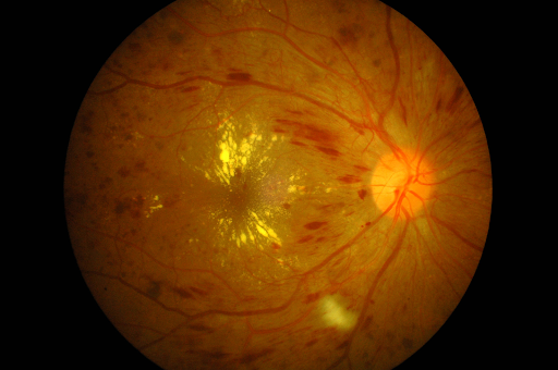
Figure 4
Glaucoma
Glaucoma is an ocular disease characterized by the progressive optic degeneration with subsequent loss in retinal nerve fiber layer (RNFL) and retinal ganglion cells (RGCs). It is the leading cause of irreversible blindness worldwide with no definitive cure. However, if caught early in the disease process, treatments are available to preserve vision and maintain quality of life.
Patients do require consistent and close monitoring for any changes in RNFL and RGC health. These tests include tonometry, humphrey visual field, retinal photography, stereo optic disc photography, OCT, gonioscopy and pachymetry. Routine retinal screening can help clinicians better detect changes in the retinal nerve fiber layer thickness. Treatment protocols for glaucoma can also be modified based on changes seen via retinal screening and imaging—adding additional IOP lowering drops or considering surgical intervention.
In Figure 5, an optic nerve image shows glaucomatous optic neuropathy.

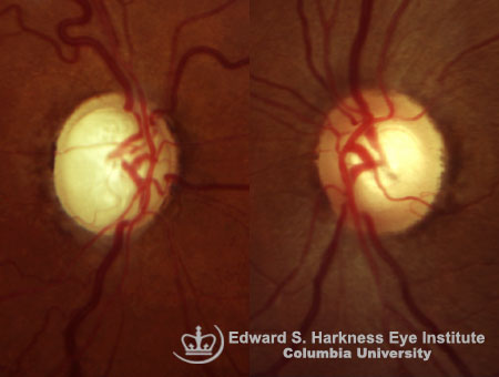
Figure 5
Age-related macular degeneration
Age-related macular degeneration (AMD or ARMD) is a macular condition that most often occurs in patients over the age of 50 years old and is characterized by pigmentary abnormalities, geography atrophy, hemorrhage, sub/intraretinal fluid, and/or choroidal/macular neovascularization. These changes all occur within the macula.
With retinal imaging, progression of this condition can be monitored more closely. Imaging tests such as OCT can be used to monitor changes within each specific retinal layer as well as the accumulation of drusen and any fluid buildup in the macular region. With the advancement of OCT-A, microvascular structures of the retina and choroid can be observed noninvasively—without the need of dye injection.
OCT-A can detect, localize and define neovascularization thus allowing easier monitoring of disease progression and helping determine the level of treatment required. OCT-A can detect early choroidal neovascularization caused due to dry and exudative AMD and can determine the exact blood flow patterns.
Fundus photography in Figure 6 shows Dry vs Wet AMD with associated OCT images showing retinal atrophy Dry AMD and fluid/blood build up in Wet AMD.

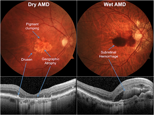
Figure 6
Retinal detachment
A detached retina is when the retina lifts away from the back of the eye. Usually, it begins off as vitreous sticking to the retina and creating traction - leading to a retinal tear. Fluid can then pass through this tear leading to a retinal detachment. Patient’s usually complain of flashes and floaters, shadows appearing in the peripheral vision or a gray curtain covering the respective part of the visual field. These are usually the early signs for retinal detachment/tear/holes.
Retinal screening would be able to detect these retinal changes and confirm the diagnosis in addition to dilated fundus examination. Optos is a great tool to be used to capture 200 degrees of field of view of the retina in a single capture and helps to determine changes with any peripheral retinal condition.
Figure 7's Optos image shows a large retinal tear associated with retinal detachment.

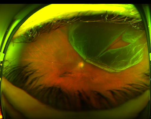
Figure 7
Hypertension
Hypertension or high blood pressure tends to affect the veins and arteries in the eye. Observing these vessels in real time can help indicate whether or not a patient’s hypertension is affecting their eyes.
Typical signs of hypertension in the eye include blood vessels becoming more narrow and harder thus having copper or silver wiring appearance. Advanced cases can also lead to bleeding/hemorrhaging, macular edema, and papilledema in the eye. Thus, retinal screening over time can be used to determine any changes.
The fundus photography in Figure 8 shows signs hypertensive retinopathy.

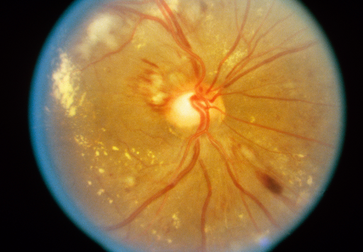
Figure 8
Cancer
Choroidal nevus and other suspicious lesions can occur in the retina and other parts of the eye, thus retinal imaging can significantly increase the chances of catching, monitoring, and treating these suspicious lesions. The longer it takes to find and diagnose signs of cancer, the more challenging it becomes to treat them. This can end up putting the patient’s well being at risk, if the cells metastasize to other regions.
Fundus photography can show such lesions in the posterior pole while Optos can image these lesions located more peripherally. OCT can be used to monitor these lesions, determining changes in elevation or subretinal fluid.
In Figure 9, an Optos/Optomap image shows intraocular melanoma.

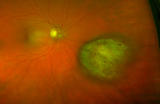
Figure 9
Incorporating retinal screenings
Retinal screening should become a part of your comprehensive eye examination to provide patients with highly detailed information about their ocular health. Patient education is the most important aspect to get the best out of retinal screening. The staff should be trained to provide detailed yet simple explanations of each test (retinal photography vs. OCT vs. Optos) so patients understand what these tests will offer them. Staff should be provided with a model of the eye so they can use them for patient education and explain what is being imaged/scanned.
These are some phrases that your staff can use to explain additional retinal imaging tests to patients:
Fundus/Optos photography: “This test will provide the doctor with high-resolution images of the back of your eye. The images produced by these screening tests will allow your doctor to assess your central vision, the optic nerve (which connects your eye to your brain) as well as your retina (for optos) and blood vessels.”
OCT: “This test is like an ultrasound of the back of your eye. It takes cross-sectional images of the layers of your retina and can determine the thickness of these layers, which can be affected by certain conditions. The doctor can use that information to assess and detect signs of diseases like macular degeneration, glaucoma or diabetes.”
Dilated fundus examination vs retinal screening
Dilated fundus examination (DFE) is the standard of care for comprehensive examination of the retina but is retinal screening enough to provide such care?
There has been a large increase in the use of retinal screening with telemedicine, especially during the COVID-19 pandemic. One such study found that teleophthalmology/retinal screening is an effective screening and management tool for a range of adult and pediatric acute and chronic ocular conditions such as retinopathy of prematurity, diabetic retinopathy, and age-related macular degeneration. This was implemented to overcome barriers created due to the COVID-19 pandemic.
Another study implemented retinal screening in primary care settings which increased the rate of evaluation for diabetic retinopathy for patients in rural and underserved settings. This can also be implemented in areas with minorities or areas with limited access to medical facilities.
Another use of retinal screening was done to assist primary care facilities to triage patients to better triage patients with acute ocular complaints and promptly refer those most in need to an eye specialist. This would widen the diagnostic and therapeutic options of retinal screening and teleophthalmology for ocular emergencies beyond the management of chronic conditions.
Now can retinal screening replace dilated fundus examination?
Regardless of how good the retinal imaging scans can be, they are not the doctor. Thus dilated fundus examination should always be the standard of care. With improvements in imaging technology and processing, and better integration of the patient’s medical record, teleophthalmology/retinal screening should become a more accepted modality with assisting the provider with enhanced ability to diagnose, treat, and manage the condition. Retinal screenings can provide enhanced care and better monitor the progression of certain conditions such as glaucoma, diabetic retinopathy, choroidal nevus, and many more.
Although there are certain limitations of all types of retinal screening tools that limit their ability to get a high-quality image. First is the skill of the technician to capture the image, considering the person taking the image is not in an eyecare facility then they might not be skilled enough to navigate any difficulties to get a good image.
Secondly, there are ocular pathologies limiting the view of the retina such as cataracts or corneal diseases which would distort the images thus requiring a physical examination with either binocular indirect ophthalmoscopy or fundus biomicroscopy with condensing lenses.
Another is the limits of the technology in providing a full view of the retina such as fundus photography only provides a 30-degree field of view, whereas Optos/Optomap images 82% or 200 degrees of field of view of the retina thus limiting view of the far peripheral conditions of the retina. Lastly, poor patient compliance or a patient with mobility problems can be an issue when getting a good image quality or in fact getting an image at all. Patients with limited mobility would require handheld retinal screening tools thus limiting their options for the type of scans available for them.
Billing for retinal screening
The current procedure billing code for Fundus Photography is 92250 and for OCT (scanning computerized ophthalmic diagnostic imaging “SCODI”) it is either 92134 (retina/macula) or 92133 (optic nerve). Typically in the United States, billing for any of the above imaging modalities is only permissible for medically-necessary reasons (e.g., primary open angle glaucoma, diabetic retinopathy, non-exudative age related macular degeneration, etc.). Consistently billing images of the eye for “screening purposes” is not done, as these images are typically captured during the routine workup of every patient in most practice settings (even healthy ones).
Cons of incorporating retinal screening
Although retinal screening is mostly beneficial and can enhance your treatment, diagnosis, and management of a patient's ocular health, there are some cons. Let's talk about the elephant in the room - the price of these machines. The price of a fundus camera can range from $5,000 to $40,000, although there are handheld ones available that can be available at a lower cost. Optos, on the other hand, can go much higher as the full purchase price of the Optos Daytona is around $85,000. Optos is a strong competitor to the OCT in terms of pricing, as its range is from $50,000 and $120,000.
The decision to buy these is not an easy one. However, in the long run, they can end up being a large portion of your practice’s profit, depending on how they’re being used. There are monthly loans available to finance these, or you can always consider paying cash on a used unit. There are also options to rent these machines.
Conclusion
Retinal screenings can enhance the care you provide to your patients and allow you to practice your profession to its highest standards. Retina screenings are a great asset in rural or underserved areas and are highly beneficial with the current pandemic-related restraints to reduce the need for physical examination to monitor a condition. The process can also be used to enhance your own charts as you can write more detailed descriptions of lesions or changes you see in the retina. This can also allow other healthcare providers to have a better understanding of the patient's condition when viewing your charts (e.g., for co-management care).
“The long-term benefit of having retinal screening capabilities can far outweigh the initial setup cost.”
Consider adding ophthalmic imaging modalities to your office’s repertoire of clinical tools. Don’t miss the opportunity to enhance patient care and education, while also increasing your practice’s profitability.
References
- https://okeyecare.com/why-retinal-imaging-is-an-important-part-of-your-eye-exam/
- https://www.coburntechnologies.com/2021/06/24/retinal-imaging-diseases/
- https://link.springer.com/article/10.1186/s12886-017-0484-5
- https://www.msdmanuals.com/home/eye-disorders/diagnosis-of-eye-disorders/tests-for-eye-disorders
- https://www.optos.com/About/
- http://kardter.narod.ru/cd_27_4_140.pdf
- https://www.nei.nih.gov/learn-about-eye-health/eye-conditions-and-diseases/retinal-detachment
- https://visionaryeyecentre.com/retinal-imaging-how-it-works-why-its-important/
- https://link.springer.com/article/10.1007/s00417-020-04879-2
- https://jamanetwork.com/journals/jamaophthalmology/fullarticle/2627936

