


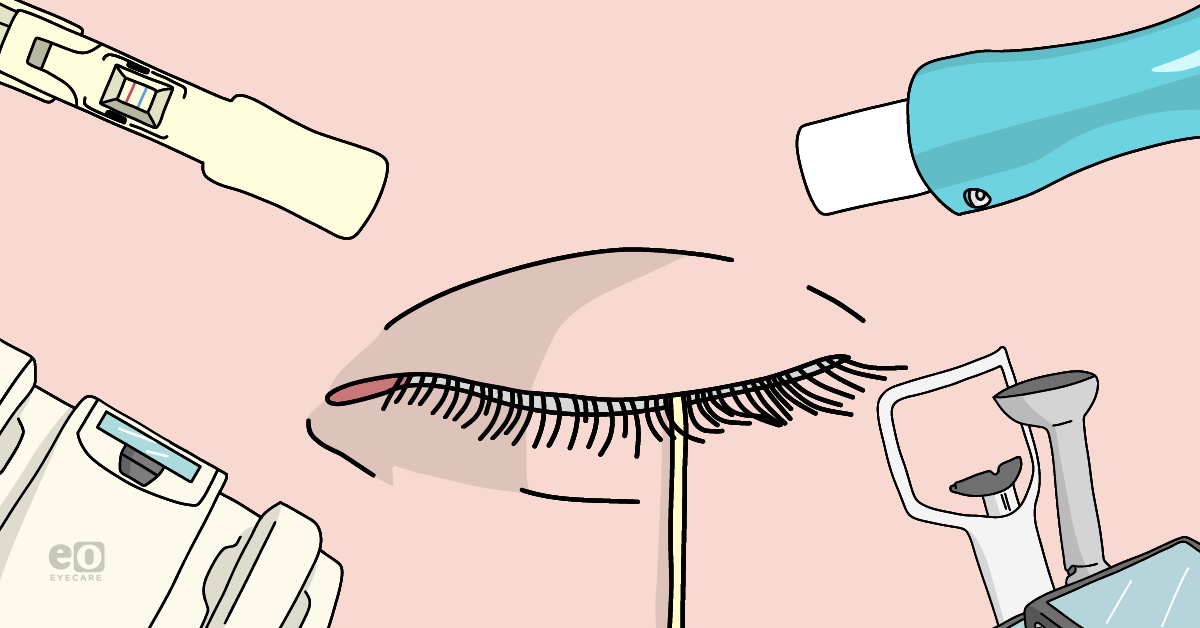
Dry eye disease (DED) is a multi-factorial disease characterized by the loss of homeostasis of the tear film by tear film instability, hyperosmolarity, and ocular surface inflammation.1 DED is frequently encountered in every optometric practice, and today, there are a number of different diagnostic tools that can help diagnose, manage, and treat this complicated condition. Even though these diagnostic tools can provide a quicker diagnosis and treatment of DED, they are supplementary tests meant to compliment a comprehensive eye exam.
LipiScan™
LipiScan noninvasively images the meibomian glands in 10 seconds using an infrared light. Eyecare professionals rely on Meibography as a critical tool to help diagnose, treat, and manage meibomian gland dysfunction (MGD) (Figure 1).
LipiScan can help establish a patient’s level of gland degeneration and atrophy and help clinicians determine whether conservative treatments such as lipid-based artificial tears and warm compresses will be sufficient enough for improving dry eye symptoms or if the MGD has progressed to an advanced stage where alternative treatments such as radiofrequency, Lipiflow, or IPL are likely needed.2
In Figure 1, Pult’s ‘meiboscale’ describes a five-grade scale with meibography images to further assist in the diagnosis in the MGD grading process. Upper and lower lids are assigned a grade from 0-4, with higher grades indicating higher gland atrophy and degneration.2

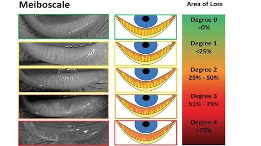
Figure 1
Meibomian Gland Evaluator™
The Meibomian Gland Evaluator is a hand-held instrument that noninvasively evaluates patients meibomian gland expressions and over all gland function. The instrument provides a quick and efficient method for applying a consistent, gentle pressure to the lower eyelid while assessing the meibomian gland expressions through a slit lamp.3
SPEED™ Questionnaire
The Standard Patient Evaluation of Eye Dryness (SPEED) questionnaire was designed to quickly and efficiently screen patients for the severity of dry eye disease over time. The questionnaire is a summed up score from 0-28 where the results are tallied from eight items that assess the frequency and severity of dry eye symptoms, this includes assessing dryness/ grittiness/scratchiness, soreness/irritation, burning/watering, and eye fatigue. The average score for mild dry eye is 5.0, moderate dry eye 6.6, and 10.0 for severe disease.4
InflammaDry
InflammaDry is a disposable, low-cost test that can be quickly administered and interpreted. It specifically tests the tears for matrix metalloproteinase-9 (MMP-9), an enzyme involved in promoting inflammatory pathways, and accurately identifies elevated MMP-9 levels in tear samples taken from the palpebral conjunctiva in the lower lids. Elevated MMP-9 levels have been shown to significantly correlate with decreased visual acuity, decreased fluorescein tear breakup time (TBUT), corneal and conjunctival fluorescein staining, and abnormal superficial corneal epithelia.5
InflammaDry has also been shown to provide highly accurate results that gives 85% sensitivity and 94% specificity.5
In order to administer the test, a tear sample must be collected, where it is then activated with a buffer solution for 10 minutes. The test uses 40 µg/ml of MMP-9 as a cutoff point meaning results below this value will show a single blue line, indicating a negative non-inflammatory result, and results above this value will show a blue line and a red line, indicating a positive pro-inflammatory result.5
JUST RELEASED: The 2025 Dry Eye Report
Get insights from hundreds of optometrists in the highly-anticipated 2025 Dry Eye Report! Explore trends, treatments, tools, and advice to help you stay ahead in dry eye management, all for FREE! Click here to download!
TearLab™ Osmolarity
Tear osmolarity assesses the severity of ocular surface inflammation. An increase in tear osmolarity can reduce tear film homeostasis and stability due to inflammation and cell apoptosis. The TearLab Osmolarity Test Card and the TearLab Osmolarity System provides a quick method for measuring tear osmolarity using nanoliter volumes of tear fluid collected from the eyelid margin.
The Test Card is held in place by the Osmolarity Test Pen for accurate and safe collection, then the TearLab Osmolarity System calculates the overall tear osmolarity by displaying a numerical value. A reading of >300 mOsm/L, or an inter-eye difference of >8 mOsm/L can indicate instability of the tear film and ocular surface inflammation.
The Tearlab Osmolarity System, as seen in Figure 2, has a positive predictive value of 89%.6

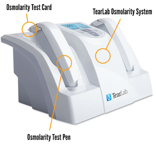
Figure 2
The Schirmer Test
A Schirmer’s Test noninvasively measures reflex tear secretions and determines if enough tears are being produced to keep the ocular surface moisturized and healthy. A Schirmer’s Test is performed by placing a piece of filter paper inside the lower eyelid of both eyes and the patient closes their eyes for 5 minutes. If the results show more than 10 mm of moisture on the filter paper after 5 minutes, this is a sign of normal tear production. Also, numbing drops may be administered to prevent the patient’s eyes from watering in reaction to the test strips.7
The Phenol Red Thread Test
The Phenol Red Thread Test (PRT) is similar to the Schirmer test, except red strips of thread are used instead of paper strips and the test takes only 15 seconds. The PRT measures the volume of the tear film in the inferior conjunctival sac of the eye. The test is conducted by inserting the 3 mm folded portion of the thread into the palpebral conjunctiva of the lower eyelid. The thread is originally yellow in color and when it comes into contact with tears, it changes to a light red color.
After 15 seconds, the thread can be removed and the entire red colored portion can be measured. A reading of less than 10 mm indicates dry eyes, less than 20 mm indicates minimal dry eyes, and more than 20 mm indicates normal tear volume.7
Slit lamp
Careful examination of the external eye, focusing on the ocular surface is performed with a slit lamp. A number of dyes are used with special filters to highlight any corneal surface abnormalities. These can include fluorescein and lissamine green stains. Using a slit lamp, eyecare professionals can diagnose dry eye, a poor TBUT, poor tear film quality, a defect on the surface of the cornea or conjunctiva, the presence of demodex mites, and many other dry eye related conditions.
TBUT and epithelial staining
Performing TBUT assesses for the stability of the tear film and evaporative dry eye disease. To measure TBUT, sodium fluorescein is introduced to the patient's tear film. While the patient tries not to blink, the optometrist observes the tear film under cobalt blue illumination. TBUT is measured as the time taken for the first dry spot to appear on the cornea after a complete blink. A TBUT under 10 seconds is indicative of tear film instability that is associated with evaporative dry eye disease.
Fluorescein staining helps assess ocular surface damage, particularly highlighting cells with a defective glycocalyx and/or tight junctions. This kind of staining can show severe DED from the appearance of defective corneal epithelial cells, and poor stability of the tear film.8
Incorporating dry eye diagnostics into your clinical practice
Dry eye diagnostic testing is not always easy to incorporate into a regular clinic day. Ways to help with time and efficiency when trying to successfully implement these diagnostic tools include reserving dedicated blocks of time to see dry eye patients exclusively. Another key to building a successful dry eye clinic is getting staff on board and this means educating staff on dry eye procedures.
Testing will also need to be performed systematically due to some dry eye tests not being able to be used after instilling staining agents. It is also necessary to delegate staff on particular dry eye tasks so that technicians can easily shift their job responsibilities to dry eye work-ups on clinic days devoted to such care.
Once dry eye diagnostic testing is complete, it is critical to educate these patients on what is occurring with their eyes so they can begin to comprehend why their eyes feel the way they do. Patient education is an important step of any dry eye clinic and can easily be done by showing patients images of their eyes that reflect their dry eye or other ocular surface disease conditions.
Including animations and drawings can help explain tear treatment recommendations, instructions, and when they should schedule their next visit. Handouts are also very helpful in providing patients further instructions on how to appropriately apply warm compresses or take certain medications formulated for dry eye disease.
Adding the latest dry eye diagnostic tests to your daily clinic flow is not an easy mission but is definitely worth the time to figure out the process. Your dry eye patients will be impressed with the technology but will also be thankful for the accurate diagnoses and treatments that will help resolve their specific dry eye problems.
References
- TFOS DEWS II Definition and Classification Report. Craig JP, Nichols KK, Akpek EK, Caffery B, Dua HS, Joo CK, Liu Z, Nelson JD, Nichols JJ, Tsubota K, Stapleton F Ocul Surf. 2017 Jul; 15(3):276-283.
- Johnson + Johnson, Lipiscan Dynamic Miebomian Imager (2021): https://www.jnjvisionpro.ca/products/lipiscan-dynamic-meibomian-imager
- Johnson + Johnson, Meibomian Gland Evaluator (2021): https://www.jnjvisionpro.ca/products/meibomian-gland-eye-exam-tool
- Andrew D. Pucker, Bradley E. Dougherty, Lisa A. Jones-Jordan, Justin T. Kwan, Carolina M. E. Kunnen, Sruthi Srinivasan; Psychometric Analysis of the SPEED Questionnaire and CLDEQ-8. Invest. Ophthalmol. Vis. Sci. 2018;59(8):3307-3313.
- Labtician Thea, InflammaDry (2021): https://www.labticianthea.com/product/inflammadry
- TearLab, TearLab Osmolarity Test System (2021): https://www.tearlab.com
- Saleh, T., McDermott, B., Bates, A. et al. Phenol red thread test vs Schirmer's test: a comparative study. Eye 20, 913–915 (2006). https://doi.org/10.1038/sj.eye.6702052
- Review of Optometry, When Dry Eye Compromises Corneal Integrity (2017): https://www.reviewofoptometry.com/article/ro1117-when-dry-eye-compromises-corneal-integrity
