


An open globe injury (OGI) involves a full-thickness defect in the eyewall, comprised of the sclera and cornea, resulting from penetrating or blunt trauma. An open globe is a vision-threatening ophthalmic emergency and should be immediately examined by an ophthalmologist.
Open globe injuries can be categorized into lacerating injuries and blunt ruptures. Lacerating injuries involve full-thickness disruption of the eyewall from a sharp object. These injuries can be subdivided into penetrating, perforating, and intraocular foreign body. A perforating wound enters and exits the eye, whereas a penetrating wound enters the eye but does not exit. An intraocular foreign body is a penetrating wound where a foreign material is retained within the eye. A globe rupture occurs when blunt force causes the intraocular pressure (IOP) to increase dramatically and leads to an inside-out rupture of the eye at a weak point in the cornea or sclera.
In eyes that have not previously undergone intraocular surgery, the most likely location of compromise is posterior to the extraocular muscle insertions, where the sclera is often thinnest and weakest. In eyes that have previously undergone intraocular surgery, incision sites are frequently breached in open globe injuries.
Epidemiology and presentation
The worldwide incidence of open globe injuries is around 3.5 cases in 100,000 people per year. Males suffer approximately 80% of open globe injuries. Males between 10-30 are in the highest risk category, with males under 40 twice as likely to sustain an open globe injury than males over 40.
In patients over 75, a ground-level fall is the most frequent cause of a globe rupture. The likelihood of a globe rupture increases after ocular surgery due to weakness in the cornea or sclera from the surgery.
Diagnosing open globe injuries
Because of the vision-threatening nature of open globe injuries, clinical suspicion for the possibility of an open globe injury should be high in all trauma cases. However, an ophthalmic examination should be deferred until the patient’s airway, breathing, and circulation are stabilized. Patients presenting with a history of severe ocular and/or periocular trauma should be considered to have an open globe until ruled out. A detailed history of the mechanism of trauma is essential in the initial evaluation of ocular trauma.
For example, IOFBs are most commonly secondary cases of metal-on-metal grinding or hammering without protective goggles. The timing of the trauma is also important as repairs should ideally be performed within 24 hours of the injury. It is also imperative to determine the ocular surgery history, tetanus status, and time of last meal.
Patients presenting with an open globe injury usually complain of severe eye pain and blurring of their vision after undergoing some sort of trauma. Patients may complain of diplopia due to a concomitant orbital fracture with/without extraocular muscle entrapment.
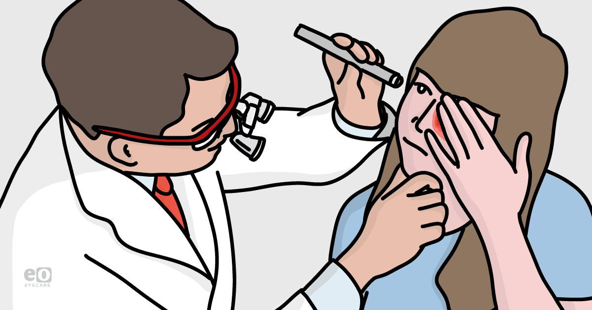
A detailed ophthalmic examination is critical in the evaluation of a suspected open globe injury. The clinician should take care to minimize manipulation of the globe and avoid any maneuvers that may apply pressure on the eye to decrease the risk of extrusion of the intraocular contents. For this reason, tonometry should be avoided until an open globe injury is ruled out. That said, low intraocular pressure is often associated with an open globe injury.
The physical exam should begin with an inspection of the head, face, periocular region, and eyelids to evaluate for lacerations, ecchymosis, and edema. If protruding foreign bodies are present, they should not be removed until the time of surgery. Measurement of visual acuity (VA) and testing for a relative afferent pupillary defect (RAPD) should be performed. Poor presenting VA and the presence of an RAPD are significant prognostic factors for a poor outcome.
A meticulous, comprehensive slit lamp examination should be performed to evaluate for eyewall defects and evidence of trauma. The sclera and conjunctiva should be carefully inspected for lacerations, foreign bodies, and defects. The cornea should also receive a detailed inspection. A Seidel test with fluorescein should be carried out, which may reveal anterior chamber fluid leakage from a corneal wound. Any extrusion of intraocular contents, such as uvea or vitreous, is pathognomonic of an open globe injury.
The anterior chamber (AC) should be assessed, with special attention to depth, hyphema, presence of vitreous, and cells. A deep AC may be present in an occult, posterior, open globe injury. The pupil should be examined for shape and reactivity. A teardrop-shaped pupil may be present in a penetrating corneal injury that passed through the iris and is often considered pathognomonic for an open globe injury.
The lens should be examined for clarity and position. Fundoscopy may be difficult, but should be attempted, and the posterior segment should be examined for retinal detachment, IOFB, and hemorrhage.
A non-contrast orbital CT can help with the diagnosis of an open globe injury and evaluate for IOFBs. The following CT findings are suggestive of an open globe injury:
- eye wall and globe contour irregularities
- intraocular air
- IOFB
- vitreous hemorrhage
- retinal detachment
- orbital and facial fractures
- orbital volume loss
- decreased anterior chamber depth
A CT scan can be very useful in diagnosing an occult rupture. An ultrasound of the orbit is relatively contraindicated due to the direct pressure of the probe on the globe.
Open globe injuries are anatomically divided into three zones of injury. The zone of injury is important in determining prognosis:
- Zone 1 injury involves the cornea and the limbus
- Zone 2 injury describes a full-thickness scleral laceration or rupture in the anterior 5mm of the sclera and does not extend into the retina
- Zone 3 injury is a rupture or laceration more posterior than 5mm from the limbus and involves the retina
Differential diagnosis
- Subconjunctival hemorrhage
- Orbital blowout fracture
- Corneal abrasion/ulceration
- Retrobulbar hematoma
- Traumatic glaucoma
- Traumatic iritis
- Vitreous hemorrhage
Management and treatment of open globe injuries
Once an open globe is suspected, urgent ophthalmic consultation is essential in order to protect vision. Other potentially life-threatening injuries should be treated first, and the patient should remain nil per os (NPO). The patient’s affected eye should be protected with a shield. Foreign body removal should not occur until the patient is in surgery. Any maneuvers that may increase IOP should be avoided to avoid potential extrusion of the intraocular contents. These maneuvers include tonometry, lid retraction, ocular ultrasound, and unnecessary manipulation of the eye.
Open globe injuries are tetanus-prone wounds and patients should receive a booster if they are not fully immunized or their immunization history is uncertain. Antibiotic drops should be started prophylactically to decrease the risk of endophthalmitis. Usually, systemic antibiotics are started as well; this may include cefazolin and/or fluoroquinolone.
In patients requiring intubation, high doses of ketamine or succinylcholine are relatively contraindicated due to the risk of increasing IOP. However, there is conflicting data regarding this effect on clinical outcomes. Regardless, rocuronium is currently generally preferred in open globe cases as it does not increase the IOP. Patients should also receive IV antiemetic therapy with ondansetron.
Patients and family members should be realistically counseled regarding the ocular prognosis of an OGI, including possible permanent vision loss, loss of the eye, need for secondary surgery(ies), and risk of sympathetic ophthalmia.
Once the patient is stable and prepared for surgery, they should undergo rapid surgical primary closure of their open globe. Delays in surgery can lead to worse visual outcomes and increase the risk of endophthalmitis. Surgical management of a globe rupture includes corneal and/or scleral wound repair, with the treatment of any secondary injuries in a comprehensive or staged surgical approach.
Postoperatively, patients should wear a protective shield at all times and avoid rubbing or touching their eyes.
Summary
An open globe injury is a vision-threatening ophthalmic emergency.
Trauma patients should receive stabilization of life-threatening injuries before attention is turned to the eye. Patients often complain of eye pain, blurred vision, or double vision. A thorough ocular examination is essential to evaluate trauma patients for an open globe, with special attention to visual acuity, presence of RAPD, teardrop pupil, low IOP, extrusion of vitreous, uveal tissue, and Seidel’s test status.
Patients should receive prophylactic antibiotics for endophthalmitis, tetanus prophylaxis if required, and should undergo primary surgical repair within 24 hours.
References
- Andreoli MD, C. M. (n.d.). Open globe injuries: Emergency evaluation and initial management. Retrieved May 02, 2021, from https://www.uptodate.com/contents/open-globe-injuries-emergency-evaluation-and-initial-management?source=history_widget
- Blair, K. (2020, December 19). Globe rupture. Retrieved May 02, 2021, from https://www.ncbi.nlm.nih.gov/books/NBK551637/
- Chronopoulos, A., Ong, J. M., Thumann, G., & Schutz, J. S. (2018). Occult globe rupture: Diagnostic and treatment challenge. Survey of Ophthalmology, 63(5), 694-699. doi:10.1016/j.survophthal.2018.04.001
- Jung, H. C., Lee, S., Yoon, C. K., Park, U. C., Heo, J. W., & Lee, E. K. (2021). Intraocular foreign body: Diagnostic protocols and treatment strategies in ocular trauma patients. Journal of Clinical Medicine, 10(9), 1861. doi:10.3390/jcm10091861
- Pieramici MD, D. J. (2005, June 15). Open-globe injuries are rarely hopeless. Retrieved May 02, 2021, from https://www.reviewofophthalmology.com/article/open-globe-injuries-are-rarely-hopeless
- Serrano, F. (2020, December 12). Traumatic eye INJURY management principles for the Prehospital Setting. Retrieved May 02, 2021, from https://www.jems.com/patient-care/traumatic-eye-injury-management-principl-0/
- Tirakunwichcha, S., & Pongsachareonnont, P. (2021). Factors associated with Visual outcome after primary repair of open-globe injury by ophthalmology residents in training in a Tertiary eye center. Clinical Ophthalmology, Volume 15, 1173-1181. doi:10.2147/opth.s300753
- Upaphong, P., Supreeyathitikul, P., & Choovuthayakorn, J. (2021). Open globe injuries related to traffic accidents: A retrospective study. Journal of Ophthalmology, 2021, 1-5. doi:10.1155/2021/6629589
- Wang MD, D. (2021, January 31). Open globe injury: Assessment and preoperative management. Retrieved May 02, 2021, from https://www.aao.org/eyenet/article/open-globe-injury
