


Since the invention of the first ophthalmoscope and slit lamp over 100 years ago, no device or technique has been as important to eyecare diagnostics as optical coherence tomography (OCT). Though the clinical exam is still an indispensable portion of any eye exam, OCT imaging has revolutionized how we examine and think about ocular disease. OCT technology has moved clinicians from a “how does it look” diagnostic model to a “what layer is affected and how thick/thin is it” clinical paradigm. What started as a grainy image of the retina has evolved into high-resolution imaging of the retina layers, optic nerve head, and anterior segment; all of which now have bundled quantitative analytics.
Be sure to check out the latest course on OCT Angiography!
The goal of any OCT is to acquire detailed, accurate, and repeatable images of ocular tissues. A major contributor to an instrument’s ability to accomplish this is the scan speed, measured in A scans per second. This is especially important in our patients where the rate limiting factor in image acquisition is not the OCT, but the person. In everyday practice, we often reach for our OCT to manage our glaucoma, AMD, and diabetic retinopathy patients. Unfortunately, these patients tend to be of more advanced age, fatigue quicker, and have poorer fixation, all of which can decrease the quality of your OCT images. With faster acquisition time, many of these patient-induced errors are diminished, providing better data for the clinician with less patient and technician frustration.
Wider and Deeper Scans
The ZEISS CIRRUS OCT allows clinicians to visualize vitreo-retinal disease over a 12mm wide scan using the High-Definition Raster scan. This scan protocol is especially important for conditions affecting the vitreo-retinal interface such as vitreomacular traction and diabetic neovascularization. The 12mm wide scan size in case 1 reveals that the central epiretinal membrane is deforming the central macula but also extends into the peripapillary area. Mild vitreal degeneration is noted anteriorly but there is no significant vitreo-retinal interaction. In contrast, OCT imaging in case 2 reveals early retinal-vitreous separation (in the context of diabetic neovascularization elsewhere) throughout the posterior pole and including the peripapillary zone. Anteriorly, significant vitreal degeneration can be visualized due to the deep 2.9mm scan size.

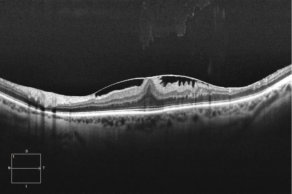
Case 1

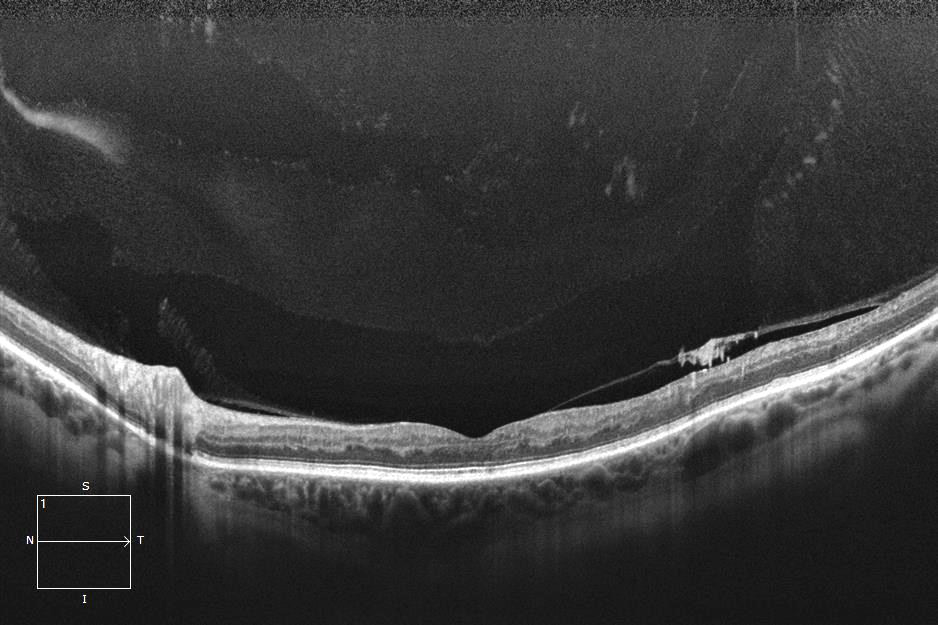
Case 2
OCT Angiography: Dye-less Vasculature Analysis
Though OCT angiography (OCTA) is still a relatively new technology, researchers and clinicians have already incorporated OCTA imaging into the management of numerous ocular conditions. Wide scan protocols are especially useful in cases of diabetic retinopathy where disease can present at the disc, macula, and/or vascular arcades. Studies have shown that OCTA is superior to clinical examination/color photography and similar to widefield fluorescein angiography images in the detection of proliferative disease. This high detection rate with a non-invasive imaging modality can greatly elevate the clinical exam in expediting referral and treatment.

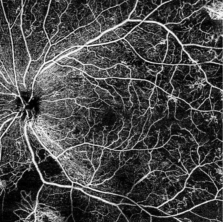
Case 3

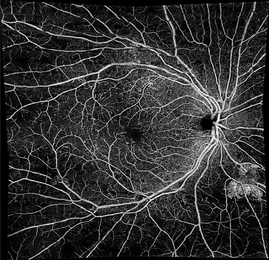
Case 4
Age-related macular degeneration (AMD) is another condition where changes in management are being driven by new technologies such as OCTA. Using OCTA, a new subtype of wet AMD was elucidated: nonexudative CNV. This entity describes CNV that is quiescent and does not leak on fluorescein angiography. Though there is active blood flow, there is no leakage on fluorescein angiography because no dye is escaping the blood vessels. These lesions can be seen in both treatment naïve patients and those treated with anti-VEGF. On OCT these lesions present as vascularized pigment epithelial detachments without associated serous fluid. OCTA will demonstrate blood flow but often these lesions are asymptomatic and not a detriment to vision. The treatment for this recently described phenomenon is still not well defined but it is thought that these lesions can be monitored without treatment.
Though a small study, 11 patients with neovascular AMD in one eye and intermediate AMD in the other eye were imaged with OCTA, fluorescein angiography, and indocyanine green angiography. With OCTA, the physician detected neovascularization in 3 of 11 intermediate AMD eyes. All cases were asymptomatic and had neovascularization confirmed with indocyanine green angiography. Of interest, OCTA performed better than fluorescein angiography, which is routinely used to detect neovascularization.
Big Data and Eye Wellness
In the era of big data, eye care has been late to the table. Now with ever-improving data management software like ZEISS FORUM, clinicians are able to analyze large amounts of multimodal data quite rapidly. In many chronic eye conditions such as glaucoma or diabetic retinopathy, minuscule changes eventually snowball into detectable clinic disease. The next generation of big data in eye care will be gathering long-term (decades worth of) data on patients throughout their “healthy” years so that any pathological changes can be detected earlier. Many practices have already begun collecting “ocular wellness OCT data” on their patients, which will likely pay dividends as these patients age and begin to develop various ocular conditions. The ocular wellness report assists the physician since many posterior segment conditions such as vitreomacular traction, epiretinal membrane, and glaucoma may be difficult to detect clinically in their early stages. With this type of seamless transition from CIRRUS generation to generation, ophthalmic big data is easily achievable.
ZEISS CIRRUS: The New Standard
The OCT has become the workhorse of the medical eyecare practice. The breadth of OCT technology and clinical utility has supplanted standard fundus photography in many disease management paradigms and continues to alter how clinicians care for their patients. Where fundus photography provides information that overwhelmingly overlaps with funduscopy, OCT imaging offers additional diagnostic data that cannot be ascertained by the clinical exam. With increasingly wider OCT scan sizes and indeed Ultra Widefield fundus cameras such as the ZEISS CLARUS, together with advancements in vascular OCTA technologies, the traditional 45-degree fundus photograph may soon be a relic of the past. A top-of-the-line OCT is not just about keeping up with technological advancements but about staying in line with medical progress. OCT technology unlocks the ability to provide true full scope care through the management of a wide variety of conditions, which would otherwise need to be referred out.

