The three optometrists featured in this paper have one specific thing in common: not one of them can imagine going back to practicing before they incorporated the
CLARUS 500 into their clinics. Each has found reasons to rely on CLARUS’s capabilities, day in and day out, to help diagnose and monitor disease progression, screen for pathology, and ultimately improve efficiency and workflows within their practices.
Designed for optimal color, clarity, comfort, and confidence
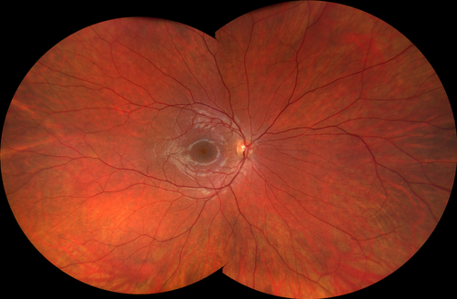
Since its launch in September of 2017, the CLARUS 500 has become an indispensable piece of equipment in many practices, largely due to its true color capacity, imaging clarity, and ease of use. This ultra-widefield fundus imaging system enables clinicians to capture high-resolution images from macula to the far periphery, all in true color.
Color
Across the board, Patricia Fulmer, OD, FAAO; Joan Miller, OD; and Aaron Lech, OD, FAAO appreciate CLARUS’s color accuracy and identify it as a key feature that separates it from the other imaging systems on the market. These doctors laud its ability to capture images that closely duplicate the natural coloration of the fundus as it appears through direct observation, which they say helps to remove uncertainty regarding what is actually being captured.
For Dr. Patricia Fulmer, true color imaging is one of her favorite aspects of the instrument because of how easily it enables image analysis and patient education. Dr. Fulmer, who practices in Huntsville, Alabama at Legacy Vision Center (which she opened as a cold-start practice in December 2020), also credits CLARUS with simplifying patient education. For example, after using the instrument to take both anterior and posterior segment images, she can then manipulate the images in the exam room while presenting them to and reviewing them with the client.
Dr. Fulmer stated that the CLARUS increased her confidence in diagnosing and treating patients, and she believes that what she is seeing in the images is indeed what is representative of her patients’ retinas.
Clarity
Optometrists have also expressed that they are impressed with how accurately the retina is depicted when they use the CLARUS. They compare the view to what they see during a dilated fundus exam. For them, this level of clarity often means there is far less uncertainty regarding the interpretation of the images, thus removing the subjective factor when managing disease—which can be especially pertinent in a practice where multiple doctors may see one patient.
Dr. Aaron Lech stated that he uses the CLARUS because of a feature that essentially offers several vivid images in one and allows for manipulating those images in real time. The owner of ClearVue Eye Care in Roseville, California said that he appreciates the machine’s ability to offer incredible detail not only for objects 1.5mm to 2mm, but all the way down to small fibrous tissues that are just fractions of a millimeter.
These three clinicians have also found that ultra-widefield retinal imaging acts as an indispensable tool when used in conjunction with dilated fundus examination (DFE), especially when examining the peripheral retina for pathology.
Comfort
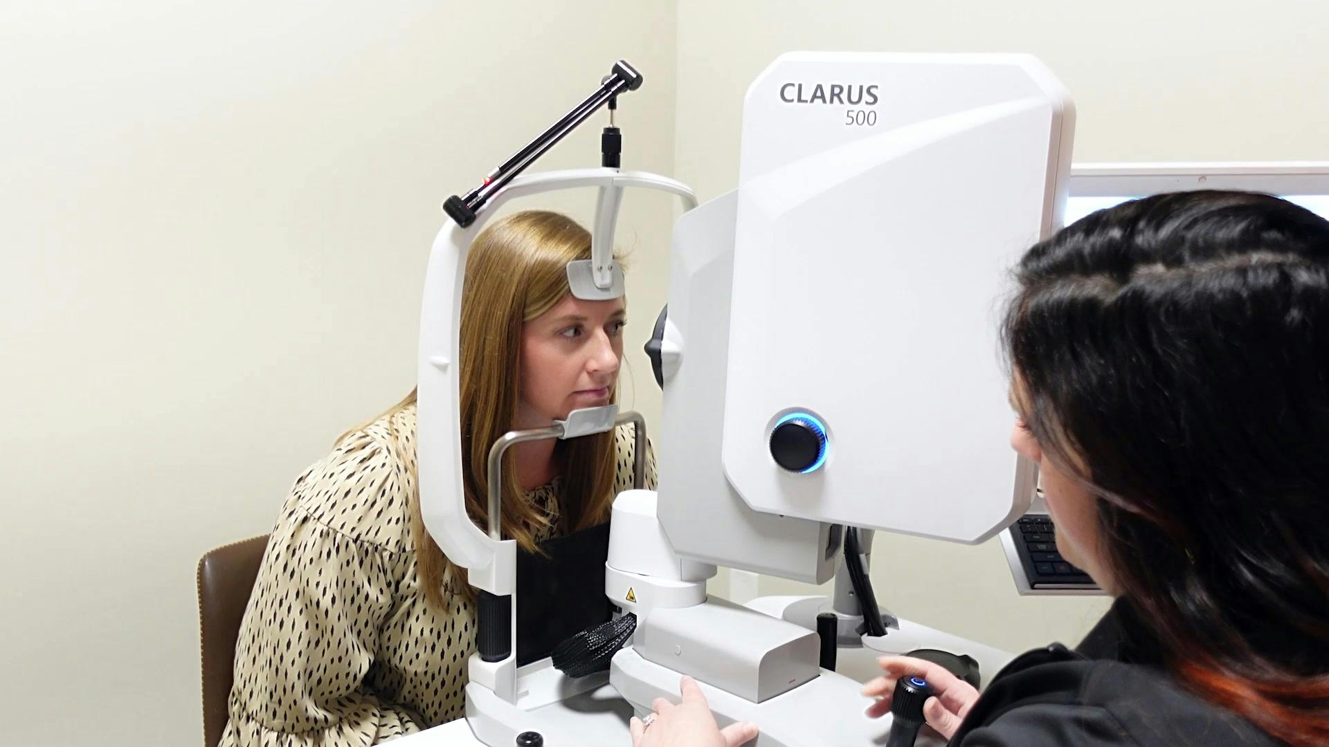
The CLARUS was designed with the comfort of both the patient and the operator in mind. Utilizing a chin rest and joystick approach make it easy to image patients ranging from young to old as well as accommodating those with neck or back issues. Operators find the acquisition head easy to maneuver and line up for the best possible image. Dr. Fulmer has found that by being able to image an increased retinal area, the CLARUS has really aided in documenting peripheral disease without putting the patient through multiple image acquisitions and flashes.
Confidence
The CLARUS ultimately provides a level of confidence in both the optometrist and the patient. Dr. Lech has found that having the ability to not only pick chronic conditions up on a screening basis but to closely track and monitor conditions over time adds a level of certainty as to when to hold onto a patient or when to refer. In turn, he believes that having the ability to deliver a diagnosis while showing the patient their diagnosis on the screen vastly improves their understanding—and subsequently their confidence—that they are receiving the right treatment.
Doctors use CLARUS for screening and diagnostics
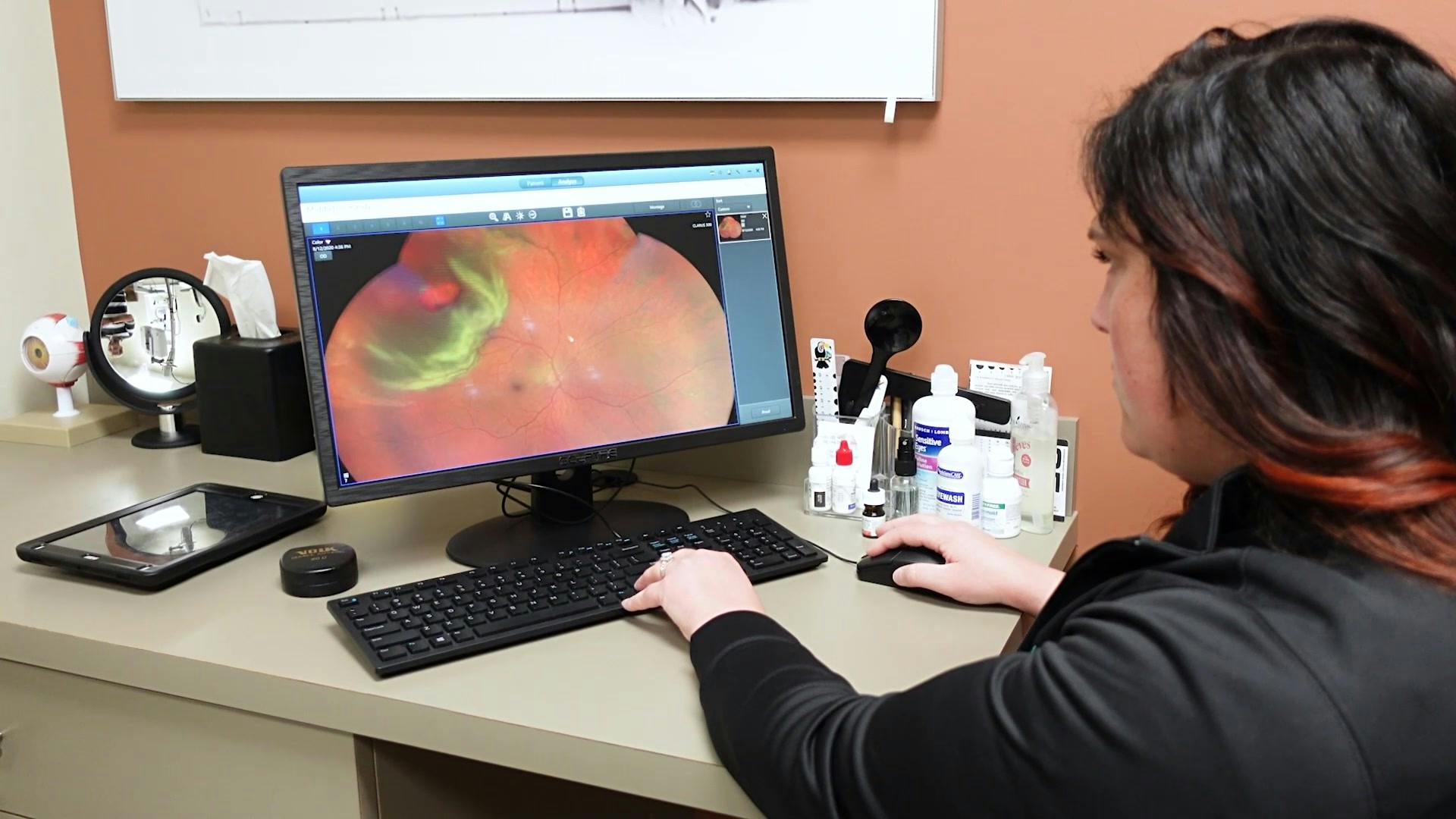
With its true color capabilities and capacity for capturing 133 degrees in a single image or 200 degrees in a montage of two sequential images, the CLARUS can also serve as a crucial component for screening for pathology. Factor in its amazing auxiliary modalities—including fundus autofluorescence, stereoscopic photography and external imaging—and this one piece of equipment can vastly improve diagnosis and monitoring disease progression.
For Dr. Fulmer, one of the most important ways that she and her staff use their CLARUS in-office, every single day, is in fact, in a screening modality. When her patients present for a comprehensive eye exam, they’re offered retinal imaging as a screening. This allows her to see any conditions that might be present and become prepared to review those results with the patient and educate them appropriately upon arrival in the exam room. Once her patients were given the option of retinal imaging, many became motivated to request the photos, which has helped to grow her practice’s revenue.
“For us, our CLARUS was a huge tool in making sure that we were being as efficient as possible. We’ve increased revenue through more medical billing and being able to collect private payments on our screening photos. Our patients have really gotten excited about the photos. They ask about them, they want to see them, and they tell their friends and family about them, generating more business and more foot traffic through our doors.”
Dr. Patricia Fulmer
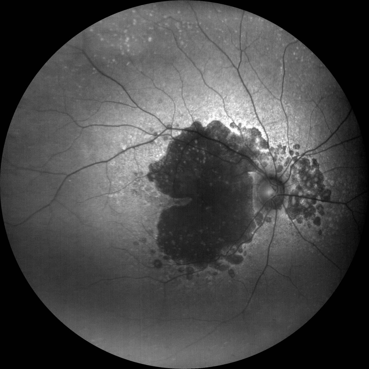
Dr. Fulmer often takes advantage of the fundus autofluorescence feature using FAF blue and green to screen for retinal findings like choroidal nevus, which she has found very easy to miss in a traditional exam if the patient isn’t fixating well or dilated properly. She’s found with CLARUS photos, on the other hand, even the faintest of nevi are detected.
Dr. Lech echoed this by saying that the CLARUS offers a host of tools to tease out layers within the retina to spot retinal bleeds or hemorrhages and identify where the tissue is most disruptive. For instance, in a patient with toxicity associated with other therapeutics that they are undergoing systemically, the images offer the detail to assess whether other diagnostics are required and which is the appropriate referral.
Detecting and monitoring ocular and general health conditions
Dr. Joan Miller, owner of Baseline Vision Clinic in Hillsboro, Oregon deems the CLARUS 500 critical in finding and monitoring ocular and general health conditions, including diabetes, macular degeneration, retinal pathologies, and glaucoma.
Recently, Dr. Miller witnessed the CLARUS 500’s lifesaving abilities firsthand when a close friend presented with a right eye floater. Upon imaging both eyes with the CLARUS 500, a malignant melanoma was discovered. Further examination confirmed the diagnosis and a prompt referral to a retinal oncologist led to a successful treatment and an outstanding prognosis. If undetected, this could have become a life-threatening situation.
“I believe the CLARUS is the most important screening tool in my practice to detect and follow potentially sight-
threatening, and even life-threatening, conditions. It really helps me explain conditions to patients, to explain what I’m concerned about, and to help them understand the things that they need to do to maintain good eye health.”
Dr. Joan Miller
How can optometrists benefit from technology?
As far as Dr. Lech is concerned, the primary reason optometrists should invest in technology is to get more care to more patients in an effective and efficient manner. The second is because patients expect it. Today’s patients want to be able to participate in their care and gain insight into their condition. In addition, patients are mobile and demand access. CLARUS provides both tangible information and a digital footprint they can take with them.
"It’s really important that the investments we make from a technology standpoint aren’t just about us and the clinic but the patient’s long- term health and viability.”
Dr. Aaron Lech
Early detection and patient care
Due to its array of technical features and multi-use functionality, CLARUS has emerged as a powerful tool within the ZEISS imaging suite. For clinics of all sizes, expanding on advanced diagnostic tools and technology may offer a huge return on investment because of the potential to streamline workflows, elevate efficiency and potentially lead to practice growth—but it is in the realm of early detection and patient care where eyecare practitioners state the CLARUS has the most value for their practices
Perhaps Dr. Miller said it best: “When we take care of patients, they take care of us. And having the right equipment makes all the difference in the world. In my practice, the CLARUS 500 makes optometry a joy.”