


When it comes to vision, most people are familiar with and have heard of nearsightedness, farsightedness, and even astigmatism. Both we and our patients aim for 20/20 vision, believing that this is the best standard of care.
Optometrists have been measuring these types of visual imperfections, also known as the lower-order aberrations (LOAs), for centuries without much difficulty. But what are we actually measuring when we measure a patient's vision to be at 20/20 or even 20/15? We are only measuring the quantity of a patient's vision when we report their acuity, and nothing more.
How often have we heard a patient complaining of their blurry vision, only to measure them at a perfectly “normal” 20/20? For the seasoned eyecare provider, quite often. They understand that the variety of patients who measure at 20/20 will see the 20/20 line very differently with varying quality.
While LOAs can be corrected easily with glasses or contact lenses, higher-order aberrations are more challenging to correct for and require specialized equipment and materials. With the use of an aberrometer, we can measure the quality, not quantity, of vision, also termed higher-order aberrations (HOAs).
In clinic, I will call HOAs “visual noise” as an easier way to understand aberrations. Visual noise such as ghosting, light scatter, haloes, glare, streaking, starbursts, and poor night vision are all symptoms a patient with HOAs will experience. Even though they may be able to read the letters on the chart, they can also understand that there are varying resolutions to seeing the letters more vividly.
Types of higher-order aberrations
Although there are other types of higher-order aberrations, coma, spherical, trefoil, and quadrafoil are the ones that are of most clinical relevance, with coma and spherical aberration being the most dramatic.
Each produces a unique aberration, such as:
- Spherical aberration: A rounded halo around a point of light or image, which also reduces contrast sensitivity.
- Coma: Distortion resembling a comet-like blur, where a point of light appears to have a blurry tail. This type of HOA produces the most image degradation.1
- Trefoil: A triangular halo and/or glare around objects.
- Quadrafoil: Distortion that appears elongated in a cross-like or cloverleaf-like pattern.
Figure 1: Types of HOAs demonstrated via a wavefront map, simulated image, and point spread function.


Figure 1: Courtesy of Nick Brown, Ovitz Corporation.
These aberrations can result from irregularities in the corneal shape, scarring, previous eye surgeries, LASIK/refractive surgery, those with large pupils, or certain medical conditions affecting the eye, such as keratoconus or other corneal dystrophies. They can also be induced by the aging process as the eye's internal structures change. By far, the most common group of patients to be impacted by higher-order aberrations are those with irregular corneas.
No eye is free from aberrations—we are all human and our eyes are soft, changing tissues with changing tear films that can fluctuate our visual quality frequently. Thankfully, the vast majority of higher-order aberrations are subclinical, not impacting our day-to-day visual duties.
For the irregular cornea, a well-fitted gas-permeable, hybrid, or scleral lens will suffice by masking irregularities of the front surface of the cornea. But for those who continue to experience visual noise from internal structures, a wavefront scleral lens is their best option.
Diagnosis and testing for HOAs
Because of evolving technology, we are now able to recognize, measure, and correct for HOAs. In practice, it has been quite validating to show my patients that what they are experiencing can be measured.
There are several aberrometers used in research, but very few are available for practical application. There are also multiple devices and topographers on the market and in our offices that can measure aberrations, but are of minimal applicable value as the resulting information cannot be applied to a contact lens.2
For the purposes of this article being clinically relevant, we will be using the Ovitz aberrometer system to describe how we diagnose, test, and manage patients with HOAs, as it is a commercially available system not restricted to certain practices.
How to measure HOAs
The first step is to take an aberration screening to provide us with a wavefront map and detect candidacy for a wavefront scleral lens. HOAs are highly dependent on pupil size, with higher amounts of HOAs being present as the pupil dilates. This explains why some patients have good daytime vision but poor night vision, as more HOAs are being uncovered when the pupil dilates in the evenings.
This is also why HOAs continue to be subclinical as we age; although HOAs do increase with time, they are balanced out by the increase in pupil miosis as well.1,4 In order to mimic this, we take our aberration screening in a dark room.
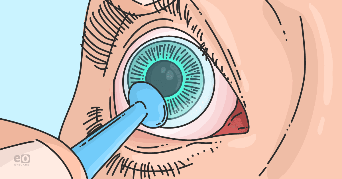
The only source of light is the light emitted from the aberrometer itself. We also cover the eye not being measured with an occluder to further limit the amount of light perceived to maximize natural pupil size. The scan is quick and drop-less.
Using RMS to screen for HOAs
There are a few clinically relevant data results from the aberration screening with the most useful being the root-mean-square (RMS). This is an easy way to determine at a glance what the combined amount of HOAs the patient is experiencing is and whether they are a candidate for a wavefront scleral lens.
The RMS provides a single value to determine if the patient has significant HOAs. Although we like to categorize the different types of aberrations into coma, trefoil, and such, the actual eye is much more complex, having varied amounts of each type to produce its own aberrated image.
Candidacy based on HOA RMS values:
- RMS under 0.2μm: Aberrations are low (Green). Only performance vision patients (i.e., athletes) are likely to benefit significantly from HOA correction.
- RMS between 0.2 to 0.5μm: Aberrations are moderate (Yellow). Vision distortion will start to have an impact. A patient will likely benefit from HOA correction, although other treatment factors may come into play (e.g., travel distance).
- RMS over 0.6μm: Aberrations are high (Red). The patient is very likely to benefit from HOA correction unless there are significant complicating factors.
Figure 2: A point spread function of a patient with keratoconus, demonstrating the combined effect of multiple HOAs on a single point of light.


Figure 2: Courtesy of Noha Seif, OD.
Using a wavefront map to assess HOAs
A wavefront map is also produced from this screening, using Zernike coefficients as the unit of measurement of the deviations with + and - describing directionality. The manufacturing lab then creates the inverse of the measured wavefront error and adds this to the optics of the scleral lens to neutralize the HOAs.
In order for the wavefront correction to work, precise alignment and orientation of the scleral lens on the eye is mandatory. Scleral lenses are the vehicle for this vision correction due to their lack of mobility and enhanced stability on the eye compared to other lens modalities.3
Figure 3: A marked scleral lens to determine the orientation and, therefore, the eventual location of the wavefront correction optics.


Figure 3: Courtesy of Noha Seif, OD.
Case report #1: Corneal transplants secondary to keratoconus
A 24-year-old male who had corneal transplants secondary to keratoconus came to our office wearing standard scleral lenses. He reported vision quality degradation and sub-optimal vision with shadowing of letters. His visual acuity was measured at 20/20 OD and 20/40 OS.
Aberrometry was performed over a refit of their scleral lenses to ensure stability and decreased fogging. His OD RMS was measured at 1.42μm, with vertical coma being the highest offender. The wavefront measurement was sent to the lab for manufacturing and dispensed to the patient.
The patient reported a significant overall improved quality of vision, noting that his vision was “smooth” and that his family members appeared to be glowing. He also reported that the world appeared to be more 3D and bold. Objectively, the HOA RMS measured on the OD wavefront lens was reduced to 0.39μm. Visual acuity was measured to be 20/20 OD and 20/25 OS.
Table 1: The before and after OD RMS values of a standard scleral lens and a wavefront correcting scleral lens being reduced from a severe 1.42μm to a moderate 0.39μm.
| Measurement 1 | Measurment 2 | |
|---|---|---|
| Sphere | -2.00D | -2.50D |
| Cylinder | -2.25D | -2.00D |
| Axis | 25° | 24° |
| HOA RMS | 1.42μm | 0.39μm |
| Original Analysis Diameter | 6.33mm | 7.37mm |
Table 1: Courtesy of Noha Seif, OD.
Figure 4: On the left, we have the wavefront map of the standard scleral lens, and on the right, we have the wavefront map of the wavefront scleral lens.


Figure 4: Courtesy of Noha Seif, OD.
Figure 5: HOA magnitudes as measured by Zernike coefficients comparing the aberration screening (in blue) from their standard scleral lens to their wavefront scleral lens (in red). Note the significant reduction in vertical coma.


Figure 5: Courtesy of Noha Seif, OD.
Case report #2: Post-refractive case
A 62-year-old male who underwent refractive surgery decades ago complained of difficulty with night vision and haloes around lights. He had no issues during the daytime, but he was looking to improve his night vision, if possible.
Visual acuity with glasses was measured to be 20/25 OD and 20/20 OS with minimal prescription. We took an aberration screening that measured an HOA RMS of 0.67μm OD and 0.86μm OS, with coma and spherical aberration being the highest offenders.
After proceeding with the wavefront scleral lens fitting process, we reduced the RMS value to 0.45μm OD and 0.32μm OS. The patient reported that his night vision was “dramatically better,” with no haloes or flare around lights seen any longer.
Figure 6: HOA magnitudes as measured by Zernike coefficients comparing the aberration screening (in blue) from their standard scleral lens to their wavefront correcting scleral lens (in red). Note that many of the HOAs initially measured in blue have been completely eliminated altogether, and a severe reduction in the vertical coma was initially measured.


Figure 6: Courtesy of Noha Seif, OD.
Figure 7: On the left, we have the wavefront map of the standard scleral lens OS, and on the right, we have the wavefront map of the wavefront scleral lens OS.


Figure 7: Courtesy of Noha Seif, OD.
Conclusion
Higher-order aberrations are a complex and fascinating topic, previously a measurement that only research labs could provide.
With the continued emergence and improvements in wavefront-guided scleral lenses, consider higher order aberrations are at play the next time that your 20/20 vision patient reports blurry vision.
Wavefront scleral lenses have provided the consistent ability to reduce and, in some lucky cases, eliminate the visual noise that many suffer from.

