


The Early Treatment Diabetic Retinopathy Study (ETDRS) classification system has evolved and, in a congruent manner, the approach for assessing and grading diabetic retinopathy severity has been modified in attempt to describe diabetic retinal disease with both accuracy and precision. Here, we review the evolution of the ETDRS classification system and the Diabetic Retinopathy Severity Scale (DRSS) and provide insights for future directions.
Historical perspective
While it began as the O’Hare Classification, the Airlie House Committee on diabetic retinopathy classification developed a comprehensive system to critically describe diabetic retinopathy and to evaluate the effect of treatment modalities.1 Specifically, the Airlie House Classification of diabetic retinopathy qualitatively and quantitatively described the status of diabetic retinopathy with fundus photography to permanently record the locations of these lesions in certain predesignated areas of the fundus1 (FIGURE 1).

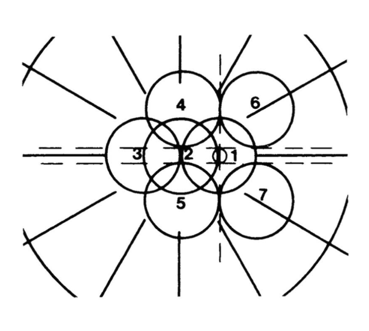
FIGURE 1: Seven standard fields of the modified Airlie House Classification of diabetic retinopathy. Using the right eye as an example, Field 1 is centered on the optic nerve, Field 2 is centered on the macula and Field 3 is temporal to the macula. Field 4, 5, 6 and 7 capture the retinal mid-periphery.
In 1971, the Airlie House Classification was modified for use in the first landmark clinical trial, the Diabetic Retinopathy Study, which demonstrated significant reduction in the rates of severe vision loss in eyes treated with panretinal photocoagulation compared to untreated control eyes.2 The grading system was further modified for use in the ETDRS randomized prospective study which found the rate of moderate vision loss in eyes with clinically significant macular edema treated with macular focal laser had a relative risk reduction of 50% compared to untreated eyes.3
The ETDRS, by use of fundus photography, defined and detailed microaneurysms (MA), venous beading and narrowing, intraretinal microvascular abnormalities (IRMA),
diabetic macular edema (i.e., clinically significant macular edema). Moreover, ETDRS definitions compared with standardized fundus photographs produced the DRSS for the description of non-proliferative diabetic retinopathy (NPDR) and proliferative diabetic retinopathy (PDR). TABLE 1 and FIGURE 2 summarize the ETDRS DRSS.

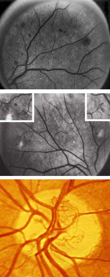
FIGURE 2: Early Treatment of Diabetic Retinopathy (ETDRS) standard photos 2A (top), 8A (middle) and 10a (bottom).
| Disease Severity Level | Clinical Findings | ETDRS Definition |
|---|---|---|
| Mild NPDR | At least 1 MA and definition not met for any other disease severity level | MA defined as red spot 125μm (longest dimension) with sharp margins |
| Moderate NPDR | MA and hemorrhages May include soft exudates, venous beading, IRMA with definition not met for any other disease severity level | Hemorrhage defined as red spot with irregular margins and a dimension that exceeded 125μm Hemorrhages and/or MA ≥ standard photograph 2A |
| Severe NPDR | Cotton wool spots, venous beading and IRMA in at least two of fields 4-7 Two of cotton wool spots, venous beading, IRMA in at least two of fields 4-7, with hemorrhages in all fields 4-7 (consistent with photo 2A) IRMA in all of fields 4-7 and definition not met for any other disease severity level | IRMA in fields 4-7 must be ≥ photograph 8A |
| Non High-Risk (Early) PDR | Neovascularization with definition not met for high-risk PDR | |
| High-Risk PDR | New vessel growth on or within 1 disc diameter of optic disc with or without vitreous or preretinal hemorrhage Vitreous or preretinal hemorrhage with NVD or NVE | Neovascularization ≥ photograph 10A (one-quarter to one-third disc area) NVE ≥ than one-quarter disc area |
TABLE 1: Summary of the diabetic retinopathy severity scale (DRSS) with associated disease severity, clinical findings and Early Treatment of Diabetic Retinopathy (ETDRS) standard photos. (NPDR, non-proliferative diabetic retinopathy; PDR, proliferative diabetic retinopathy; MA, microaneurysms; IRMA, intraretinal microvascular abnormalities; NVD, neovascularization of the disc; NVE, neovascularization elsewhere.)
The ETDRS and DRSS evolution
The ETDRS DRSS was subsequently updated to provide a coherent but simplified approach to retinopathy level and disease severity. The summary of the ETDRS Final Scale of Diabetic Retinopathy Severity is provided in TABLE 2 and FIGURE 3.

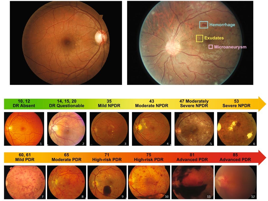
FIGURE 3: Photographic depiction of the abbreviated summary of the Early Treatment of Diabetic Retinopathy (ETDRS) Final Scale of Diabetic Retinopathy Severity. (NPDR, non-proliferative diabetic retinopathy; PDR, proliferative diabetic.)
| ETDRS Level | Disease Severity | Definition |
|---|---|---|
| 10 | No retinopathy | Diabetic retinopathy absent |
| 20 | Very mild NPDR | MA only |
| 35 | Mild NPDR | MA plus hard exudates, soft exudates (cotton wool spots) and/or mild retinal hemorrhages |
| 43 | Moderate NPDR | MA plus mild IRMA or moderate retinal hemorrhages |
| 47 | Moderate NPDR | More extensive IRMA. Severe retinal hemorrhages or venous beading in 1 quadrant only |
| 53 | Severe NPDER | Severe retinal hemorrhages in 4 quadrants, or venous beading in at least 2 quadrants, or moderately severe IRMA in at least 1 quadrant |
| 61 | Mild PDR | NVE < 1/2 disc area in 1 or more quadrants |
| 65 | Moderate PDR | NVE ≥ 1/2 disc area in 1 or more quadrants or NVD < 1/4–1/3 disc area |
| 71-75 | High-Risk PDR | NVD ≥ 1/4–1/3 disc area and/or vitreous hemorrhage |
| 81-85 | Advanced PDR | Fundus partially obscured |
TABLE 2: Abbreviated summary of the Early Treatment of Diabetic Retinopathy (ETDRS) Final Scale of Diabetic Retinopathy Severity. (NPDR, non-proliferative diabetic retinopathy; PDR, proliferative diabetic retinopathy; MA, microaneurysms; IRMA, intraretinal microvascular abnormalities; NVD, neovascularization of the disc; NVE, neovascularization elsewhere.)
The future of diabetic retinopathy grading
With the advent of vascular endothelial growth factor (VEGF) inhibitors and widespread widefield photography systems, the ETDRS DRSS classification of diabetic retinopathy has been criticized to be less clinically relevant.4 The ability of anti-VEGF agents to both promote anatomic resolution of diabetic macular edema and regress many of the features of diabetic retinopathy require a classification system for assessment and grading with high sensitivity and reproduceability.5
Moreover, the need to account for technological advances in imaging with respect to optical coherence tomography (OCT), OCT angiography (OCTA) and ultra-widefield imaging will create significant selective pressure to evolve the ETDRS system further.6 Concomitantly, new insights into diabetic retinal disease will require both clinicians and researchers to adapt so as to best characterize retinal changes in diabetes.
References
- Goldberg MF, Fine SL. Symposium on the Treatment of Diabetic Retinopathy. Arlington: US Department of Health, Education, and Welfare; 1968. Public Health Service Publication No. 1890.
- The Diabetic Retinopathy Study Research Group. A Modification of the Airlie House Classification of Diabetic Retinopathy. DRS report #7. Invest Ophthalmol Vis Sci. 1981;21:210–26.
- Early Treatment Diabetic Retinopathy Study Research Group. Grading Diabetic Retinopathy from Stereoscopic Color Fundus Photographs - An Extension of the Modified Airlie House Classification. ETDRS Report Number 10. Ophthalmology. 1991 May; 98 (5): 786-806.
- Wang K, Jayadev C, Nittala MG, Velaga SB, Ramachandra CA, Bhaskaranand M, et al. Automated detection of diabetic retinopathy lesions on ultrawidefield pseudocolour images. Acta Ophthalmol. 2018 Mar;96(2):e168–73.
- Arcadu F, Benmansour F, Maunz A, Willis J, Haskova Z, Prunotto M. Deep learning algorithm predicts diabetic retinopathy progression in individual patients Digital Medicine (2019) 2:92.
- Abramoff MD, Fort PE, Han IC, Jayasundera KT, Sohn EH, Gardner TW. Approach for a Clinically Useful Comprehensive Classification of Vascular and Neural Aspects of Diabetic Retinal Disease. Invest Ophthalmol Vis Sci. 2018;59(1):519-527.

