


As primary eye care physicians, we all can agree on the importance of the Humphrey Visual Field Analyzer (HFA) and visual field testing in our practices to both diagnose and manage ocular conditions. Visual field defects can be due to damage anywhere in the visual system from the retina through to the brain's visual cortex.
Here are 5 major ocular conditions in which visual field testing is warranted.
1) Glaucoma Suspects

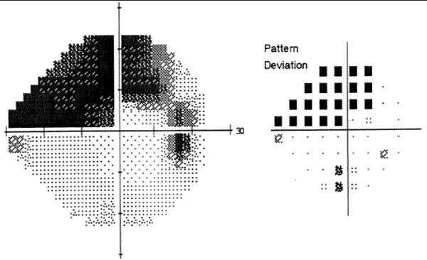
Image Source: Department of Ophthalmology and Visual Sciences – The University of Iowa
Glaucoma has plagued eye care physicians for years! The most common form of glaucoma has been called the “sight stealing disease” because it is asymptomatic to the patients. It’s extremely important to control this condition at its early stage before the downward spiral of damage begins.
Since glaucoma primarily damages the Retinal Nerve Fiber Layer’s ganglion cells that are responsible for visual processing, the HFA is excellent and is the standard in detecting for damaging changes. Aside from determining if the patient has glaucoma, you use the HFA to monitor and track the progression of the condition and treatment efficacy.
Read more on major risk factors in primary open angle glaucoma.
2) Optic Neuritis -MS
Optic neuritis is an inflammation of the optic nerve due to various conditions with multiple sclerosis (MS) being a common systemic cause. Optic neuritis is a presenting sign in patients with MS, and so we are generally the first ones to detect it. Patients usually complain of eye pain (especially with movement), vision blur or “fog,” with a pretty sudden onset too.

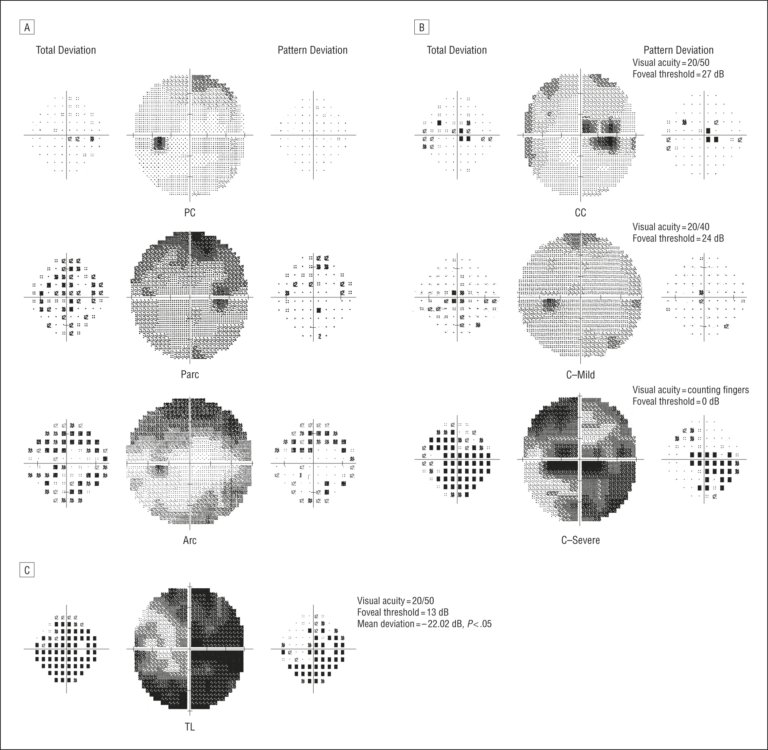
Optic Neuritis Visual Field Profile – Image source: JAMA Network
A HFA is used to determine the visual defects of the optic nerve inflammation BEHIND the optic nerve (retrobulbar neuritis). The typical visual field defect of an optic neuritis is a central scotoma, but it can also be arcuate, altitudinal, hemianopic, or cecocentral.
3) Plaquenil/Hydroxychloroquine Use and Toxic Retinopathy
The use of hydroxychloroquine and plaquenil has long been used for treatment of malaria but now also successfully is used for systemic inflammation such as lupus and rheumatoid arthritis. When the cumulative dosage of these medications are too high, it initially causes cell toxicity while their VA’s are usually normal and no retinopathy may be noted, meaning no bull’s eye maculopathy can be seen funduscopically (premaculopathy).

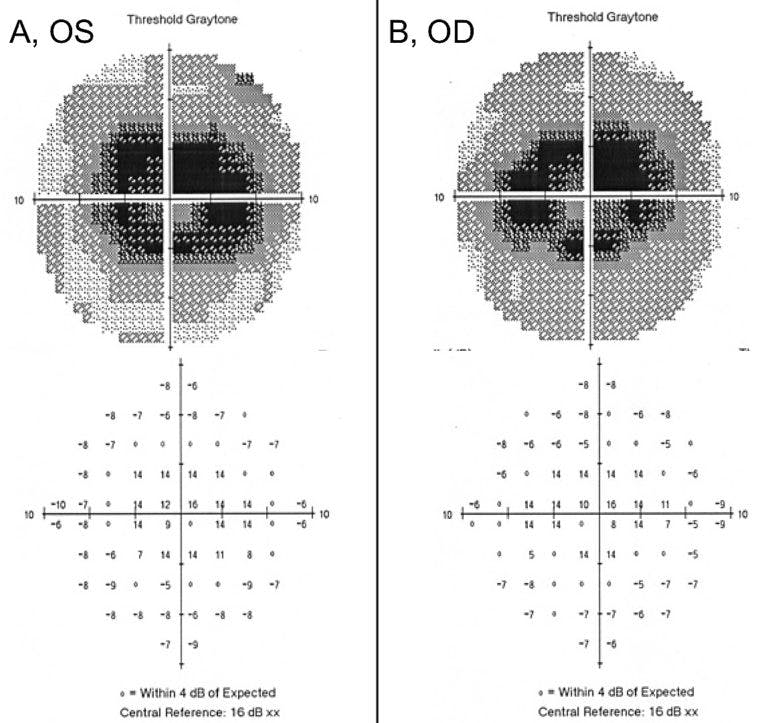
Image Source: University of Iowa Department of Ophthalmology
As we know, early changes can be detected with OCT and we can also utilize clues from diminished visual fields. Changes here mean a phone call to the rheumatologist and/or pcp is warranted because this is when vision loss can be stopped. Once we see the maculopathy (funduscopically or on the visual field), it is generally irreversible due to severe cell toxicity and death and the patient is left with permanent vision loss.
Therefore, any slight changes in fields is a definite cause for concern.
Check out this in-depth guide to using OCT in the diagnosis and management of ocular disease.
4) Ischemic Optic Neuropathy (ION)
Non-arteritic ischemic optic neuropathy (NAION) aka a “stroke in the eye” is a common form of ION. We are generally the first health professional patients call due to sudden loss of vision that results.
Cardiovascular disease is the common culprit in causing NAIONs, so patients with those family of conditions should undergo periodic visual field testing along with their comprehensive eye exams.

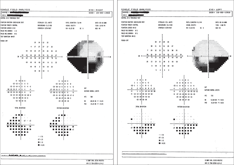
Image Source: Review of Optometry
Sometimes these conditions are undiagnosed and unfortunately are found out the hard way. Visual outcomes are generally poor and irreversible. The lack of blood flow to the nerve generally affects a specific network of blood vessels thus creating a typical altitudinal visual field.
5) Lesions
With the above mentioned ocular conditions, we always always (always!) want to rule out masses. The HFA is one of the diagnostic tools we can use to assist in ruling out these potentially compressive lesions. But confirming the final diagnosis usually requires neurological imaging. Depending on where the lesion is located along the optic tract, it will often produce a certain visual field defect.

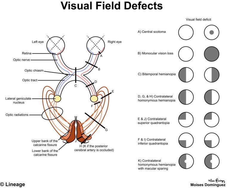
Image Source: Medbullets.com
One of the most common types of intracranial lesions we encounter are pituitary adenomas. It produces the classic bitemporal hemianopsia due to the location of the lesion compressing on the visual fibers in the optic chiasm.
In Summary
If you haven’t noticed with the conditions above, we are generally the patient's first responder and are able to reveal possible underlying systemic conditions. Having a HFA is an important piece of diagnostic equipment to assist in our patient’s ocular well-being and maybe even saving their lives!
