


An estimated 450 million people worldwide have undiagnosed visual field defects and lack adequate follow-up for chronic diseases such as glaucoma and age-related macular degeneration (AMD), which remain leading causes of blindness and vision loss globally.
According to the World Glaucoma Association, the number of people with glaucoma worldwide has surpassed 80 million and is expected to exceed 111 million by 2040.1 Over 3 million Americans suffer from glaucoma, but only about half even know they have it.2 Caused by damage to the optic nerve, glaucoma leads to loss of the visual field. Because early detection is key to successful patient outcomes and glaucoma often has no symptoms during the early stages, regular testing of all patients is critical.
AMD affects about 11 million Americans,3,4 although a recent study demonstrated that 25% of AMD in the primary eyecare setting remains undiagnosed.5 A progressive disease that worsens over time, AMD affects central vision and the ability to see fine details—and is the most common cause of severe loss of eyesight among people 50 and over.10


Heru AR/VR technology enables early detection of visual field defects
Visual field defects can be due to damage anywhere in the visual system from the retina through to the brain’s occipital cortex, including the optic nerve, and it’s something for which I routinely test. My practice is a tertiary referral center, meaning that 80 to 90% of the patients I see are referred to us. We specialize in cataracts, cornea, refractive surgery, and glaucoma. About 35 to 40% of my patients are new evaluations, 35% come for postoperative care, and another 30% of my practice consists of patients who need long-term glaucoma care. I work closely with my patients’ primary optometrists, primary care physicians, and ophthalmologists according to the medical model of optometry. Many times a patient isn’t just a vision patient but also a medical patient with other caregivers involved—and it’s important to me that I collaborate in the care of my glaucoma patients and suspects to the extent that supports the best possible outcomes.
I tend to manage more moderate to severe glaucoma patients, because in our area, primary optometrists are very comfortable and do a great job in the earlier stages of the disease. We also do a lot of research in our practice, so I’m commonly a sub-investigator and occasionally a primary investigator for clinical trials related to cataracts, cornea, refractive surgery, and glaucoma. At any given time in our practice, we’re doing between 15 to 20 FDA or industry-sponsored clinical studies.

How Heru wearable technology improves our practice efficiency
Technology is one of the keystones of our practice, and whenever we adopt new technology, it needs to meet the following criteria:
- Clinically Validated: We demand proven results that any technology we use is equivalent to or elevates our current standard of care. We evaluate the testing, science, and data that backs any new equipment very closely.
- Seamless Integration: Any technology we adopt has to be functional, easy to integrate into our practice, and easy on our patients.
- Improve Practice Efficiency: We see a lot of patients and need to take care of them thoroughly in the most efficient manner possible.
Why our practice chose the Heru AR/VR platform for visual field testing
Because we specialize in visual field defects, our practice has very high standards in terms of the technology we choose to adopt. We currently use the gold standard for visual field testing—a Humphrey 24-2C visual field test—which takes about 3 to 5 minutes while incorporating 10 additional test points in the central 10 degrees of vision.6 We also require our visual field testing to be highly accurate, reliable, and repeatable in order to help our patients achieve the best possible outcomes.
The Humphrey Field Analyzer (HFA) by ZEISS has become the most universally used static perimetry system to measure and examine visual fields, especially for glaucoma management.7
However, with a machine this big, it has to have a dedicated space. And you don’t just have it in a room and say, “Hey, we’ll get it when we need it.” You learn to respect the equipment and the process, and as a result, the visual field test becomes something that’s scheduled.
Quite honestly, visual field testing can be a bottleneck in our practice because we have multiple providers, optometrists, and ophthalmologists seeing patients at any given time with a few clinic schedules needing to access visual field testing. For example, on a day I’m seeing new glaucoma patients, one of my partners might be doing glaucoma follow-ups or another doctor may be using it for oculoplastics. So it’s not unusual that visual field testing can be a logjam—and it’s not because of patients who didn’t have it scheduled. It’s every day.
As a result of the pain points we were experiencing, we decided to bring in Heru to help our workflow run more efficiently. We evaluated all wearable technology and there were several factors that validated our decision to choose the Heru platform.
- The platform has over 10 years of scientific research and development led by Heru’s founder and CEO, Mohamed Abou Shousha, MD, PhD, Associate Professor of Clinical Ophthalmology, Bascom Palmer Eye Institute.
- Heru technology has been upheld by more than 1,000 patients in clinical studies.
- Heru has over 40 U.S. patents.
- Preliminary data from an R&R study at two sites demonstrate a repeatability standard deviation of 1dB and a reproducibility standard deviation of 2dB for the median value on 26 subjects.*
How Heru performs against traditional HFA visual field testing
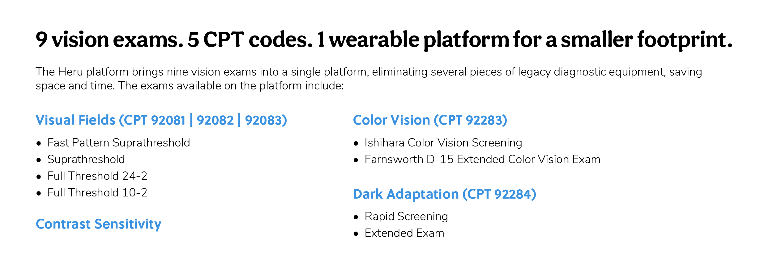
The Heru platform’s visual field exams have excellent repeatability and reproducibility while demonstrating strong correlation to the current gold standard of care. The Heru wearable headset enables physicians to rapidly screen their patients for visual defects, which can indicate the presence of glaucoma or AMD. The platform’s Fast Pattern Suprathreshold exam is a 40-point screener that can be done in as little as 40 seconds per eye–without refractive correction for most patients.
There are several key differences between the HFA and Heru visual fields, which are summarized in Table 1 below:

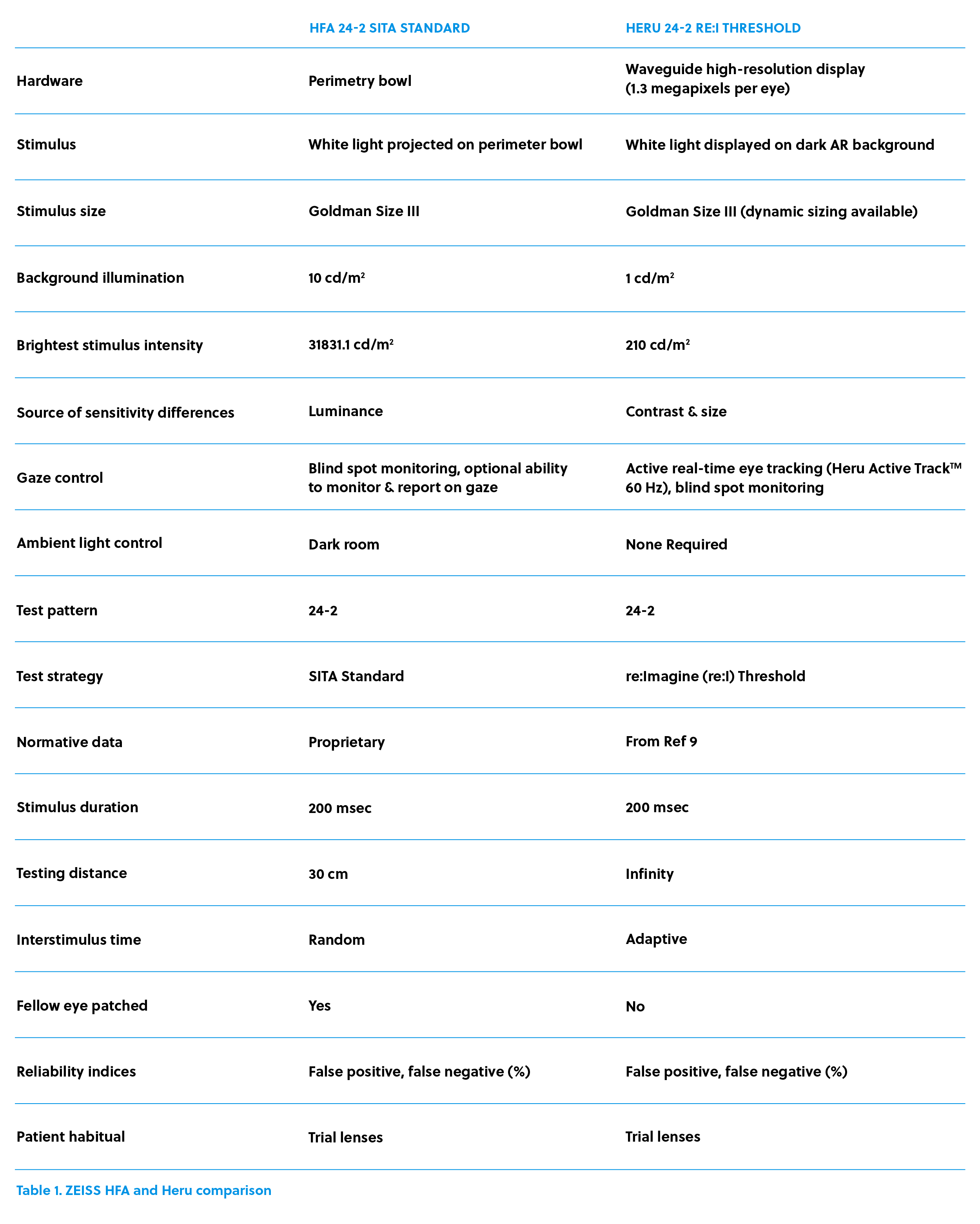
The visual field report layout and metrics are similar between Heru and HFA, which significantly decreases the learning curve of interpreting results and allows for easy comparison between the two systems. In addition, Heru’s portal has the option to allow for colored reports, which overlay the deviation maps with the threshold values.
Unlike the traditional HFA testing distance of 30cm (approximately 3.3 diopters of accommodative demands), which require optical correction changes based on the patient’s age and presbyopic correction, Heru technology presents the stimuli at optical infinity so the patient’s distance correction is always used and testing is not affected by presbyopia or its associated image magnification differences due to changes in trial lens power.
To further evaluate visual field measurements between Heru technology and traditional HFA analysis, a study was conducted comparing the Heru multi-platform visual field testing application downloaded onto two separate headsets against a standard perimetry platform. Both normal patients and those with visual field defects were evaluated to determine the reproducibility of Heru technology. The study included 81 eyes, 40 of which were normal and 41 of which were patients with glaucoma and neuro-ophthalmic diseases (hemianopic, altitudinal, and ring scotomas). All patients underwent standard 24-2 test patterns with 3 testing devices.8
Mean deviation (MD) and Threshold values obtained using Heru technology on two wearable devices showed strong correlations with those of Humphrey Field Analyzer (HFA) (R=0.91 and R=0.80, P <0.001, respectively). The Heru VF software was reproducible with ICC of 0.974 (95%CI 0.940-0.987) for MD and 0.8 (95%CI 0.813-0.845) for threshold values in normal eyes and in patients with glaucoma and neuro-ophthalmic diseases.

Applications of multi-modal Heru technology beyond visual field deficiencies
While I primarily treat glaucoma and cataracts and am most interested in peripheral vision loss, the Heru platform has applications in other areas. In fact, one of the things that impresses me about Heru technology is that it can be used for applications beyond visual field deficiencies, including:
Dark adaptation
Globally, there are 196 million people living with age-related macular degeneration (AMD), and this number is expected to reach 288 million by 2040.9 Up to 78% of patients with AMD do not seek help until it is too late, which is why early detection is needed to reduce these numbers.
The leading cause of irreversible blindness in adults over 50,10 AMD is a complex, multifactorial disease in which central retinal photoreceptors are lost by atrophic or neovascular processes, resulting in progressive loss of central acuity. Typically characterized by a thickening of Bruch’s membrane and the accumulation of drusen in the macula, AMD makes it difficult for patients to drive, read, and conduct normal activities.11
Until recently, AMD was diagnosed based on morphological changes, including pigmentary mottling, geographic atrophy, choroidal/macular neovascularization12 and drusen formation. Associated with several retinal conditions, drusen refers to the round lesions noted on fundoscopy that are thought to represent extracellular accumulations of debris.13 However, a recent study demonstrated that 25% of AMD in the primary eyecare setting goes undetected.1
This has been validated by other studies which demonstrate that despite normal retinal morphology, dark adaptation tests can identify functional abnormalities that are missed when only looking at retinal morphology. For example, approximately 25% of older adults recruited from primary eye clinics had abnormal results on dark adaptation tests, which are associated with risk factors for AMD, including elevated C-reactive protein.14
At its core, dark adaptation measures the amount of time it takes to recover scotic, or night vision, after exposure to bright lights. In individuals with healthy retinal function, this generally takes 5-10 minutes, which is when the modulation of vision switches from cones (more sensitive in brighter illumination) to rods (more sensitive to dark illumination).
"The amazing thing about Heru's dark adaption test is how it's a fraction of the time of traditional testing methods. And they've got the data behind them that shows it's maintaining high sensitivity even with rapid testing speed."
— Milan Lockhart, OD, Spectrum Eye Care
In 2014, the Alabama Study on Early Age-Related Macular Degeneration (ALSTAR) study began assessing dark adaptation in 325 adults ages 60 and up, who had all been deemed in good macular health after a clinical exam. After 3 years, subjects who showed impaired dark adaptation were twice as likely to develop AMD compared to individuals who displayed normal adaptation time.14
Historically, dark adaptation testing has been regarded as too cumbersome for widespread clinical application, but the ease of conducting these tests using a portable, wearable platform such as Heru, is shifting this outdated belief.
I don't see a tremendous amount of AMD patients in my practice and have not used the dark adaptation testing on my patients, but for those who do, Heru’s rapid and extended dark adaptation exams may be used as a functional test for impaired dark adaptation. Heru’s dark adaptation takes either 4.5 minutes or 20 minutes to complete depending upon if it is a rapid or extended exam.
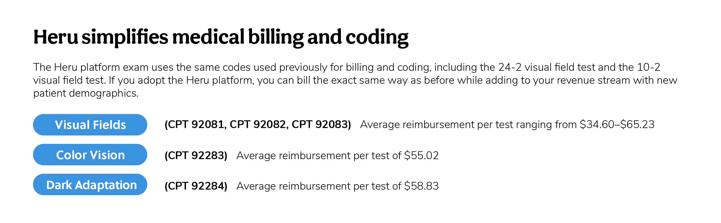
Dark adaptation exam is billable to insurance with a national reimbursement average of $58.83 and is co-billable with visual fields, OCT, fundus imaging, and/or office visits. Dark adaptation has multiple supported ICD-10 codes and may generate additional practice revenue from auxiliary testing, plus nutraceutical and optical sales.

Color vision deficiency
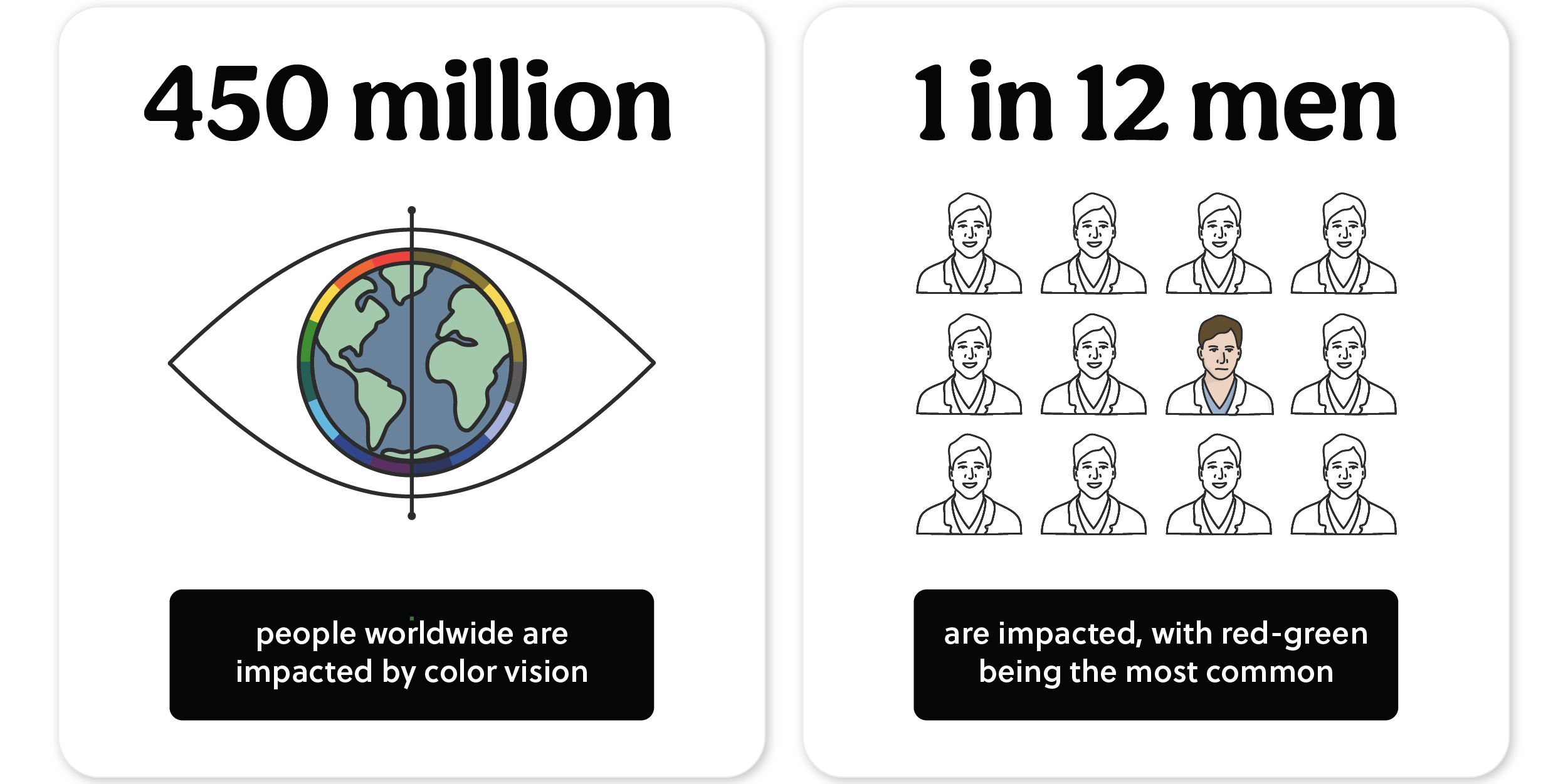
Currently, an estimated 300 million people in the world are impacted by color vision deficiency (CVD), which affects 1 in 12 men (8%) and 1 in 200 women (0.5%) with color blindness. Traditional color vision tests are routinely administered in the clinic to screen for CVD, which is caused by an X chromosome mutation that overwhelmingly affects males.16 Printed pseudoisochromatic plates such as the Ishihara and Hardy-Rand-Rittler tests are commonly used to detect and classify the different types of color vision deficiencies: protan, deutan, and tritan defects.
While screening may be routine for the congenital form of color vision deficiency, it is important to remember that color vision testing also plays an essential role in detecting and classifying acquired color vision deficiency, as the result of a systemic or ocular disease or the side effect of certain medications. For example, in older patients, color vision deficiency can be an indicator of serious medical conditions, including diabetes, glaucoma, macular degeneration, Alzheimer’s disease, Parkinson’s disease, leukemia, sickle cell anemia, and chronic alcoholism.17 Studies have also shown that color vision testing may also aid in early detection of optic neuropathy, multiple sclerosis, and Duchenne muscular dystrophy.18 Additionally, medications commonly prescribed to older adults contribute to color vision loss, including lanoxin, hydroxychloroquine, sildenafil, and tamoxifen.
Using traditional methods like printed color plate tests can be complicated because of the precise illumination requirements under Standard Illuminant C. Additionally, the printed tests have to be stored with strict requirements to avoid sunlight and finger oils.
The Heru platform allows doctors to easily test for both the Farnsworth D-15 Extended Color Vision exam as well as the Ishihara Color Vision Screening, using the same, traditional, 12 numbered plates, without worrying about operator error, because the Heru platform is completely patient-guided. Additionally, the Heru platform controls both background illumination and ambient light, ensuring proper testing conditions to maximize clinical accuracy.
Heru’s digital Ishihara color vision screening exam takes less than 30 seconds per eye, and if 3 or more plates are missed during the test, the platform will AutoFlow™ into the Farnsworth D-15 extended color vision exam which is billable to insurance. The national reimbursement average for the Farnsworth D-15 exam is $55.02 and is supported by multiple ICD-10 codes, generating more revenue opportunities for your practice.

Contrast sensitivity
The ability of the human visual system to perform under real-life conditions is often missed in visual acuity tests. While visual acuity tests measure vision in a controlled and well-lit environment, the eyes must also be able to qualify, and not just detect, visual stimulus, which can be better measured in contrast sensitivity tests.19 While the Snellen visual acuity test can diagnose the inability to detect an object from its background, it can really only inform the quantity of vision. In order to detect a true account of a patient’s vision—which is necessary to determine if a patient has the vision needed to drive safely at night, see curbs and steps, and enjoy TV and near work like reading without eye strain—a contrast sensitivity test is required.
As with other conditions like color vision deficiency, a lack of contrast sensitivity can indicate more serious concerns, such as AMD, glaucoma, cataracts, aberrations in the corneal surface, diabetic retinopathy, and even dry eye syndrome. For example, even in patients with a normal Snellen acuity, contrast sensitivity testing can serve as an early predictive marker for diabetic retinopathy.20 Given that half of all Americans aged 65+ have prediabetes with higher than normal blood sugar levels, a contrast sensitivity test can be an important aspect of the medical model of optometry and can give a patient’s primary care physician valuable insight into a patient’s health.
A Snellen test also can miss early signs of cataracts. For example, a person with posterior subcapsular cataracts might display sufficient visual acuity but have significant problems with glare, which can be detected by a contrast sensitivity analysis. It’s also been reported that decreased contrast sensitivity can be correlated with the optical density of cataracts, meaning that this test can potentially be used for early detection and screening.21
Based on the current standard of contrast sensitivity testing using the Pelli-Robson test, Heru’s patient-guided contrast sensitivity exam leverages a tumbling E presentation with a shrinking staircase threshold testing strategy that advances from darker to lighter contrast. When the patient answers incorrectly, the contrast is then increased until the patient provides a correct answer. Like all Heru tests, the contrast sensitivity exam can be performed out of the exam lane and outside of traditional points of care, including assisted living centers and remote locations, as long as there is access to a secondary device (such as a smartphone or tablet) and internet connection.
For patients who complain of glare, difficulty with dim lighting, and difficulty driving at night or in fog, a contrast sensitivity test is easy to administer on the Heru platform. It can be added seamlessly to a visual field test and can easily integrate into your practice workflow without necessitating the need to schedule any other equipment or ask the patient to return for another appointment. From initial set-up, the Heru test takes less than 2 minutes with the contrast sensitivity exam lasting less than 40 seconds. This is largely due to the fact that the Heru contrast sensitivity test uses a shrinking staircase algorithm to quickly measure a patient’s ability to detect background from an object rather than having to rely on the fixed increments inherent in a more traditional Pelli-Robson test.

How we have adopted Heru in our practice
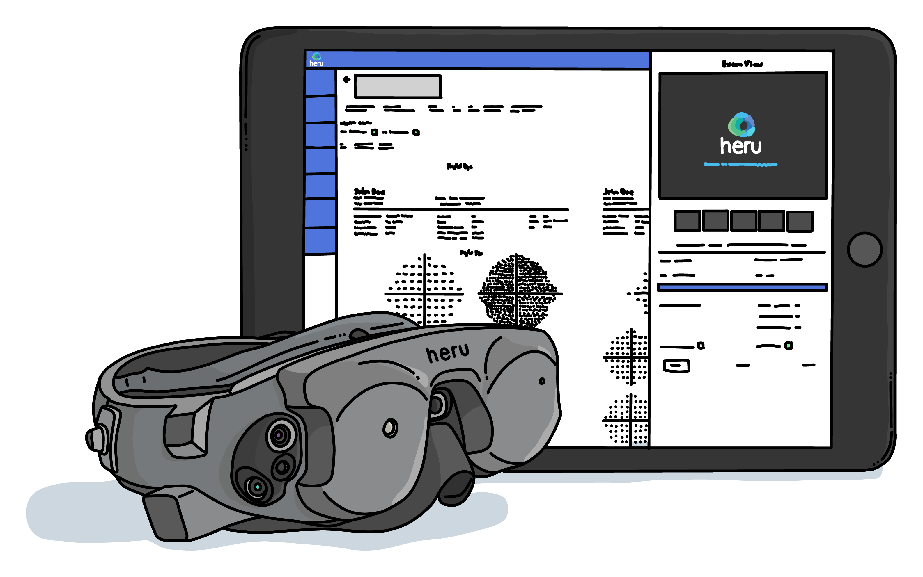
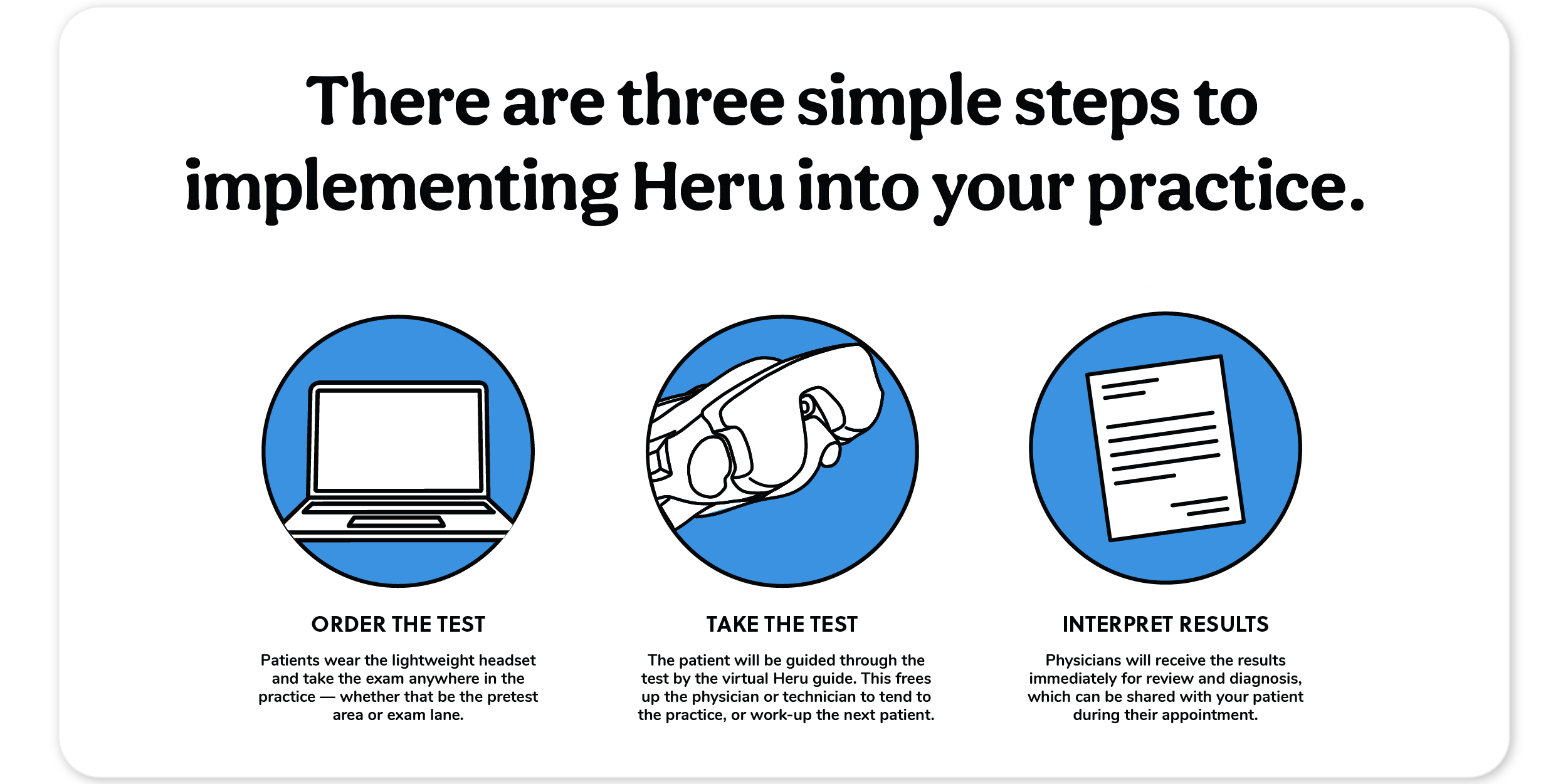
The Heru device is portable, lightweight, and affordable. Patented autonomous software is downloaded on a wearable device, which has its own controlled lighting environment, enabling the performance of a visual field test at any time during a patient’s visit, anywhere within the practice with clinical results available in real-time.
For patients who we didn’t think needed a visual field test but then they did, Heru has been a big win for our practice. More importantly, the Heru platform is functional and easy on our patients. Additionally, we have easily worked it into our evaluation and testing process while providing the data we need, rely on, and expect from visual field testing.
Heru adds another perimetry tool in the glaucoma space. For practitioners or practices who currently don't have perimetry tools in their practice due to space or economic considerations, adding this device allows access to perimetry for more patients, which is beneficial to both the patient, and the provider.
We’ve participated in Heru’s clinical trials, and have used the Heru wearable headset in our clinical practice, so we really trust in the technology as well as the results. For me personally, Heru is ideal for testing the patient who has ocular hypertension, a glaucoma suspect, or a first-time patient showing signs of glaucoma.
What is Heru wearable AR/VR technology like for patients taking perimetry exams?
Most patients don’t come in saying, “Hey doc, I’d love to be able to do a visual field test today where you cover my eye and I sit in that dark room and listen to a lot of beeping.” For our patients, Heru is more like a game than a test. In order to take the test, they wear a comfortable headset that features the Heru virtual guide, which instructs and monitors them throughout the exam, ensuring they are engaged.
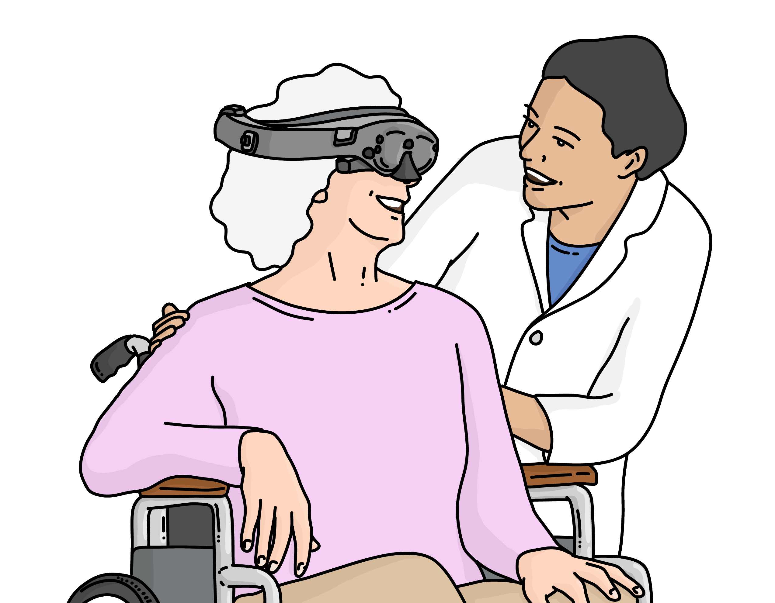
Where we really see a difference is for patients who struggle with mobility or having to position themselves in front of a traditional visual field exam, which can be uncomfortable for them. With the Heru headset, we can meet those patients where they are and do a visual field test without disrupting their comfort or asking them to make special accommodations.
How the Heru platform enhanced our practice workflow
The advantages of adopting new, wearable technology were very attractive to us, not just because it didn’t require a designated dark room, but because some of our patients suffer mobility challenges, are elderly, or suffer from developmental delays that prohibit them from being positioned for a traditional visual field test. Other patients have anxiety placing their head inside of a bowl for prolonged periods of time, and do not like having one eye closed.
We don’t plan on removing the HFA from our office, as it adds value to have other visual field technologies. For example, if we have a first-time patient who may be a glaucoma suspect, or we are dealing with a new patient with mild-to-moderate glaucoma, it is beneficial to perform a same-day visual field test, even in situations where we didn’t have this scheduled. There are instances where a patient comes in whom we didn’t originally think had glaucoma, but we want to make sure. Maybe we dilated a patient and looked at the optic nerve and had concerns or checked ocular pressure and it didn’t make sense. With Heru, we can do an instant visual field test rather than have to schedule a test for when the HFA room is available. From a flow standpoint, that’s a huge win for our practice. Heru empowers us to offer the test in an exam room in an efficient manner rather than trying to reroute our entire schedule. There are so many patients who you assume don’t need a visual field test—but if they do, Heru technology makes that really easy to accomplish.
Advantages of mobile AR/VR vision testing for optometry practices
One of the biggest advantages of Heru technology is that it’s mobile. What this means is that you don’t have to keep it in a designated dark room or testing space. With Heru, vision exams can be accomplished in the pre-test area, or exam lane. Maybe it’s a dedicated space, but with Heru, there’s no need to drastically decrease or control the lighting. But maybe the biggest advantage is that it’s a very user-friendly platform that conforms to the patient and doesn’t ask them to try to lean into or be positioned in front of a device. Heru fits the patient rather than asking the patient to conform to standard equipment.
For many patients, the Heru platform is similar to the augmented reality/virtual reality headsets for gaming, so there’s a very easy adoption. Heru technology makes room for a different option and a better patient experience. Patients are open to new protocols, and my experience has been really positive with Heru for a couple of reasons:
- You can use Heru technology in any room from the waiting area to the exam lane.
- The Heru platform makes testing quick and easy.
- The patients can keep both eyes open.
Features and benefits of the Heru AR/VR technology wearable platform
The Heru platform includes the following features:
- Heru’s proprietary threshold algorithm adapts and predicts an optimized full-threshold testing workflow without compromising clinical performance, ensuring continuity of care with clinical results correlated to the Humphrey perimetry test.9
- Autoflow™ minimizes the time needed to detect disease and can improve staff efficiency.
- The Heru virtual guide is multilingual. English, French, Italian, German, and Spanish languages are offered.
- ActiveTrack™ real-time gaze tracking confirms the patient’s fixation is always appropriate, improving data quality while keeping the patient engaged and focused throughout the exam. By eliminating the need for fixation loss monitoring, it enhances clinic workflow and reduces repeat testing.
- During the VF test, infrared LEDs and cameras in the device illuminate the eye and provide individual gaze coordinates (60Hz).
- Stimuli are only presented when the patient is properly fixating.
- Patients are prompted to regain fixation when they lose focus.
- ActiveTrack minimizes fixation loss errors.
- It ensures the reliability of each stimulus presentation in real-time and may improve reliability and repeatability of the test.
- There’s no need to interpret a “gaze report.”


How Heru meets global vision care needs for remote populations
While I appreciate how the Heru platform helps our practice enhance efficiency, and the patient experience, the Heru wearable advice has widespread applications that have the potential to dramatically impact unmet eyecare and vision needs worldwide. Portable technology holds great promise for improving access to glaucoma care, especially in remote locations around the globe, where mobile transportation of visual field testing democratizes access for those living without reliable vision care.
People without regular access to healthcare don’t have the privilege of routine glaucoma testing. Because glaucoma is often asymptomatic until it is too late, it remains a leading cause of blindness, especially in areas with limited resources and eyecare.22 The lightweight, Heru wearable platform holds great promise for these remote geographic locations and may be pivotal in bringing eyecare to those who need it most.**
What excites me most about Heru technology is its ability to make a meaningful difference in global unmet needs for vision care—especially in the detection and management of sight-threatening eye conditions such as glaucoma and age-related macular degeneration—which remain among leading causes of blindness in many places without reliable access to eyecare practitioners. With a lightweight headset that can fit into a backpack or be strapped onto a bicycle, the number of people whose vision can be saved with this technology is astounding on a global level. Sight remains our most prized human sense, and with Heru's easy-to-use platform, any place with internet access is capable of providing early detection to those who can’t afford vision care. While I love what Heru does for my practice in terms of efficiency, patient satisfaction, and increased revenue, what Heru can do for the world is truly mind revolutionary.
References
- Kang JM, Tanna AP. Glaucoma. Med Clin North Am. 2021;105(3):493-510.
- Friedman DS, Wolfs RC, O'Colmain BJ, et al. Prevalence of open-angle glaucoma among adults in the United States [published correction appears in Arch Ophthalmol.] 2011; Sept;129(9):1224.
- Neely DC, Bray KJ, Huisingh CE, et al. Prevalence of undiagnosed age-related macular degeneration in primary eye care. JAMA Ophthalmol. 2017;135(6):570-5.
- https://www.aarp.org/health/conditions-treatments/info-2019/macular-degeneration.html
- AMD: Facts & Figures. (2019). https://www.brightfocus.org/glaucoma/article/glaucoma-facts-figures
- Yamane MLM, Odel JG. Introducing the 24-2C Visual Field Test in Neuro-Ophthalmology. J Neuroophthalmol. 2021;41(4).
- Delgado MF, Nguyen NTA, Cox TA, et al. Automated perimetry: A report by the American Academy of Ophthalmology. Ophthalmology. 2002;109(12):2362-2374.
- Goldbach A, Shousa M, Duque C, et al. Visual field measurements using Heru Visual Field Multi-platform application downloaded on two different commercially available augmented reality devices. ARVO Annual Meeting Abstract. June, 2021.
- Keenan TDL, Cukras CA, Chew EY. Age-Related Macular Degeneration: Epidemiology and Clinical Aspects. Adv Exp Med Biol. 2021;1256:1-31.
- Pennington KL, DeAngelis MM. Epidemiology of age-related macular degeneration (AMD): associations with cardiovascular disease phenotypes and lipid factors. Eye Vis (Lond). 2016;3:34. Published 2016 Dec 22.
- Mangione CM, Gutierrez PR, Lowe G, et al. Influence of age-related maculopathy on visual functioning and health-related quality of life. Am J Ophthalmol. 1999;128(1):45-53.
- Gheorghe A, Mahdi L, Musat O. Age-Related Macular Degeneration. Rom J Ophthalmol. 2015;59(2):74-77.
- De Jong PTVM. Elusive drusen and changing terminology of AMD. Eye (Lond). 2018;32(5):904-914.
- Owsley C, Huisingh C, Jackson GR, et al. Associations between abnormal rod-mediated dark adaptation and health and functioning in older adults with normal macular health. Invest Ophthalmol Vis Sci. 2014;55(8):4776-4789.
- "Facts About Color Blindness." NEI. February 2015. Archived from the original on 28 July 2016. Retrieved 29 July 2016.
- Colour Blind Awareness. www.colourblindawareness.org. 1/22.
- Color blindness. www.mayoclinic.org/diseases-conditions/poor-color0vision/symptoms-causes/syc-20354988.
- Color Me Curious: Did you know that color vision testing has the potential not only to uncover optic neuropathy, but also glaucoma? Article in Review Of Optometry; February 15, 2013.
- Jindra LF, Zemon V. Contrast sensitivity testing: a more complete assessment of vision. J Cataract Refract Surg. 1989;15(2):141-148.
- Subhasish P, Subhankar C, Upasana G, et al. Visual contrast sensitivity could be an early marker of diabetic retinopathy. Heliyon, Volume 6, Issue 10, 2020.
- Shandiz JH, Derakhshan A, Daneshyar A, et al. Effect of cataract type and severity on visual acuity and contrast sensitivity. J Ophthalmic Vis Res. 2011;6(1):26-31.
- Bourne RRA, Stevens GA, White RA, et al; Vision Loss Expert Group. Causes of vision loss worldwide, 1990–2010: a systematic analysis. Lancet Glob Health. 2013;1(6):e339-e349.
*Preliminary data from a study currently in review, authored by Dr. Mitch Ibach.
** A trained optometrist must interpret results and make any diagnosis.
