


Can you imagine if your child lost their vision before first grade? In 1990, Optos founder Douglas Anderson’s 5-year old son Leif—who was born with high myopia—lost all sight in his left eye. Despite regular exams, a retinal detachment went undetected. Because of its location in the periphery, it could not be seen by the imaging equipment available at that time.
Grief for his son propelled Anderson to take action, with the mission to protect the vision of other children and save parents from what he and his family had to endure. First, Anderson sought to determine if any current technology could be enhanced to provide a more complete image of the retina and posterior pole without dilation.
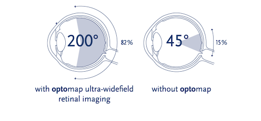
When he couldn’t find a technology that gave the visibility of the peripheral retina he wanted, he decided to create it himself. Two years later, he developed a prototype of what is now known as the optomap® image—the only single-capture, ultra-widefield retinal image to reveal up to 200 degrees (82%) of the retina.
Given that an indirect ophthalmoscope captures about 30 degrees of the retina, and a fundus camera captures about 45 degrees, a 200-degree view of the retina offers far greater visibility of the retina—including the periphery—which is essential for early and accurate diagnosis of diseases, including diabetic retinopathy (DR), age-related macular degeneration (AMD), vascular disease, pediatric retinal disease, inflammatory disease, retinal detachments, and geographic atrophy.
Since its first prototype in 1992, Optos has expanded its ultra-widefield imaging devices to include multiple modality options, called an optomap. Depending on physician needs, the Optos devices have color, autofluorescence, and dye-based angiography options as well as integrated OCT. To date, optomap imaging is able to capture the widest field of view in a single capture of any imaging technology, with more vortex veins visualized1 as well as statistically significantly more retinal surface area.2,3
Perhaps even more meaningfully to its founder, the Optos system has been featured in published studies that cite the instruments’ abilities to detect pediatric retinal diseases in children without the need for anesthesia. Additionally, these studies validate Optos’ ability to capture images of the far peripheral retina in young children as this is typically where pediatric retinal diseases first exhibit pathology.4
optomap high-resolution retinal images cover more than 82% of retinal surface area or 200 degrees of the retina in a single capture. To gain even greater visibility, optomap auto-montage can be used to visualize up to 97% of the retina can be imaged with the multi-capture montaging functionality.
What is ultra-widefield imaging?
The necessity for consistent nomenclature for widefield and ultra-widefield imaging was recognized by the The International Widefield Imaging Study Group, which defined ultra-widefield as images that make the retinal anatomy anterior to the vortex vein ampullae visible in all four quadrants, for a 110- to 220-degree field of view.
Following the agreement on retinal imaging terms, the group then reviewed 100 images on the Optos California. While the consensus group was device-agnostic, it was determined “that images from the Optos California most consistently provide a complete view of the vortex veins and retinal periphery without the need of a montage. Furthermore, the accuracy and precision of quantification of the images have been validated and published.”5
Widefield – centered on the fovea and includes the retina in all four quadrants posterior to and including the vortex vein ampullae
Ultra-widefield – images showing retinal anatomy anterior to the vortex vein ampullae in all four quadrants
Pan-retinal – ora to ora image of the retina either in the horizontal or vertical direction
Why ODs choose Optos technology for early detection of retinal disease
optomap ultra-widefield retinal imaging provides information not readily available with traditional imaging techniques, such as limited field fundus photography (which only captures 45 degrees of the retina, on average) or a montage of seven-standard-field (7SF) stereoscopic fundus photography (which about captures 90 degrees of the retina.)
Additionally, because optomap ultra-widefield imaging collects data with a single capture, patient comfort and clinical efficacy are increased, which is helpful with young, compromised, or noncompliant patients.6
Other benefits include:
- Non-contact
- Images through most cataracts and small pupils (2 mm)
- Single-capture
- 3-in-1 color depth imaging
- 82% coverage of the retina (200 degrees) in less than half a second
- Proven clinical value and practice efficiency are proven in over 2,000 published studies
- Imaging can be done at a safe distance
- Comfortable for the patient
- Non-mydriatic (no dilation needed)

Why is the periphery so important?
Over the last decade, many large studies have highlighted the importance of visibility in the periphery of the retina for early detection and management of diseases including, diabetic retinopathy (DR), age-related macular degeneration (AMD), vascular disease, pediatric retinal disease, inflammatory disease, retinal detachments, geographic atrophy, retinal tears, and some systemic diseases as well.
Since many eye conditions may first appear in the outer periphery and influence disease progression, their early identification and timely management are critical to the patient’s vision and systemic health. Early diagnosis based on peripheral findings enables more individualized treatment and monitoring.
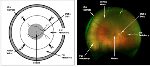
How is the retina defined geographically into regions?
Retinal regions were defined in 1961 by Duke-Elder, who categorized the peripheral retina into four distinct zones,7 which correspond to:
- Near periphery, a 1.5mm ring adjacent to the macula
- Middle periphery, the next 1.5mm ring
- Far periphery, measuring the next 9-10mm on the temporal and 16mm on the nasal side
- Ora serrata, or extreme periphery, measuring an additional 2.1mm in the temporal and 0.7mm on the nasal side

The launch of the optomap made it possible to see a more comprehensive view of the retina than ever before for a greater chance of detecting retinal pathologies in locations that might be missed with previous technologies.8 Additionally, the devices from Optos reduce the number of images required to map the fundus while also decreasing patient exposure to light as well as waiting times, which can be important in a busy clinic.
What are some common retinal diseases that benefit from greater resolution and visibility of the periphery?
Diabetic retinopathy (DR), glaucoma, dry age-related macular degeneration (AMD), geographic atrophy (GA) retinal vein occlusions, (RVO), retinopathy of prematurity (ROP), uveitis, and inherited eye disorders are chronic, progressive diseases that may first show signs in the periphery of the retina. Therefore, in these instances where early diagnosis is key to effective disease management and successful outcomes, images of the periphery are of utmost importance.
Diabetic retinopathy
Diabetic retinopathy (DR) is one of the most common causes of visual impairment in working-age adults worldwide,9 according to the American Academy of Ophthalmology. Nearly 80 percent of people with type 1 diabetes develop diabetic retinal disease. Duration of diabetes is a major risk factor associated with the development of DR, which is why all patients with diabetes should be monitored regularly. The care process for diabetic retinopathy includes a medical history, a regular ophthalmologic examination or screening of high-quality retinal photographs of patients who have not had previous treatment for DR—or other eye disease—and regular follow-up. The goal of treatment is to improve or stabilize visual function, improve vision-related quality of life; and, through close communication with the patient's primary care physician, achieve optimal control of blood glucose, blood pressure and other metabolic risk factors.
Peripheral lesions in patients with DR have been linked with an increase in retinal nonperfusion area and worsening DR. They have also been associated with a more than four-fold increased risk of disease progression within four years in patients with nonproliferative DR (NPDR).10
Age-related macular degeneration
The most common cause of age-related macular degeneration, AMD (dry/atrophic) usually progresses slowly over years and results in a degeneration of the macula that can end in blindness. Because early dry AMD doesn’t usually result in symptoms, it’s crucial that eyecare providers have tools to identify the early signs. In fact, until these new advances in ultra-widefield imaging, our understanding of peripheral retinal abnormalities to disease in general and AMD in particular was actually limited by a lack of detailed peripheral imaging studies.11
Geographic atrophy
An advanced form of dry AMD, geographic atrophy (GA) is characterized by localized, sharply demarcated atrophy of the peripheral retinal tissue, retinal pigment epithelium, and choriocapillaris.
In November, 2016, the Association for Research in Vision (ARVO), together with the Foundation Fighting Blindness (FFB), organized the National Eye Institute (NEI)-Food and Drug Administration (FDA) endpoints workshop on AMD and inherited retinal diseases. The aim of the workshop was to assemble stakeholders from academic and regulatory spheres to discuss clinical data on potential primary and secondary clinical trial endpoints, patient stratification for disease monitoring, interventions, and evaluation of treatment response.
Dr. Frank Holz provided data on the utility of fundus autofluorescence (FAF) in detecting loss of the retinal pigment epithelium (RPE). He presented additional studies demonstrating that RPE loss detected on FAF correlates with photoreceptor loss observed using spectral-domain optical coherence tomography (SD-OCT). Geographic atrophy (GA) lesions are often complex and multifocal, and Dr. Holz described semi-automated image processing software to detect and quantify such lesions,12 driving home the need for powerful imaging and software that empowers clinicians with early detection in order to preserve vision health.
Glaucoma
More than 3 million people in the United States have glaucoma. The National Eye Institute projects this number will reach 4.2 million by 2030, which is a 58% increase. Approximately 120,000 Americans have gone blind from glaucoma, accounting for 9-12% of all cases of blindness. Grading of ultra-widefield imaging has high reproducibility in evaluating glaucoma and may be an important tool for glaucoma diagnosis.13
Retinal degeneration
Peripheral retinal degenerations like lattice degeneration, retinal tufts, retinal tears, retinal holes, and paving-stone degeneration, can eventually lead to retinal detachments. Over the past decade, several key studies were able to use the information gleaned from ultra-widefield imaging to provide clear characterization of microstructural details in the peripheral retina as well as important information about the vitreoretinal interface.14
Retinal Vein Occlusions (RVO)
Blockages in small blood vessels that carry blood away from the retina, RVOs can cause increased pressure in the eye, which can lead to blurry vision, bleeding, swelling, and if not treated—vision loss. Risk factors may include a blood clot, atherosclerosis, diabetes, or high blood pressure.
Retinopathy of Prematurity (ROP)
Retinopathy of Prematurity causes blood vessels in premature infants to grow abnormally in the retina. It’s not yet understood what causes this condition, but the more premature the baby and the lower the birth rate, the greater the risk.15
Uveitis
Uveitis occurs when the middle layer of the eye gets inflamed (red and swollen). This layer, called the uvea, has many blood vessels that nourish the eye. Uveitis can damage vital eye tissue, leading to permanent vision loss. It’s not certain what causes uveitis, but it’s correlated to shingles virus, herpes simplex virus, Lyme disease, some parasites, and inflammatory diseases
How imaging relates to the medical model of optometry
The eye is the only place in the body that offers an unobstructed view of blood vessels, nerves, and connecting tissue. As the medical model of optometry continues to advance and we learn new ways to correlate structural and functional changes observed in the retinal vasculature with early signs of stroke, heart disease, hypertension, Alzheimer’s disease, and some cancers,16 medical professionals will rely even more heavily upon the clarity and resolution of ultra-widefield imaging.
Termed oculomics, the identification of ocular biomarkers of systemic disease demands high-resolution imaging modalities. For example, retinal hemorrhages are associated with leukemia, and the detection of a hemorrhage in the retinal periphery may be an early warning sign. Similarly, the association between the structure of the neurosensory retina and prevalent neurodegenerative disease, in particular, Alzheimer’s disease, is also well-established. Advances in retinal imaging may provide new and potentially important insights into cerebrovascular neurodegenerative processes involved in stroke and dementia.17
How color retinal imaging modalities impact visibility
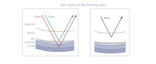
Unlike full-spectrum white light used in conventional devices, optomap technology incorporates low-powered laser wavelengths that scan simultaneously. Each capture results in a three-in-one color depth image, which allows review of the retinal substructures in their individual laser separations.
- Green laser (532 nm) scans from the sensory retina to the pigment epithelial layers
- Red laser (633 nm) scans from the RPE to the choroid
- Blue laser (488 nm) used in fluorescein angiography procedures
- Infrared laser (802 nm) used in indocyanine green angiography procedures
Optos instruments enable eyecare professionals to assess the retina with two color images (red plus green; and red, green, and blue) as well as fluorescein angiography, indocyanine green angiography, fundus autofluorescence, and red- and green-free images.18
The visible spectrum is divided into short (blue), intermediate (green), and long (red) wavelengths. In general, the shorter the wavelength, the greater the light scatter, which can sometimes impede an image’s clarity or get in the way of crisp, clear visibility. Chromatic filters greatly improve the contrast and resolution of the retinal tissue for better localization and more accurate diagnosis.19 For example, the red-free, or “green” filter, blocks red wavelengths and allows for greater contrast between structures and tissues by blocking out “visual noise” that can occur as shorter wavelengths scatter.
The red/green/blue (color rgb) modality, launched in 2023, captures images simultaneously to the optomap color rg image, to provide additional retinal visualization, including identifying holes or deterioration in the periphery. Because blue wavelengths are more likely to be scattered, the blue light unable to reach the back of the retinal is reflected by the internal limiting membrane and additional anterior retinal layers to provide another method of visibility, especially for cases of proliferative diabetic retinopathy or epiretinal membranes.20,21

In 1968, an international consortium of leaders in ophthalmology, internal medicine, and neurosurgery gathered in Virginia to address a growing concern about DR as a leading cause of blindness. What came out of the Early Treatment Diabetic Retinopathy Study (ETDRS) was a set of standards to critically describe stages of diabetic retinopathy and evaluate the effect of treatment modalities.
The standards were used in a landmark clinical trial: the Diabetic Retinopathy Study (DRS), which demonstrated a significant reduction in the rates of severe vision loss in eyes treated with panretinal photocoagulation compared to untreated control eyes.21 Today, the ETDRS remains the gold standard for grading the severity of a patient’s diabetic retinopathy, but it’s important to acknowledge that this grading system only covers 90 degrees of the retina. It should also be noted that these grading standards are currently being re-evaluated as part of the Mary Tyler Moore Vision Initiative Moonshot project.
Studies show optomap is equivalent to ETDRS
In 2018, a study of over 700 eyes at 38 sites demonstrated that UWF imaging has moderate to substantial agreement with determining diabetic retinopathy severity within the area covered by the ETDRS 7-standard field.22 There were 737 gradable eyes on both ETDRS 7-field images and ultrawide-field images masked to contain the same 7 fields after adjudication; 435 eyes (59.0%) had exact agreement, and 714 eyes (96.9%) were within 1 step of agreement.22
The results of additional studies comparing optomap images indicated there is substantial agreement with ETDRS 7-standard film photographs and dilated fundus examination in determining diabetic retinopathy severity.23,24, 25 A recent, large, multi-center trail also found that UWF is superior to ETDRS at identifying high-risk PDR.26
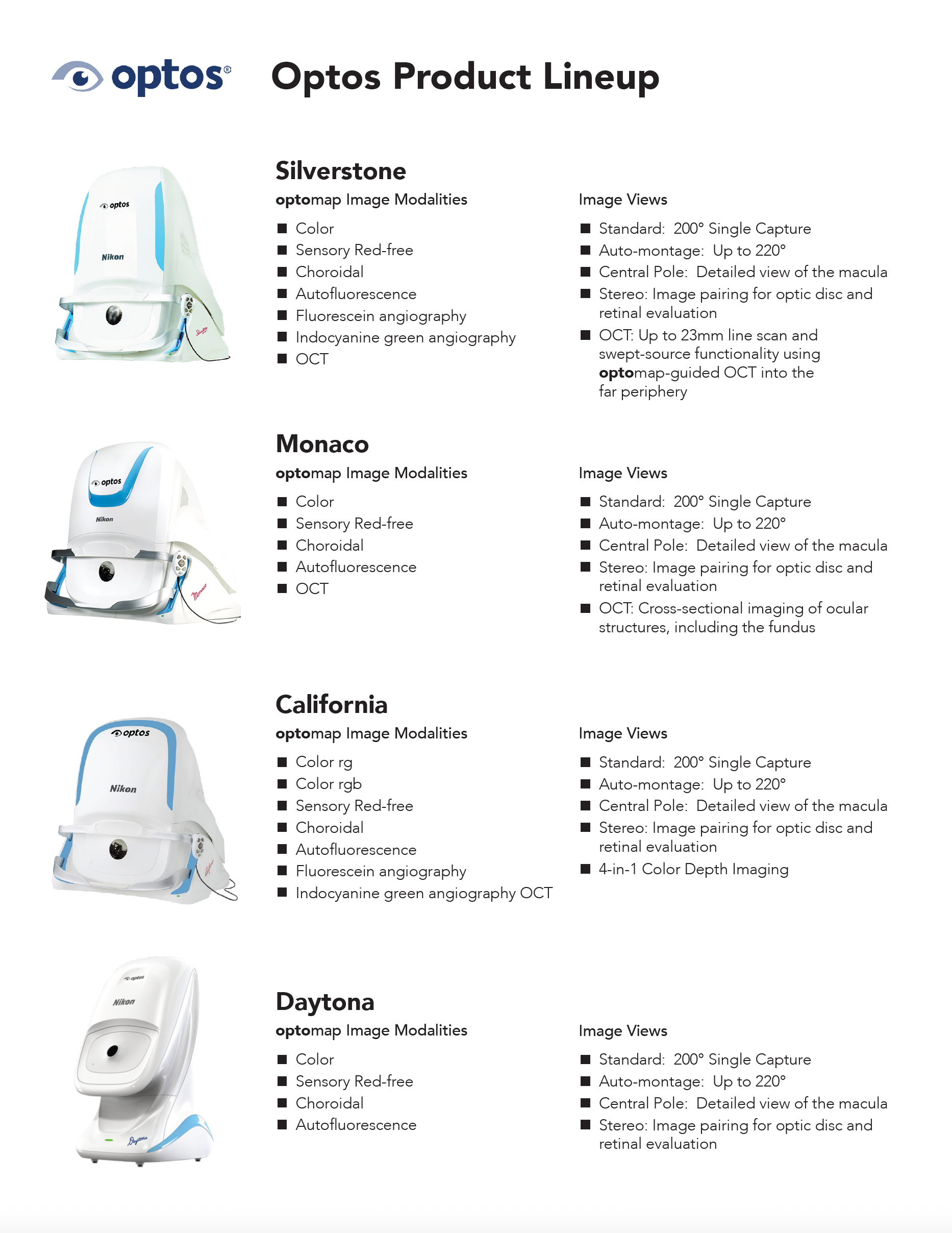
Optos product line
For the past three decades, optical coherence tomography (OCT) has been a crucial tool for capturing the peripheral retina and unlocking visibility of the retinal anatomy and vasculature. Until recently however, OCT could provide high resolution but didn’t have the ability to capture lesions in the far periphery of the retina.
Optos’ Silverstone and Monaco models leverage the power of the ultra-widefield view combined with the detailed resolution offered by OCT to enhance pathology detection and disease management effectively and efficiently.
Today, Optos includes a full product line that includes the following models:

Optos products can reduce cost and increase efficiencies in clinical settings
In many recent studies, ultra-widefield imaging proved superior at identifying lesions in the periphery at reduced cost, improved efficiencies, and with greater patient satisfaction:
- A recent large, multi-center trial found that UWF is superior to ETDRS at identifying high-risk proliferative diabetic retinopathy.27
- A 2021 study demonstrated that significantly more lesions were seen on ultra-widefield imaging compared to the seven-standard ETDRS fields. Additionally, the study found that the current DR severity scales do not incorporate retinal periphery, neuroretinal, or pathophysiologic changes and may not be well suited for documenting progression or regression eyes with very early or advanced DR, nor in the setting of vascular endothelial growth factor inhibitors (anti-VEGF).28
- Another recent study found that implementing optomap into a screening program with patients previously treated for Diabetic Macular Edema (DME) helped improve efficiency and reduce cost.29
- UWF imaging in clinical settings not only increases the frequency of diabetic retinopathy identification nearly two-fold, but also reduces acquisition time, ungradable image rates, and image evaluation time compared to nonmydriatic fundus photography.30
- A study conducted at a prominent research university found a 28-minute (33%) reduction in patient visit duration after implementing centralized optomap imaging.31 The study also showed that improving clinic efficiencies can be accomplished by reducing bottlenecks in clinic workflow.
- The use of ultra-widefield retinal imaging resulted in a practice seeing 220 additional patients in the first year of implementation, an increase of 4.4% over the pre-UWF period or an average of 1.5 additional patient encounters per day.
Learn more about the Optos product line
Fill out the form below to see what Optos product is right for your practice.
