


Since 2003, iCare has been innovating how optometrists and ophthalmologists visualize, monitor, and manage sight-stealing conditions, including glaucoma and retinal diseases like diabetic retinopathy, age related macular degeneration, and geographic atrophy. Since introducting the first rebound tonometer over 20 years ago, they have expanded their product line to include the iCare EIDON family, iCare DRSplus imaging devices, and the iCare COMPASS perimeter. These retinal imaging devices and the retina tracked perimeter establish iCare as a trusted partner to ophthalmologists, optometrists, and physicians passionate about fast and reliable diagnoses.
Learn how the iCare EIDON product family delivers excellent image quality, clarity, and sharpness
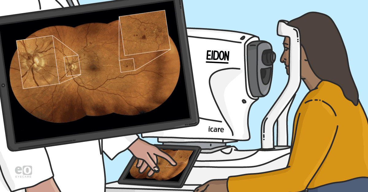
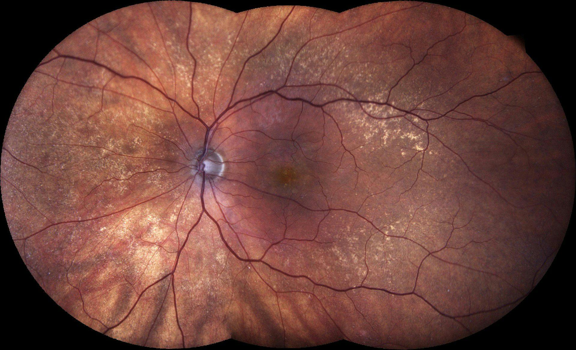
With the iCare EIDON product family, iCare has proven that it has advanced beyond tonometry with the first TrueColor Confocal system for high-resolution, ultra-widefield imaging. The advantage of TrueColor Confocal technology is the detail-rich images captured using white LED light to provide superior color fidelity while confocal imaging blocks scattered light for sharper, more informative images with greater contrast.
In 2021, iCare received FDA 510(k) clearance for the EIDON Ultra-Widefield Module, which combines high-definition image quality with a 200° view of the retina. Specifically, the Ultra-Widefield Lens Module can capture 120° images of the retina in a single shot or up to 200° with the expanded SmartMosaic function.
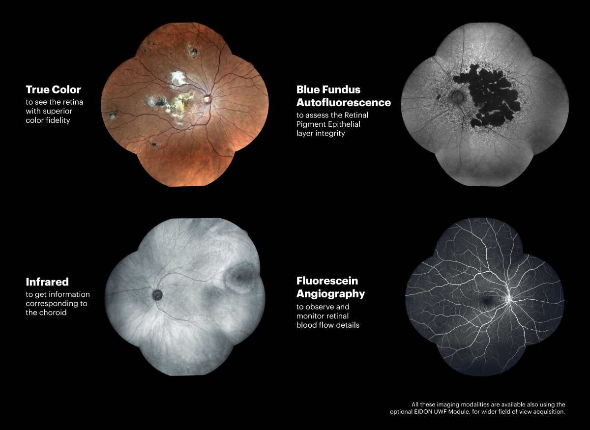
When the Ultra-Widefield (UWF) Lens Module is used with iCare EIDON devices, it provides clear visibility into the periphery. Because it enables the acquisition of ultra-widefield color, infrared, autofluorescence, and fluorescein angiography images, eyecare providers (ECPs) have come to trust and rely on the iCare EIDON family of products for optimal patient care, ease of use, and workflow efficiency—with the added benefit of 200° panoramic view.
What features can ECPs count on from the iCare EIDON family?
- High-resolution, UWF with FA and AF capabilities
- Multiple imaging modalities, including TrueColor, infrared, and red-free
- Capabilities to scan through cataracts and media opacities
- Dilation-free operation with minimum pupil size of 2.5mm (and some doctors claim even smaller)
- Fully automated for a more agile workflow
- Increased patient comfort
- All-in-one design, including software and tablet
- Flexibility in operational modes from fully automated to manual
iCare DRSplus offers a smaller platform and faster image acquisition without sacrificing quality
iCare DRSplus is an easy-to-use and intuitive imaging system that requires minimal staff training to obtain high-quality images. This TrueColor Confocal Technology promotes detailed 45° retinal images and allows scanning through media opacities. Additionally, the field of view increases to 80° using the Mosaic feature. iCare DRSplus is designed to optimize patient comfort while improving workflow and clinical efficiency.
What is confocal scanning laser ophthalmoscopy?
A confocal scanning laser ophthalmoscope can be used for several retinal imaging modalities, including fluorescein angiography, indocyanine green angiography (ICG), fundus autofluorescence and optical coherence tomography (OCT).1 The technology leverages horizontal and vertical mirrors to scan distinct layers of the retina while a confocal aperture blocks non-image-forming light to minimize scatter and chromatic aberration.2
Monochromatic laser illumination together with a confocal optical system produce high-contrast, detailed images due to the fact that illumination is solely provided by a laser beam and the remainder of the patient’s pupil is available for light collection. The laser also delivers a narrow wavelength band for more efficient excitation of fluorescence than the filtered flash illumination of a traditional fundus camera.2
How does TrueColor Confocal Technology work?
TrueColor Confocal Technology is unique to iCare imaging and perimetry and provides higher image quality, sharpness, and richer details than traditional fundus cameras. It’s based on a unique combination of white LED illumination and a confocal scanning optical engine.
- The illumination beam enters the eye in a slim line, allowing acquisition through very small pupils.
- Next, the illumination beam captures a thin line of the retina.
- An imaging beam is reflected back and passes through the confocal slit, which is able to select the light coming from the specific slice of the retina.
- The system then scans the retina, slice by slice.
- Several scans are acquired until the image on the sensor reaches the perfect brightness. Sharp and detailed images are acquired due to TrueColor Confocal Technology’s ability to suppress scattered or reflected light from outside the retina’s focal plane.
- Any light reflected by structures different from the retina (i.e. a lens with cataracts) is blocked and eliminated by the confocal slit.
Why is TrueColor confocal imaging superior to conventional retinal photography?
TrueColor Confocal Technology is the next level for image quality as it delivers increased sharpness, better optical resolution and greater contrast. Unlike other technologies, it preserves image quality even in the case of media opacities—including cataracts—and can work with pupils as small as 2.5mm—eliminating the need for dilation.
iCare imaging and perimetry devices combine a TrueColor Confocal optical engine with a true white light LED source—which includes the entire visible spectrum of light. As a result, the entire retina is illuminated and captured. The result is TrueColor retinal images characterized by colors that represent the actual appearance of the retina.
In contrast, traditional fundus cameras use a flash lamp with a broad spectrum illumination. In the absence of confocal optics, the reflected signal is derived from all tissue levels in the beam of the flash of light, and light scattering anterior and posterior to the plane of interest can greatly influence the detected signal.3
In iCare devices, white LED illumination provides high-quality, TrueColor images.This differs significantly from other imaging systems that combine different colored lights to create an artificial white light. Other features available with each iCare imaging device are stereo viewing, RGB channels and IR. The red-free filtering enhances visualization of retinal vasculature, the blue channel provides an improved view of the RNFL, the red channel allows visibility into the deep layers of the retina, and the infrared light provides detailed information about the choroid.
iCare technology enables ECPs to gain insight into the periphery and posterior pole
Many diseases that affect ocular health and threaten blindness first display pathology in the periphery, making early detection of abnormalities crucial to safeguarding patient health and well-being.
Diabetic retinopathy (DR), age-related macular degeneration (AMD), geographic atrophy (GA) retinal vein occlusions, (RVO), retinopathy of prematurity (ROP), and uveitis are chronic, progressive diseases that may first show signs in the periphery of the retina. Therefore, in these instances where early diagnosis is key to effective disease management and successful outcomes, images of the periphery are of utmost importance.
Currently, there is still no one, single test that can be used to diagnose these degenerative diseases. While structural analysis can be performed with fundus photos or optical coherence tomography imaging (OCT), other leading ultra-widefield imaging systems compromise resolution of posterior pole and may miss important vascular abnormalities or indications of retinal diseases. Conventional flash fundus cameras capture color images that are oversaturated in the red channel and washed out in the green and blue channels, resulting in a retinal picture that often looks flat and overly red.4

Flicker technology makes monitoring pathology changes even easier
Doctors have historically been able to compare images side by side in order to monitor pathology and disease progression over time. However, the proprietary iCare Flicker technology enables doctors monitor pathology and disease progression over time with features that allow users to zoom in and out on an image. However, because some differences over time are so subtle, the software now harnesses the power of Flicker technology to give users even more information. Flicker technology enables images to be superimposed over each other so eyecare professionals can better visualize the change in pathology and disease progression over a given period of time. In addition to providing more granular information about fine details, this feature can also be used to educate patients about their eyes and help engage them in their treatment.
How iCare imaging compares to other retinal technology
In a study that evaluated differences in acquisition time, peripheral extension, and chromaticity between three different commercialized ultra-wide-field (UWF) fundus cameras, the iCare EIDON FA with the UWF module demonstrated the lowest variability of acquisition time (9.5 s), compared to ZEISS Clarus (25 s) and Optos Silverstone (38.5 s).5
A statistically significant difference was found in the RGB distribution between each of the three devices (p < 0.001). iCare EIDON demonstrated an average barycenter position (RGB = [0.412, 0.314, 0.275]) that represented the best color balance of the image. ZEISS Clarus had a noticeable red shift at the expense of the blue and green channels (RGB = [0.515, 0.294, 0.191]). Optos Silverstone showed an absence of the blue channel (RGB = [0.621, 0.372, 0.007]) which results in a distortion of the color of the image.5 Overall, the researchers concluded that the iCare EIDON provided more color-balanced retinal images with greater richness of color content than the other two devices.
iCare COMPASS pairs functional analysis with TrueColor fundus imaging

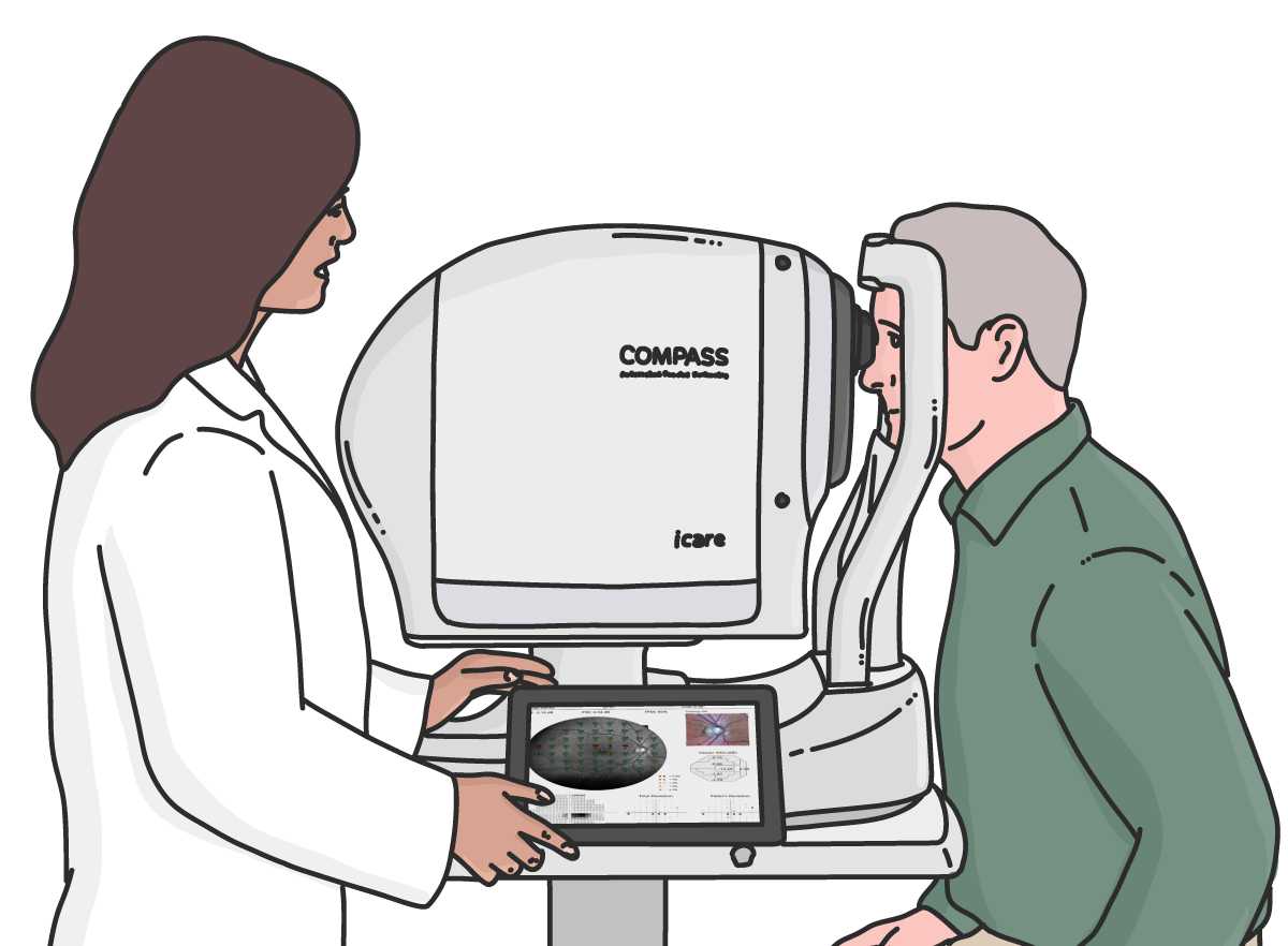
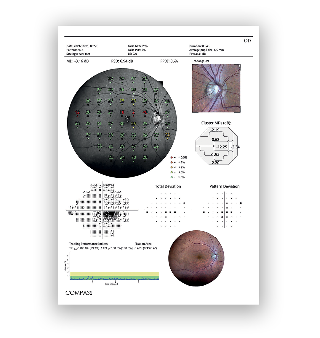
iCare COMPASS brings visual field analysis to the next level by combining Automated Tracked Perimetry—including an active retinal tracker—with TrueColor Confocal imaging. This ensures an accurate match between structure and function and informs diagnoses while reducing motion artifacts. By pairing structure with function, providers can unlock more details that lead to better disease diagnosis as well as more accurate patient monitoring.
Real-time retinal tracking actively compensates for eye movements during the visual field test for more accurate results. Additionally, the device is designed for patient comfort by allowing for natural blinks without impeding the data or results. For example, defects are delineated precisely as motion artifacts are reduced.
The iCare COMPASS perimeter is designed for a high correlation between retinal sensitivity values and retinal structure. This means that patient results will be informative about the structure and functionality of the retinal for better accuracy and patient monitoring.
The importance of pairing structural information with functional assessments
While retinal imaging combined with confocal technology can provide a wealth of information on the retina's structure, this information is even more powerful when paired with functional analysis that delivers a more complete assessment of a patient's vision.
Timely detection of glaucomatous progression is crucial in the delivery of glaucoma care, and event-based analysis like visual field tests can be incredibly valuable when predicting overall visual function6 and when monitoring glaucomatous patients. While images can provide insight into structural changes in the optic nerve and retinal nerve fiber layers, glaucoma progression can also be observed as increasing functional loss in a series of visual fields.7
Also called standard automated perimetry, (SAP), visual field tests commonly deliver sensitivity measurements in the form of light flashes at 52 test points (for 24-2) stimuli. Recent improved testing algorithms like the Swedish interactive thresholding algorithm (SITA) and the availability of progression detection software like guided progression analysis (GPA) and visual field index have solidified SAP as the preferred method for diagnosis and follow-up of functional visual field loss.8
A 2016 study that analyzed structural and functional progression of glaucoma by fundus photography and visual field tests of 249 subjects revealed that in patients with manifest glaucoma, progression was detected first in the visual field test in 163 eyes (52%) in the optic disc first in 39 eyes (12%), and in one eye it was found simultaneously in both fundus photos and visual field progression.9
Other assessments of normative databases of commercial devices used to assess visual fields showed that a large proportion of ganglion cells must be lost before a visual field defect can be detected on SAP. Conversely, later in the disease process, the opposite tends to hold true where incremental losses in ganglion cells result in detectable declines in visual function before they can be visualized by imagery.10
Most eyecare providers agree that in order to adequately detect and monitor progression of glaucoma and retinal disease, retinal imaging and fundus perimetry are both critical to providing optimal patient care.
iCare COMPASS is also ideal for busy practices
iCare COMPASS enhances practice efficiencies due to the dual capabilities of combining a visual field test with retinal imaging all in one platform. It is easy to use, does not require a trial lens, and allows remote viewing for patient education. Traditional SAP is typically performed through refractive correction with trial lenses, which can increase examination time and may cause artifacts. The iCare COMPASS removes this roadblock with its automatic refractive correction system with auto-focus.
Remote Viewer software offers embedded capabilities for network connectivity for both remote data review and data backup. The iCare COMPASS Remote Viewer is a browser-based software that allows for reviewing from any network computer on the same local area network (LAN).
Additionally, the Remote Viewer provides image comparison tools, anatomic measurements, and intuitive post-processing tools. For example, images taken at different times can be registered and superimposed using Flicker technology to facilitate detection of morphological changes over time.
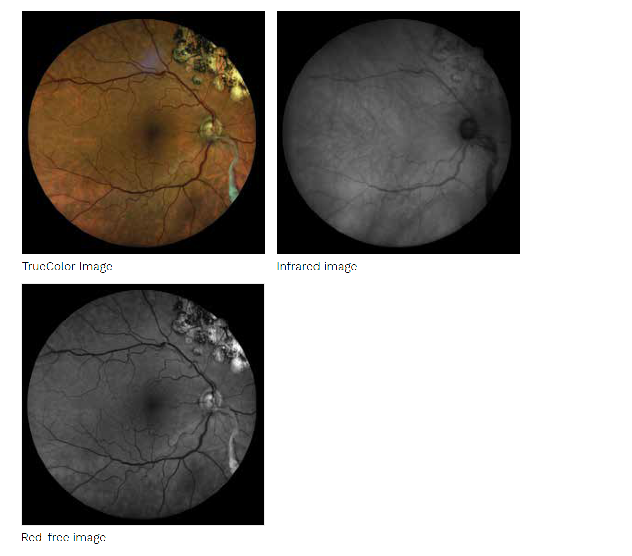
For the first time ever in a visual field test, iCare COMPASS delivers 60° confocal images of the retina in different modalities: TrueColor, infrared, and red-free.

iCare COMPASS also leverages a new SmartMosaic feature to enable acquisitions of high-definition TrueColor images from the posterior pole to the periphery and creation of a seamless 100° montage.

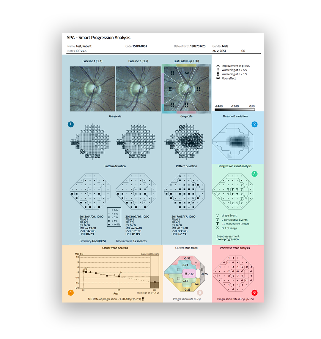
What's Smart Progression Analysis (SPA)?
The SPA report shows structural and functional changes over time. Progression assessment, rate, and prediction reliably evaluates disease stage in glaucoma management.
1. An overview of 2 baseline exams and the last follow-up
2. The Threshold Deviation map
3. The Progression Event Analysis, which is an estimation of visual field loss
4. The Global Trend Analysis, which is an expected loss over 3-5 years
5. Cluster MDs deviation and trend, for 24-2 and 30-2 grids
6. Pointwise Trend Analysis with progression rate

Reduced exam time with ZEST Fast
The ZEST Fast is a new threshold strategy to decrease exam time and boost efficiency. When compared to ZEST, this threshold is 30% faster in glaucoma patients and 40% in healthy patients.
How iCare devices can help your practice overcome workflow obstacles
While structural analysis can be performed with fundus photos or optical coherence tomography imaging (OCT), traditional instruments can be expensive and time-consuming. In some cases, technicians are required to take multiple images to eliminate views with eyelashes and to accommodate for blinks.
Other retinal imaging systems can be uncomfortable for patients who may be sensitive to a bright flash or have to contort themselves to “fit” in the machine. iCare overcomes these obstacles with their product line that allows eyecare professionals to choose the device that works best for them.
The agility of the iCare products means doctors can see more patients per day OR perform the test alone for enhanced workflow efficiency. In addition, the iCare EIDON FA does not need an experienced retinal photographer, and the fully automated device eliminates the need for specialized training.
The iCare family of products offers fully automated devices that deliver comprehensive assessments. As a result, doctors gain a more informative view of retinal health and disease progression in an easy-to-use, patient-friendly format.
The newest addition to the iCare portfolio is the iCare ST500—the world’s first slit lamp-mounted rebound tonometer.
