


Corneal dystrophies are a collection of rare, inherited, noninflammatory eye disorders that are typically bilateral and progressive over time. Most corneal dystrophies result in the abnormal accumulation of foreign material and debris within a particular layer(s) of the cornea. Corneal dystrophies are generally not associated with environmental or systemic risk factors.
Corneal dystrophies are relatively rare with an overall prevalence rate of approximately 1,300 per million in the United States (0.13% of population). These dystrophies occasionally present at birth, but more commonly become apparent in the first or second decades of life or even later into adulthood. These dystrophies can lead to myriad ocular symptoms including decreased vision, photophobia, foreign body sensation, ocular hypertension and glaucoma. In other cases, patients may be asymptomatic.
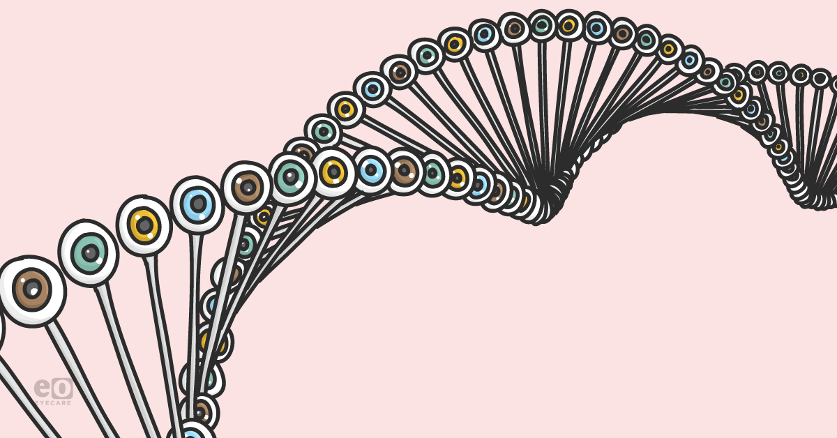
In this article, we’ll review the genetics behind common corneal dystrophies and explore how they affect each layer of the cornea. From troubleshooting unexplained vision loss and dry eye symptoms, to assessing future risk of other ocular diseases, having a thorough understanding of corneal dystrophies is essential for all eye care practitioners. Let’s have a look!
For further reading on corneal dystrophies, check out the Ultimate Corneal Dystrophies Study Guide—a complete list plus clinical photos!
Corneal Anatomy
The cornea has 5 layers, with the majority of dystrophies being named based on which layer is primarily affected. These layers are separated as epithelium, bowman’s membrane, stroma, descemet’s membrane and endothelium.
The epithelium and strong bowman’s layer both comprise the outermost protective layers. The stroma is the thickest corneal layer and consists of water, collagen fibers, and various other connective tissues. The stroma is responsible for the cornea’s flexibility and comprises 90% of the cornea’s thickness. Descemet’s membrane consists of a thin, strong inner layer, while the innermost endothelium is responsible for pumping excess H20 out of the cornea.
With over 20 specific types of corneal dystrophies, they are typically placed into 1 of 3 groups based on which layer of the cornea they affect - anterior/superficial (epithelium/bowman’s membrane), stromal, or posterior (endothelium/descemet’s membrane). Most dystrophies tend to start centrally along the visual axis and spread peripherally over time.
Genetics
Most corneal dystrophies are autosomal dominant in nature, meaning half of all direct family members are expected to be affected. Other corneal dystrophies can be autosomal recessive and/or sex-linked traits.
Based on the Cornea Society’s IC3D classification system, five types of corneal dystrophies are directly associated with mutations in the transforming growth factor, beta-induced (TGFBI) gene. These dystrophies are lattice corneal dystrophy type I and its variants, granular corneal dystrophy types 1 and 2, Thiel-Behnke corneal dystrophy and Reis-Bückler corneal dystrophy. Epithelial basement membrane dystrophy (EBMD) has also been linked to mutations in TGFBI.
The TGFBI gene encodes for the adhesion protein keratoepithelin which is produced by the corneal epithelium. Mutations in TGFBI result in the formation of abnormal deposits of keratoepithelin within the cornea. In addition to affecting the epithelium, these mutations are also involved in other dystrophies that affect bowman’s and stromal layers as well.
Mutations in the keratin encoding genes KRT3 and KRT12 are involved in the pathophysiology of Meesman’s dystrophy. These genes are responsible for maintaining the strength and integrity of the corneal epithelium. Mutations cause the abnormal accumulation of subepithelial filaments that result in an unstable epithelium that is more susceptible to even the smallest amount of trauma.
The gelsolin gene (GSN) encodes for a specific filament protein which, when mutated, is responsible for abnormal amyloid deposition throughout the body, including the corneal stroma, and is responsible for lattice dystrophy type 2.
Keratin sulfate is a protein responsible for the production of proteoglycans which help the cornea maintain its structural integrity. Numerous mutations in the CHST6 gene have been shown an association with macular dystrophy.
The COL8A2 gene is responsible for encoding collagen synthesis throughout the body. When mutated, this gene may be responsible for the breakdown of the structural integrity of descemets membrane leading to corneal guttata (focal thickening of descemets membrane) and FECD. Corneal guttata can be seen in over 10% of women aged 55 and older. While some patients with guttata may be asymptomatic, this ocular finding is classically associated with Fuch’s endothelial dystrophy.
The VSX1 gene is also a protein coding gene responsible for ocular development, particularly the cornea. In addition to the COL8A2 gene, VSX1 is also associated with posterior corneal dystrophies. Mutations in this gene are responsible for the development of PPD, and possibly keratoconus.
Treatment
Most corneal dystrophies are treated topically based on signs and symptoms (eg. dry eye, corneal edema, photophobia, ocular hypertension, etc). Some patients may also experience blurred vision due to the development of astigmatic refractive error. Corrective lenses can improve vision in some of these patients. In more advanced cases, patients may need a penetrating keratoplasty as a means of improving their vision and comfort.
With the ongoing discovery of new genetic mutations, patients with corneal dystrophies may one day have gene therapy options or other non-surgical treatment modalities at their disposal.
Common Dystrophies
The most common reported corneal dystrophies tend to affect the endothelial or anterior corneal layers. These include epithelial basement membrane dystrophy (EBMD) and Fuch’s endothelial corneal dystrophy (FECD). Women tend to be more affected by corneal dystrophies, with Fuch’s being the most common (approximately 60% of all dystrophies). It is also the most common corneal dystrophy that ultimately warrants a corneal transplantation.
Corneal dystrophies can also have varying prevalences among certain populations and demographics. For example, one study showed a very low incidence of corneal guttata and subsequent Fuch’s endothelial dystrophy among the Japanese demographic. Another study showed Posterior Polymorphous dystrophy (PPD) to be the most common type within the Czech Republic, while Macular dystrophy was shown to be more common in Iceland.
Epithelial and Subepithelial
- Epithelial basement membrane dystrophy (EBMD)
- Associated with TGFB-I/5, BIGH3 genes
- Autosomal dominant inheritance in some cases
- Not a true dystrophy given that similar changes reported in >75% of population >50 y/o
- aka: map-dot-fingerprint dystrophy, anterior basement membrane dystrophy, Cogans microcystic dystrophy
- Poor adhesion of basal epithelial cells can lead to accumulation of basal laminar/cytoplasmic material
- Rarely seen in children
- Signs/symptoms
- Dry eye/foreign body sensation, RCE
- Treatment:
- Aggressive lubrication, bandage contact lenses, in severe cases, phototherapeutic keratectomy (PTK), or corneal debridement
- Associated with TGFB-I/5, BIGH3 genes
- Meesmann corneal dystrophy(MECD)
- Mutation in keratin genes - KRT3/KRT12
- Associated with chromosomes 12q13 and 17q12
- Autosomal dominant inheritance
- Diffuse clear corneal microcysts
- Intraepithelial vesicular lesions that contain keratin
- Typically present in 4th or 5th decade of life
- Signs/symptoms
- Dry eye/foreign body sensation
- Photophobia and tearing if cysts rupture
- Mutation in keratin genes - KRT3/KRT12
Bowman’s Layer
- Reis–Bu ̈cklers corneal dystrophy (RBCD)
- Associated with TGFB-I gene
- Associated with Chromosome 5q31
- Variants
- Thiel–Behnke corneal dystrophy (TBCD)
- Grayson –Wilbrandt corneal dystrophy (GWCD)
- Central corneal opacities that appear ring shaped
- Gray/white in color
- Compromised epithelial basement membrane/bowman’s layer
- Often presents in 1st decade of life
- Signs/symptoms/treatment
- RCE, may require corneal debridement or PTK
- Associated with TGFB-I gene
Stromal
- Lattice corneal dystrophy (type 1)
- LCD type 1 variants (III, IIIA,I/IIIA, IV)
- Associated with TGFB-I/5 genes
- Associated with Chromosome 5q31
- Most common stromal dystrophy
- Thin lattice lines within stroma, central >peripheral
- Made of Amyloid deposits
- Early in life (before age 20 y/o)
- Signs/symptoms: decreased BCVA, RCE
- Treatment: Penetrating keratoplasty (PK) in advanced cases
- Lattice corneal dystrophy (type 2)
- Associated with TGFB-I gene and abnormalities in gelsolin protein
- Associated with Chromosome 9q34
- Thicker lattice lines within stroma, peripheral>central
- Made of Amyloid deposits
- Associated with systemic familial amyloidosis (aka Meretoja syndrome)
- Appear later in life (after age 20 y/o)
- Signs/symptoms: decreased BCVA, RCE
- Treatment: Penetrating keratoplasty (PK) in advanced cases
- Associated with TGFB-I gene and abnormalities in gelsolin protein
- Granular corneal dystrophy
- Associated with TGFB-I/5 genes
- Associated with Chromosome 5q31
- Autosomal-dominant inheritance
- Typically presents 1st - 2nd decade of life
- Central ring-shaped or crumb-like opacities
- Made of eosinophilic hyaline
- Variant: Avellino dystrophy
- Granular and lattice type opacities
- Signs/symptoms: decreased BCVA, RCE
- Treatment: PTK, PK in advanced cases
- Associated with TGFB-I/5 genes
- Macular corneal dystrophy (MCD)
- Associated with CHST6 gene/Chromosome 16q22
- Autosomal-recessive inheritance
- Type 1 - keratin sulfate synthesis absent
- Type 2 - keratin sulfate synthesis below normal
- White milky opacified lesions
- Composed of glycosaminoglycan
- Severe vision loss
- Most visually devastating dystrophy
- Treatment: Patients typically have PK by age 40 y/o
- Associated with CHST6 gene/Chromosome 16q22
Descemet Membrane/Endothelial
- Fuch’s endothelial corneal dystrophy (FECD)
- Associated with COL8A2 gene
- Some reports of autosomal-dominant inheritance
- Early signs : Corneal guttata (focal thickening of descemets membrane)
- Late signs : Stromal/epithelial edema worse in AM
- Corneal fibrosis and scarring
- Treatment: hyperosmotic agents (eg: Muro 128 ointment), hairdryer can help evaporate excess fluid/swelling in the cornea
- DLEK/DSEK in more advanced cases
- Associated with COL8A2 gene
- Posterior polymorphous corneal dystrophy (PPD)
- Associated with VSX1 gene
- Associated with chromosomes 20p11.2-q11.2
- Autosomal-dominant inheritance
- Early signs : vesicles, linear strands of thickened descemets membrane
- Late signs : corneal edema, peripheral anterior synechiae
- Treatment similar to Fuch’s
- Also risk for ocular hypertension, monitor IOP
- Associated with VSX1 gene
- Congenital hereditary endothelial dystrophy
- Associated with chromosome 20 and VSX1 gene
- Type 1 : Autosomal-dominant inheritance
- Type 2 : Autosomal-dominant recessive
- More common, more severe
- Presence of corneal edema at birth (type 2) or in early infancy (type 1)
- Nystagmus may also be present in type 2
- Treatment: PK early in life to prevent deprivation amblyopia
- Associated with chromosome 20 and VSX1 gene
Patient Education
After confirming a diagnosis of a specific corneal dystrophy, it is important for eyecare practitioners to explain to patients the ocular signs and symptoms to be aware of as well as their prognosis for future vision changes. In addition to ocular diseases like macular degeneration and glaucoma, confirming a positive family history of corneal pathology can also aid in the diagnosis. Patients should also notify other family members of their condition and encourage them to also have their eyes examined to rule out the early signs of corneal dystrophy.
Genetic testing can also aid with diagnosis and help determine the prognosis of the disease. It can also provide information on genetic counseling if patients are interested in assessing their risk of passing on the condition. Genetic testing may also help elucidate patients with corneal dystrophies who may be at higher risk for post-surgical complications from ocular procedures like LASIK, PRK, or cataract surgery.
Conclusion
It is crucial for eyecare providers to have a thorough understanding of the plethora of corneal dystrophies that may present to their offices. While less common than other ocular diseases, corneal dystrophies can have a significant impact on patients’ vision and ocular health. Explaining the signs, symptoms, and treatment options can help put patients at ease while also establishing a positive rapport with them. These dystrophies can cause myriad ocular signs and symptoms, from relatively benign to sight-threatening. Clinicians should never hesitate to have a second look at their patient’s cornea next time they’re suspicious of a corneal dystrophy.
