


Case study of 60-year-old female: routine to extreme cases in glaucoma
Case history presented by Dr. Steen
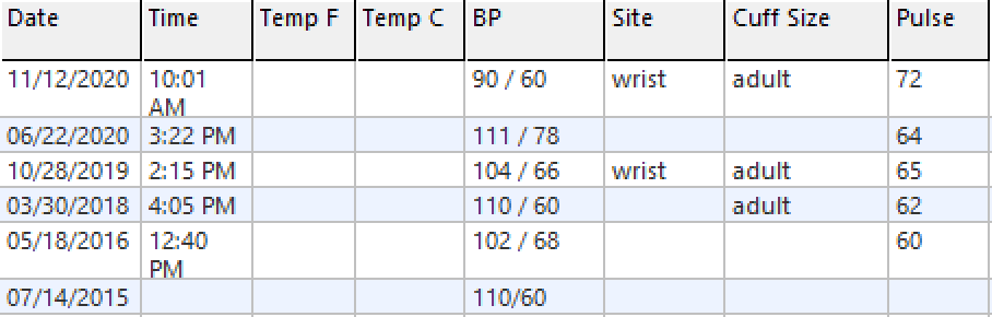
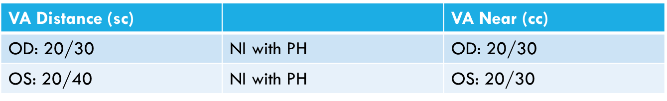
Patient is 60 years of age and was diagnosed with primary open angle glaucoma in 1998, at the age of 36. She was initially treated with timolol 0.5% in each eye and presented to our clinic about 10 years after the time of diagnosis on timolol with pressures in an 18 to 20 mmHg range. At that point we were not aware of her peak untreated intraocular pressures. She has thin corneas with a central corneal thickness of less than 500 microns in each eye. Visual acuity is 20/20. There is no afferent pupillary defect and color vision is unremarkable.
- 60-year-old hispanic female
- Primary open angle glaucoma OU
- Diagnosed in 1998 at the age of 36
- Treated with timolol 0.5% BID OU
- IOP 18-20mmHg OD and OS; peak untreated IOP not known
- CCT 477μm OD 495μm OS
Systemically, the patient has medically-treated hypothyroidism and is not hypertensive. She has no family history of glaucoma, but her mother has Alzheimer's disease.
- Hypothyroidism managed with levothyroxine
- Multivitamin, Omega-3
- Not hypertensive

- No family history of glaucoma
- Mother-Alzheimer’s disease
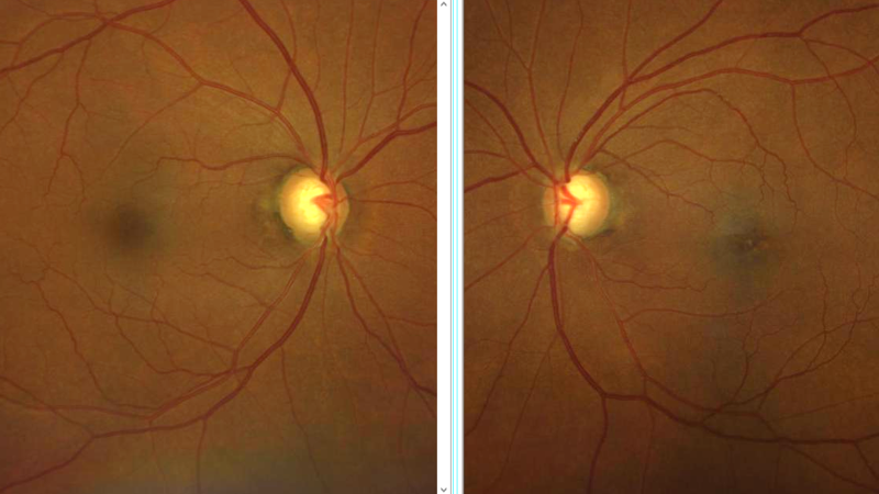
In terms of embracing and utilizing technology, I am an advocate of traditional standard fundus photography—which allows us to identify change objectively over time and manage patients objectively over many years.
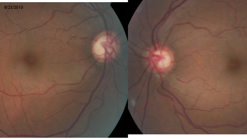
Images of her optic nerves in 2010 revealed inferior neuro retinal rim defect in each eye with a very subtle nerve fiber layer change. Superior temporal, there is also a subtle nerve fiber layer defect.

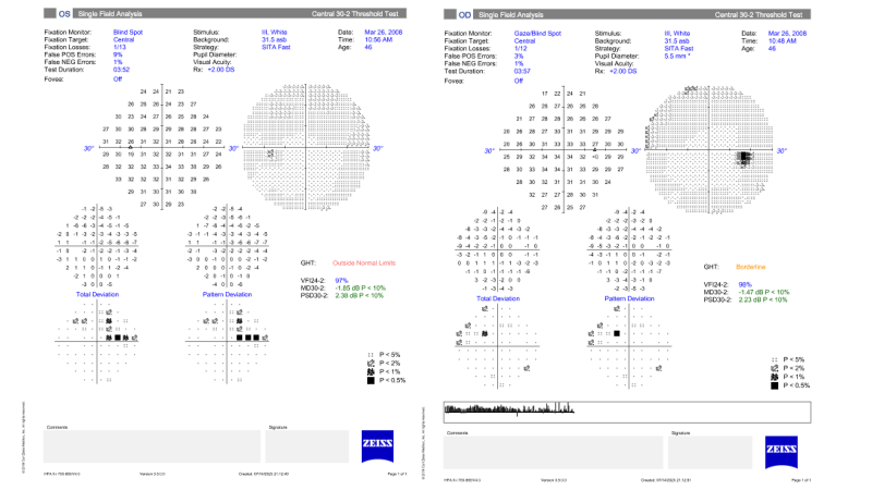
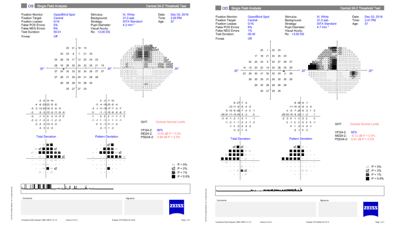
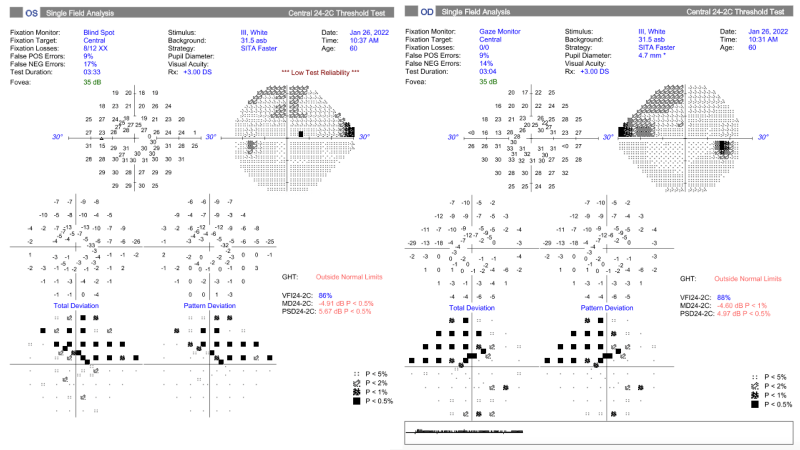
In this case, different types, strategies, and patterns of visual fields were used. This is a 30-2 SITA Fast that was performed in the right and left eyes and indicates early nasal step in each eye that's consistent with her optic disc appearance.
Nerve fiber layer imaging from 2010 provides objective, quantifiable data about this patient’s nerve fiber layer defect, which is present in the right and left eye—inferior, greater than superior.
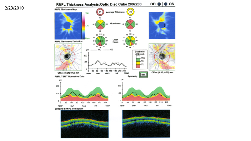
2010 didn’t have commercial, widespread availability of ganglion cell complex analysis. So at that point in time, when referring to OCT analysis, it was all about the retinal nerve fiber layer.
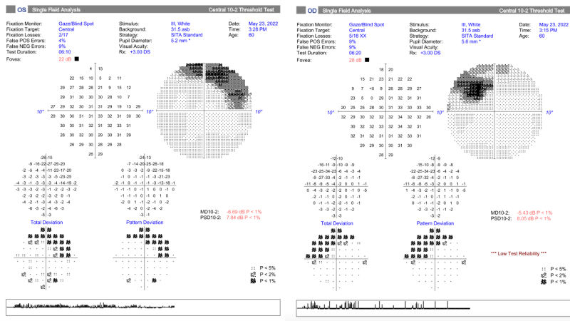
Moving forward through this patient's disease course, we see clear, functional change over time. We've lapsed about eight years here, and this is a 24-2 SITA Standard. You can observe a deepening of the visual field loss that now extends beyond the vertical midline.
If we look at the paracentral defect in more detail on a 10-2 visual field, a 10-2 SITA Standard, we start to think about functional considerations in this patient's case.
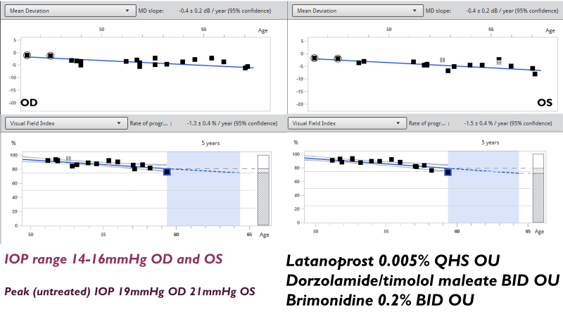
When we watch patients long enough, it’s clear that disease progresses. But when we think about this patient's early age of diagnosis and intraocular pressure readings (where progression has been identified in the 14 to 16 mmHg range) we really start to think about what else can or should be done. In this particular case, when we're looking at progression, we're focusing on the visual field mean deviation and that change in the mean deviation slope over time.
I like the visual field index progression analysis for this patient because it does weigh those paracentral points more heavily, understanding that there is more functional implication in loss of the paracentral visual field versus non-central points.
This is an ideal patient to manage and work with. She’s diligent, aware of her disease process, and really understands her central role in chronic disease management in terms of medication and medical therapy. She is as perfect as someone can be with adherence to medication. Her only lapse in medical therapy occurred when she unexpectedly left the country for longer than planned. So there was self-discontinuation of medication because she ran out. By the time she came back to our clinic, she had been off all medications for about a four- to six-week period, which allowed us to better understand her peak untreated intraocular pressure, which was still only 19 and 21 mmHg versus the 14 to 16 mmHg range on latanoprost, dorzolamide/timolol, and brimonidine.
Discussion
Dr. Panarelli:
- Are we doing too little or too much for this patient?
- Critical importance of family history—although IOP is the only modifiable risk factor for glaucoma, there are many risk factors at play.
- Too many (including eyecare professionals) believe controlling the IOP will be enough to control disease progression, but that’s not always the case.
- Even when there is no volatility in diurnal curves, disease progression may get worse over time.
- Disease progression should be confirmed by repeating visual field testing and using different types of tests.
Dr. Steen:
- Visual field testing for the patient presented was subsequently changed to 24-2C SITA Faster at least once a year, and a 10-2 at least once a year.
- Focused more on functional measurements rather than running OCTs frequently, due to her long-term robust visual field data.
- For other new and current patients, have been transitioning from 24-2 to 24-2C Sita Faster.
Dr. Laul:
- Although SITA Standard has commonly been used in the past, some research indicates that if you have long-term data over time, the variabilities from the SITA Fast and SITA Faster algorithms tend to even out.
- If you can only do one visual field, SITA Standard may provide more accurate results because of the double threshold bracketing.
Dr. Panarelli:
- This patient a perfect candidate for a 24-2C for variety of reasons.
- We need to keep patients happy—although frequent testing may seem beneficial based on available studies, it may not be practical for the patient.
- We need to find ways to keep our clinics running and to not miss things.
- We need to also consider structural testing that will capture and track early changes in the inferior hemifields.
- Software analysis may help to identify changes in the superior nerve fiber area to identify issues on the ganglion cell analysis couldn’t see before.
- The value of fundus imaging to capture changes that may be missed in an exam is important. Nothing really shows changes like the disc photograph.
- How should this patient’s glaucoma be classified? POAG? NTG? A mix? Classically, when discussing low tension glaucoma, one camp associates it with other symptoms, such as vascular abnormalities and problems with autoregulation. The other camp thinks it’s all a spectrum of one disease in which there is probably some IOP volatility. I believe there are two separate forms of NTG: the former condition referenced and the latter in which genetics play a big role.
- The only way to really fight this is to get the pressure lower. So where do you go with topical therapy? What do you think about laser? Are you thinking about trabeculectomy? How far do you take this and for how long
Dr. Steen:
- For this patient, additional treatment options explored included a consult with a glaucoma surgeon, who recommended trabeculectomy.
- However, patient was underinsured and wanted to defer surgical intervention by using any other medications possible.
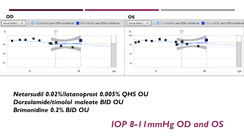
- After discussion with patient, added fixed combination of netarsudil and latanoprost, while continuing dorzolamide/timolol and brimonidine. Patient’s IOP eventually stabilized in the 8-11 mmHG range.
Dr. Panarelli:
- These new Rho kinase inhibitors seem to work really well when added to the treatment algorithm, based on some of the available phase 4 studies.
- Would hope to achieve similar reduction of IOP in this range with a trabeculectomy, which is not easy to do with traditional glaucoma medications and perhaps not with laser therapy, either.
Dr. Steen:
- Patient has remained stable as indicated by this 24-2C SITA Faster.
- Paying careful attention to the inferior visual field, which has remained unaffected or minimally affected.
In summary, a young individual who's shown progressive disease with paracentral functional damage who we’ll be watching over the next, hopefully 30 to 40 years. Certainly have a lot to consider to maintain her pressure and functional status.
Dr. Panarelli:
- It’s important to consider the patient’s age when assessing treatment options.
- Our job is to preserve a patient's functional vision in their lifetime.
- Quality conversation with the patient is so important: Ask them how they feel their vision is changing over time, because the patients know what's going on.
Dr. Steen:
- Of note, patient’s 38-year-old daughter has been seen every three to four years in our clinic with nerve fiber layer imaging and ganglion cell complex imaging with an intraocular pressure in the 20-22 mmHg range untreated.
- Just recently, her nerve fiber layer and ganglion cell complex showed a statistically- and clinically-significant change of more than 12 microns. Based on the clock-hour analysis, we made the decision to treat.
- She has a thin cornea as well, but in light of her mom's disease course and trajectory, we are being relatively aggressive.
- There is currently no visual field loss in the daughter, but we are trying to delay or prevent any functional change in this case.
Dr. Panarelli:
- In complete agreement with aggressive approach to daughter’s care.
- So many patients have IOPs of 20-24 at diagnosis, and a variety of studies indicate the significant risk of conversion.
- Must be very careful and weigh each of the risk factors when deciding on a treatment plan.
- Nothing beats your gut judgment when you're taking care of one of these family members. You get a certain feeling and think, “Now is the time.”
Case study of 59-year-old male: routine to extreme cases in glaucoma
Case History Presented by Dr. Laul
Patient is a 59-year-old male who presented with a history of chronic angle-closure glaucoma who was previously managed in an outside eye center. He had an LPI in 2018 and was previously treated with latanoprost and brimonidine. Most recently, he was using Rocklatan® twice a day in both eyes in addition to another unknown medication. However, he's been off medication for two months. He recalls that his pressures have been in the low twenties despite being on eye drops, so our thought is that his pressures are probably in the twenties without any medication. Since he feels the drops have been ineffective for lowering his IOPs, his adherence to the treatment regimen has been variable. Additionally, he was adopted, so family history is not available and we were not able to obtain much of his previous data from outside providers. The data I have is from early 2021.
- 59 y/o M with CACG OU presents for glaucoma evaluation (OCT/HVF/pachs/gonio).
- Previously managed by another ECP.
- S/P LPI OU (~2018).
- Previous history of latanoprost and brimonidine usage. Most recently, patient was taking Rocklatan® BID in addition to another unknown medication/dosage. Patient has been off meds for approximately 2 months. Recalls pressures historically have been in the low 20's range despite being on gtts and believes they were ineffective at lowering his eye pressures.
- Unknown family history of GL.
- No records of pachs, OCT RNFL/GCC or HVF OU.
- LDFE at last visit (2020.)
- Denies flashes, floaters, eye pain, diplopia.
When I saw the patient in my glaucoma service, he had good vision without any corrections. He’s not a myope or very high hyperope.

His pressures in office were about 20 and 22, and he’s been without medication for two months. I think it's always important when you're classifying disease to look at the gonioscopy readings, and as you can see, this patient does have some PAS superior nasally in both eyes.

If your patient has glaucoma or evidence of PAS, I think it's absolutely appropriate to get this patient in to get an LPI as soon as possible.


Of course, you want to assess the back portion of the eyes. On these optic disc photos, significant cupping is visible in both eyes and in general, he has large optic discs. Some nerve fiber layer reflection is visible superior in the right eye, but no nerve fiber layer reflection inferior in both eyes and superior in the left eye.
With photographs, I also assess for the presence of any other type of optic neuropathy. So I'm looking for things like pallor on the optic disc or signs of disc hemorrhage. This patient does have some peripapillary atrophy, but no signs of pallor in the discs. I also don't see any signs of vascular-type changes that would indicate a previous vascular event like BRVO, CRVO, or anything like an NAION, causing visual field defects.
When we look at this patient's angles—which are post-LPI—you can see that nasally in both eyes, it's a little bit narrow. Just to give you an idea where structures would be, if you look at the area between the clear cornea and the limbal zone, that’s the transition zone. The scleral spur is usually about a millimeter or two behind that. This was taken at the 180 mark, and as I mentioned, the PAS was at that nasal superior. So in some cases, you can get excellent images with the anterior segment OCT.
Question from Dr. Laul for the panel: How do you use the anterior seg OCT, or do you mostly focus on gonioscopy? I’ll admit that for me, gonioscopy is king and that’s what I use for most diagnoses.
Dr. Steen:
- Relies on gonioscopy.
- Finds OCT of the anterior segment to be helpful in cases in which you want to document and determine true appositional closure and appositional touch.
- Dr. Panarelli:
- Also relies on gonioscopy.
- However, OCT has gotten so much better at imaging the angle that may not use it enough.
- Uses OCT more for patient teaching, since it helps patients understand what we're going to do and why a laser might need to be done.
- When I explain with images, patients leave so much more comfortable with what's going on.
- Additional points:
- Considering the angle in the case study, patients get diagnosed with chronic angle closure, which is the appropriate term when there’s evidence of disc damage and PAS.
- But with nerves like those presented in the case study, does this patient have an open-angle component as well?
- Often, when I think of true chronic-angle closure, I'm seeing patchy PAS everywhere.
- Sometimes people develop a little PAS if it was a challenging iridotomy with post-op inflammation.
Dr. Laul: Case Study Continued
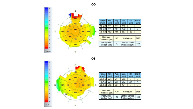
The patient’s pachymetry readings are a minimum of 441 microns in the right eye and 445 in the left, which represent a huge risk factor for this patient.
In terms of OCTs, this is the first OCT and visual field we obtained for this patient, which is an example of the combined report.
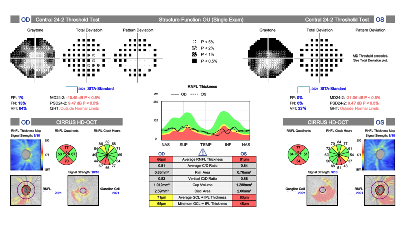
As indicated, this OCT for the right eye matches up with the disc photographs. There’s significant loss inferiorly, and in the photographs it was very thin in those places. The left eye is very similar in that you can see inferior loss. With my OCTs, I convert to an NSTIN curve, because we know that glaucomatous loss tends to happen temporally. In this case, it's easy to see significant loss inferior temporally in both eyes, which corresponds to what we see on the visual fields. In the right eye, superiorly, there's some significant loss. Although you could argue that this might be a bit of a cloverleaf pattern, I believe this patient does have significant loss in those areas. The left eye is a little worse, to the point where we can't even get a pattern deviation on them, but it makes sense based on what we see in the optic nerve.
The patient has 10-2 nasal loss in the right eye and more so in the left eye that corresponds to the inferior temporal thinning and corresponding ganglion cell.
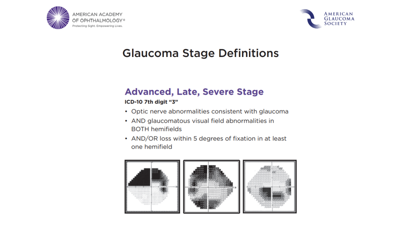
In terms of staging, this is an advanced stage of glaucoma because we are seeing loss within the central five degrees of fixation based on AGS definitions.
In terms of treatment, I started this patient on Travatan®, which is a prostaglandin, and want to consider adding additional medications in the future.
Case: plan
- Start Travatan® and consider adding Cosopt®
Once I started him on Travatan,® I was able to bring his pressures down to the mid- to lower-teens. However, they do tend to bounce back up a little bit. Unfortunately, compliance tends to be a problem for this patient, since he has never felt that drops have helped him much.

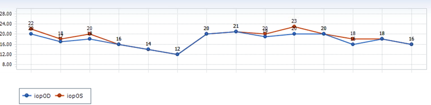
In summary, we have a young patient, just 59 years old. He’s African American with severe glaucoma in both eyes and unknown family history. Thin pachymetry in both eyes and variable drop compliance.
Case summary:
- Young (59yo) African American male with SEVERE glaucoma OD/OS
- Unknown Fhx
- Thin pachymetry OU
- Variable drop compliance (see IOP graph)
So, would it be helpful to add an additional medication?
Discussion
Dr. Panarelli:
- Use evidence as much as possible to support decision-making process, especially when aggressive treatment may be needed.
- With a younger patient, severe disease, and poor compliance, I would recommend trabeculectomies in both eyes as soon as possible.
- Recommendations are based on data from the Collaborative Initial Glaucoma Treatment Study and the new trial (TAGS trial) out of the UK: patients who presented with severe disease probably do better with earlier surgical intervention.
- Also, patient's compliance is an issue.
- Why trabeculectomies versus tubes? Starting IOP is not terribly high and some results of the PTVT study indicate trabeculectomy probably a better primary surgery.
- On the flip side, surgery includes risk—plus a patient who has compliance issues may not adhere to the extensive follow up needed for trabeculectomy in both eyes.
- That’s why building a good relationship with the patient is key. Sometimes you don’t have a lot of time to do it, but you must do it.
Question from Dr. Laul:
Would there be any benefit to other options, such as lensectomies? A clear lens exchange, potentially angle-based procedures, or is this patient too far advanced?
Dr. Panarelli:
- Angle surgery has limited utility in a patient with that kind of visual field loss, since the entire outflow pathway probably diseased.
- Although results of the EAGLE study may seem appropriate to consider, it doesn’t really apply to this situation (too often, people try to take data from big studies and extrapolate the data and make it apply to the patient sitting right in front of them).
- Patient’s already had iridotomies.
- Likely an open-angle component involved due to thin pachymetry, appearance of nerves, and how open the angle was on gonioscopy.
- Would treat as more of an open-angle glaucoma patient who has some focal PAS.
- May be some benefit to lens extraction, but not tremendous utility.
- Patient needs a low, steady, intraocular pressure.
Dr. Steen:
- Staging of glaucoma was an important component covered in the case, which is often lost in a clinical environment.
- It’s important to consider the visual fields and apply that data to correctly and appropriately stage glaucoma based on AGS, and therefore ICD-10 criteria.
Dr. Panarelli:
- Patient has true severe disease with pretty significant retinal nerve fiber layer thinning with visual field loss in multiple hemifields.
- Needs aggressive management.
Q&A: Two Case Scenarios Presented by Dr. Panarelli
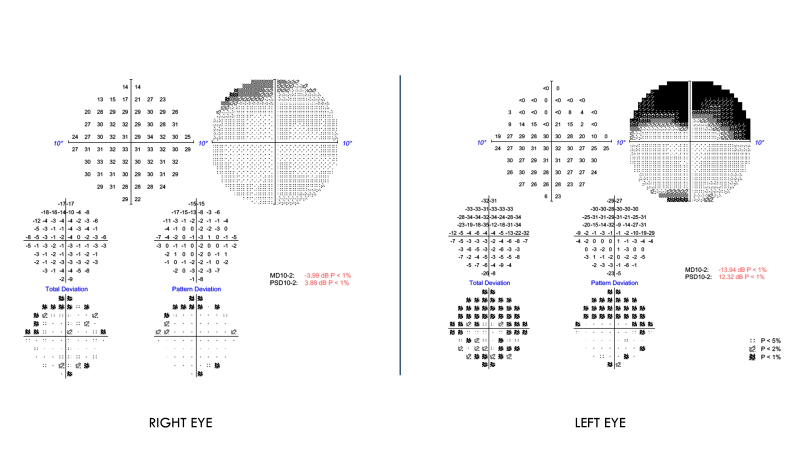
Question: What would you do with a 64 y/o patient with newly diagnosed glaucoma but not on medical therapy with an IOP: 22mm Hg OU and the following 10-2 visual fields?
- Are you going to start with laser?
- Consider medicines?
- Intracameral drug delivery?
- Microinvasive glaucoma surgery?
- Traditional surgery?
- What are your thoughts when you initially see these visual fields?

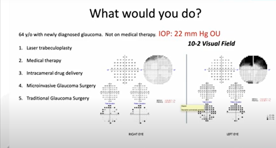
Dr. Laul:
- Since visual fields indicate severe disease, important to start with determining what the target pressure should be.
Dr. Steen:
- Important to consider potential future progression in the context of past progression.
- 10-2 visual field results with this pressure a good indicator that without aggressive therapy, patient will have poor long-term outlook.
- Need to be aggressive with targeted treatment, especially considering the patient’s age.
- More data is needed about additional risk factors.
Question: With the same scenario, but the following 10-2 visual fields, what would you do?

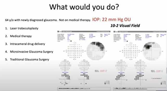
Dr. Laul:
- Similar concerns.
- Anytime the central 10 degrees is affected, aggressive treatment approach should be considered.
- Getting a 24-2 visual field would provide a more comprehensive understanding of disease severity.
Dr. Steen:
- Based on available information, no significant difference between the two cases.
Dr. Panarelli:
- Agree with all points.
- Sometimes clinicians focus so much on specific types of objective testing that the bigger picture may be missed.
- Underscores the need to get as much information as possible, including patient’s perspective about functionality.
- Polling results from participants are mixed, with some choosing medical therapy, a good number choosing laser trabeculoplasty in both cases, and some choosing more aggressive surgical interventions.
Question for the panel: What are your thoughts on laser at this point?
Dr. Laul:
- Laser should always be considered.
- LiGHT study indicated efficacy of laser and low side-effect profile (though the study included a very small cohort of patients with severe glaucoma).
- Additional benefits include potentially better IOP control at night.
Dr. Panarelli:
- Concurs that LiGHT study results may not apply in this case, since such a small number of patients with severe glaucoma included.
- It’s important to understand the methodology of a study to determine whether it applies to the patient for whom treatment is being considered.
Dr. Steen:
- Prefers treatment with medications first, since access to SLT may not be immediately available due to patient’s insurance constraints.
- With advanced disease, I think there’s an important role for the Collaborative Initial Glaucoma Treatment Study (CIGTS) and medications versus incisional surgery.
- Today’s medications are much more effective than those used in the past—including prostaglandin analogs and Rho kinase inhibitors.
- Seeks medications that increase aqueous outflow by increasing and acting on natural physiology rather than disrupting natural physiology by reducing aqueous production play a big role—even in the management of patients with advanced disease.
Dr. Panarelli:
- Concurs that all treatment options mentioned have important roles.
- Additional considerations: Laser surgery doesn’t work for everyone; certain patients may benefit from MIGS or traditional surgery sooner; and medications may carry compliance and toxicity concerns.
Question: Based on this additional information for the first patient, what would you do?
Answer still the same???
Dr. Panarelli:
- Significant amount of visual field loss with a significant amount of disc damage.
- With patient’s age, would consider trabeculectomies on both eyes early in treatment course.
- Research results support importance of being aggressive early on with severe disease.
Question: Based on this additional information for the second patient, what would you do?
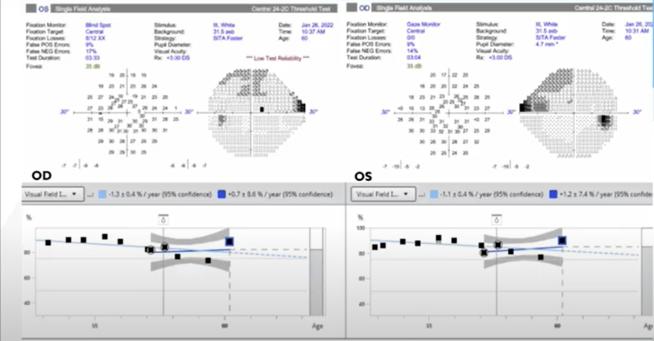
Dr. Panarelli:
- Zooming out with a new set of visual fields gives additional information.
- Has there been some stability?
- Is there progression on the visual fields?
- How aggressive should we be?
- The main point overall is how important it is to get as much information as possible to assess the big picture to inform the optimal treatment approach—and we have a lot of tools available to do it.
