


Technological advancements, including artificial intelligence (AI), have never occurred more rapidly than they do today. Everywhere we turn, our lives are seemingly enhanced through these advancements. The healthcare industry is rife with opportunity for implementing AI and other new technologies. The ultimate goal for AI within eye care is to optimize clinical decision making while also greatly reducing the burden of ocular disease and preventable blindness. Instead of fearing that technology will replace eye care providers, many proponents of AI simply see it as another tool in the diagnostic clinical toolbox, complementing what eye care providers do.
The term artificial intelligence originated in the 1950s. It is now being researched and implemented throughout healthcare, and the fields of optometry/ophthalmology are certainly no exceptions. Given the multitude of imaging modalities, the eye care field has tremendous potential for the implementation of AI.
Today, a plethora of new AI systems are being developed and implemented. By uploading patients’ retinal photos or OCT scans, eye care practitioners, trained technicians, or even AI algorithms are able to detect various ocular diseases and pathologies. In most cases, this leads to earlier intervention and treatment. With AI, innovators also hope to optimize health care costs and reduce the burden of preventable blindness on the health care system.
In this article, we’ll cover some of the latest advancements in AI and other technologies in the eye care field.
- Triaging diabetic retinopathy with IDx-DR
- Triaging glaucoma with Pegasus AI
- Continuous IOP monitoring in glaucoma - Sensimed Triggerfish
- New AI technologies in Low Vision
- Google’s “DeepMind” AI platform
1) Triaging diabetic retinopathy with IDx-DR
Regardless of practice location, most eye care practitioners will agree that a large portion of their patients’ ocular disease burden is directly related to uncontrolled diabetes mellitus. Diabetic retinopathy is the leading cause of new cases of legal blindness among US adults today. According to the CDC, between 12,000 to 24,000 cases of new legal blindness are directly related to diabetes in the US alone. Most patients with type 1 diabetes and over half of patients with type 2 will develop some form of retinopathy within the first two decades of the disease. Blindness related to diabetes, while posing a significant burden to the patient’s quality of life, also costs the US health care system $500 million annually. Through early detection and treatment, the majority of blindness related to diabetes can be avoided (approximately 90% is preventable). Unfortunately, many established diabetic patients fail to have their eyes routinely checked. According to the CDC, only half of diabetic patients typically have their eyes checked on an annual basis.
A new AI software program known as IDx-DR recently gained FDA approval. This breakthrough technology is designed exclusively for the detection of diabetic retinopathy (including macular edema). This is the first ophthalmic implementation of AI for diabetic retinopathy screening that does not require an eye care professional to interpret the results. This makes it easy to incorporate the IDx-DR system into a wide variety of health care settings unrelated to eye care.
This software uses an AI algorithm to analyze retinal images taken with a Topcon NW400 retinal camera. A growing number of primary care medical offices will begin taking this screening photo on every diabetic patient. The photos are then sent to a secure cloud-based server where the novel IDx-DR software screens the images for one of two outcomes: “greater than mild diabetic retinopathy—refer to eye care professional” or “negative for greater than mild diabetic retinopathy—rescreen in 12 months”.
While not exactly a direct comparison to a comprehensive dilated eye exam, having this screening done in any non-ophthalmic medical setting will help detect more cases of sight-threatening retinopathy in otherwise asymptomatic patients. This technology may save significant healthcare dollars through earlier detection and intervention of sight-threatening diabetic retinopathy. A recent study showed the IDx-DR algorithm has sensitivity and specificity rates close to 90% and is able to make a diagnosis within 20 seconds. It has also been shown that over 90% of acquired retinal images are of satisfactory quality to run through this AI algorithm.

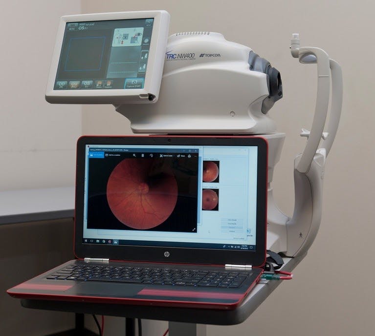
Figure 1: The IDx-DR workstation in a primary care setting.
European clinicians have had access to IDx-DR since 2014.
2) Triaging glaucoma with Pegasus AI
Similar to the IDx-DR algorithm for early detection of diabetic retinopathy, AI technology is also being developed to screen for the early detection and treatment of glaucomatous optic neuropathy. The CDC reports that approximately three million Americans are affected by glaucoma, and it is the second leading cause of blindness worldwide. Given the slow, insidious nature of glaucoma, approximately half of patients are unaware they even have the condition until significant, irreversible visual field loss has developed. With early detection and treatment, eye care professionals are able to preserve visual function.
Check out this resource for managing glaucoma patients!
London based company, Visulytix, is currently refining their AI platform “Pegasus” that assesses stereoscopic optic disc photos for glaucoma. A recent study looking at the diagnostic accuracy and repeatability of Pegasus showed results equivalent to the diagnostic accuracy of both ophthalmologists and optometrists in the early detection of glaucomatous optic neuropathy. There was no statistically significant difference in diagnostic outcomes as compared by optometrists or ophthalmologists. While yet to go mainstream, this AI platform is currently being tested worldwide in academic centers and various health care settings. High volume glaucoma practices will greatly benefit from this technology. These include VA/IHS hospitals, community health centers, and OD/MD referral centers.
3) Contact lens to supplement glaucoma management: Sensimed Triggerfish
The monitoring of intraocular pressure (IOP) is essential in the diagnosis and management of glaucoma patients. Eye care practitioners use IOP readings as an integral part of their glaucoma workup as well as determine if the patient’s current treatment regimen is sufficient in achieving a “target IOP”. Despite glaucoma patients presenting with apparently “controlled” IOP readings during office hours, those with higher diurnal IOP variability may still be at a higher risk for progression. Diurnal fluctuations in IOP have been shown to exceed 9mmHg in patients with glaucomatous optic neuropathy, whereas non-glaucomatous eyes tend to fluctuate significantly less throughout the day.
The Swiss company Sensimed gained FDA approval for marketing their new Triggerfish contact lens sensor (CLS) in the US in 2016. New technology like the Triggerfish CLS can help eye care practitioners detect continuous IOP-related changes to the eye over a 24 hour period. Using diurnal data will provide clinicians with additional information to further identify which patients may be at a higher risk for glaucoma.
With a 14.1mm diameter and a variety of base curves, this silicone soft contact lens is designed to remain on the eye for 24 continuous hours. The Triggerfish CLS contains two strain gauges that detect small spontaneous circumferential changes at the corneoscleral area over a 24-hour period. Monitoring ocular dimensional changes via the CLS has been highly correlated with accurate IOP readings. This information from the CLS is then transmitted to a portable recorder worn by the patient, which is then transmitted to a computer via Bluetooth for analysis. The Triggerfish CLS has also proved to be fairly comfortable and well-tolerated over a 24 hour period.

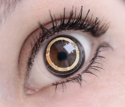
Figure 2: The Sensimed Triggerfish contact lens sensor contains two strain gauges that detect small spontaneous circumferential changes at the corneoscleral area over a 24-hour period, which correlate with IOP trends.
4) New AI technologies in low vision
Despite recent advancements in treatment for glaucoma, diabetic retinopathy and age-related macular degeneration (AMD), some of these conditions will inevitably lead to progressive, irreversible vision loss. Referring appropriate patients for low vision services can be a great way to improve patients’ functionality and restore their quality of life. By incorporating supplemental visual aids, these patients are able to reclaim some independence previously lost by their visual impairment. Over the past several years, the options for low vision patients have expanded significantly. Through various new technologies, including AI, visually impaired patients have more options than ever.
Microsoft’s new smartphone app Seeing AI turns the visual world into an auditory experience for patients with low vision. The app uses the smartphone’s camera to audibly identify objects, labels, currency, and text. The app also uses facial recognition to audibly identify names and/or physical features of people. Access to technology like this enables low vision patients to supplement their decreased vision with additional sensory input.

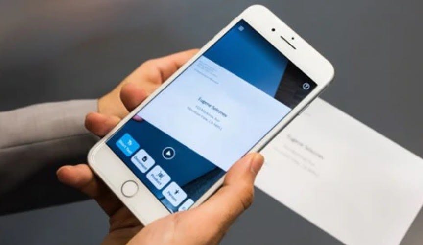
Figure 3: The Seeing AI Microsoft app also uses audio guidance to help users capture full documents.
Various wearable devices such as the OrCam (bottom left) also improve low vision patients’ quality of life by turning their world into an auditory experience. Other new portable technologies such as IrisVision (bottom right) incorporate the patient’s smartphone to enhance distance or near related tasks by enabling various magnification changes, wild field filters, and contrast settings.

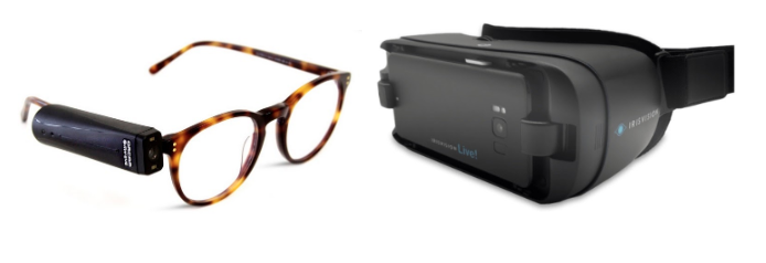
Figure 4: The OrCam and IrisVision low vision devices
5) Google’s “DeepMind” AI platform
Any discussion of AI and deep learning would be incomplete without the mention of Google’s artificial intelligence unit DeepMind. This AI technology is currently being developed to review OCT scans and has already proved a high diagnostic accuracy of 50 different ocular diseases.
The DeepMind platform was fed thousands of OCT scans of various retinal pathologies in it’s initial deep- learning process. A preclinical trial showed the algorithm was able to correctly identify specific ocular disease in over 90% of cases. By incorporating this type of technology into non-ophthalmic settings, earlier detection can occur in patients at risk for sight-threatening ocular disease.
Conclusion
Today, the eye care field is ripe with potential for the implementation of new technologies and Artificial Intelligence. In addition to these AI applications, further investigation for many other ocular conditions is also being done—including those for AMD, retinopathy of prematurity, and cataracts.
Through the high-volume screening of retinal photos and OCT scans, earlier detection and treatment will optimize clinical decision making and improve the quality of patient care. It will also reduce the cases of preventable blindness and reduce overall costs to the healthcare system. It is only a matter of time before the incorporation of AI within eyecare is a requirement for advancing the profession. The implementation of AI may soon lead to the prediction of diagnosis and management plans before the patient even sees the doctor.
How do you see AI affecting your future optometric practice?
