


Often, patients come in complaining of redness, grittiness, and a sandy sensation in their eyes. These symptoms encompass many of the defining characteristics of ocular surface disease (OSD).
Ocular surface disease occurs when there is damage to the epithelial lining covering the cornea and conjunctiva. Due to varying severity between patients and their symptoms, OSD goes undiagnosed in millions.
While ocular surface disease encompasses various pathologies, the two most common causes are dry eye syndrome and blepharitis.1
Prevalence of dry eye and blepharitis
Global epidemiology studies have found the prevalence of dry eye syndrome to be about 5 to 50%. However, in the United States, it is estimated to be about 8.1% of the population, with increasing prevalence with age. In those 18 to 34, the prevalence is 2.7% and rises to 18.6% in those over 75.2
Blepharitis, being one of the most common ocular surface diseases, has little available data on its prevalence. A study performed in 2009 selected 120 ophthalmologists and 84 optometrists and looked at their prevalence of blepharitis in practice. They found that 37% to 47% of their patients had blepharitis.3
Importance of accurate diagnostics in OSD
Undiagnosed OSD can have many effects on patients, and the importance of diagnosis cannot be overlooked. It can decrease visual acuity and quality and affect refractive measurements.
Common adverse effects include corneal “dry spots,” corneal abrasions, corneal infections, corneal scarring, corneal thinning, and corneal perforation.1 When diagnosing ocular surface disease, it is essential to identify the underlying causes to establish the best treatment plan.4
Specifically, in corneal and refractive surgery, it is essential to diagnose ocular surface disease prior to surgery accurately, as ocular surgery can exacerbate or induce OSD. This can result in worsening symptoms following surgery, worsening vision, and overall dissatisfaction after the procedure.5
With increased importance on diagnosis, there have been many new technologies developed to make the identification of ocular surface disease simpler. Below is a comprehensive review of various devices that diagnose OSD and identify underlying causes.
Ocular surface point-of-care technologies for 2023
There are several innovations in point-of-care (POC) technologies that have emerged over the past year.
ScoutPro (Trukera Medical)
This device is an innovation from Trukera Medical, launched in 2022. The ScoutPro is the first handheld compact device, making it easier for physicians to obtain osmolarity readings.
It allows for quick and efficient nanoliter volume sample collection with analysis on the go. One of the significant advantages of the ScoutPro is the increased efficiency level within a practice due to its ability to be kept in a tech's pocket.
ScoutPro collects 50 nanoliters of tear fluid directly from both eyelid margins. It runs through a temperature-corrected impedance measurement to indirectly assess osmolarity.
Additionally, it utilizes a lot-specific calibration curve, and osmolarity is calculated and displayed on the screen.6 This allows for quick readings without moving patients back and forth through the office.
Figure 1 is an image of the ScoutPro device.

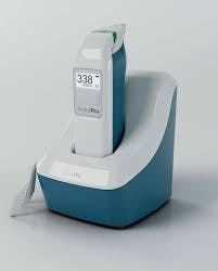
Figure 1: Courtesy of Trukera Medical.
Clinical research on ScoutPro efficacy
In a study conducted by Trukera Medical, 140 patients tested with the ScoutPro were classified as either normal or dry eye patients.7 To be classified as a dry eye subject, they must have had a positive score on the Ocular Surface Disease Index (OSDI) and two or more positive indications of tear film breakup time (TBUT), Schirmer test, corneal staining, conjunctival staining, or meibomian gland dysfunction (MGD).
From this experiment, the ScoutPro found:7
- Specificity: 71%
- Sensitivity: 64%
- Negative predictive value: 48%
- Positive predictive value: 82%
Calibration data was developed utilizing a meta-analysis performed in 2006. They found that hyperosmolarity was defined as 316mOsmol/L. Overall accuracy was determined to be superior to any other single test for dry eye diagnosis, such as Lactoplate, Schirmer test, and rose bengal staining.8
ScoutPro defines its reference values as follows:
- Normal: Mean 309.9mOsm/L ± 11.0 (288 to 331mOsm/L; 90% confidence interval [CI] 288 to 331)
- Dry eye disease: Mean 324.3mOsm/L ± 20.1 (291 to 382mOsm/L; 90% CI 284 to 392).
If there are values below or above the range, it will be displayed as such and should be redone to maintain accuracy.7
T-POC (Lactoferrin, IgE, Verséa Ophthalmics)
The underlying cause of dry eye disease often goes undiagnosed; however, with the T-POC Quantitative Testing Platform from Verséa Ophthalmics, they attempt to uncover the answer.

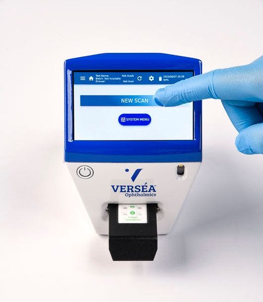
Their system, which looks at causes being allergic related, aqueous deficient, or an evaporative disease, utilizes two molecules while testing: lactoferrin and immunoglobulin E (IgE).
Lactoferrin is a glycoprotein member of the transferrin iron-binding family. It is often found on mucosal surfaces and in biological fluids, such as saliva, milk, and tears. When lactoferrin binds to iron, it prevents pathogens from obtaining sufficient stores, eliminating bacterial growth.
Along with being vital for defense mechanisms, it is a biomarker for the secretory function of the lacrimal gland.9 This system utilizes this function to determine if the underlying cause is aqueous or evaporative. Low levels of lactoferrin are directly correlated with DED caused by aqueous deficiency.
Additionally, it represents a low level of ocular immunity. On the other hand, testing for IgE helps to establish an allergic component. IgE is an antibody that binds to allergens, triggering the degranulation of mast cells, and resulting in an inflammatory response.
The presence of IgE indicates the diagnosis of allergic conjunctivitis, which has very similar symptoms to DED and a contraindication for laser in situ keratomileusis (LASIK) surgery.10
How the T-POC Quantitative Testing Platform operates
This device functions by running samples through a lateral flow reader; it is an immunoassay kit. Lactoferrin and IgE get coupled to gold nanoparticles that are conjugated with rabbit antibody for lactoferrin and mouse monoclonal antibody for IgE.
The membrane of the device contains anti-rabbit antibodies and mouse anti-human IgE antibodies; therefore, when present, they conjugate with the nanoparticles, which then bind to the membrane, producing a color formation that the reader detects and measures. This process takes about 8 minutes, and the values are displayed on the screen.11,12
The chart below, with information courtesy of Verséa, describes different diagnoses based on testing.13 Note: supporting documentation can be found on Verséa Ophthalmics' webpage and by contacting Verséa Ophthalmics, LLC.
| Tear Chemistry | Indicates | Possible Risk | |
|---|---|---|---|
| Dry Eye or Allergy Signs | Normal lactoferrin and IgE | Normal lacrimal function and no ocular allergy present | Evaporative dry eye |
| Normal lactoferrin and high IgE | Normal lacrimal function and ocular allergy present | Ocular allergy and possibly evaporative dry eye | |
| Low lactoferrin | Suppressed lacrimal function and no ocular allergy present | Aqueous deficient dry eye or evaporative dry eye | |
| Low lactoferrin and high IgE | Suppressed lacrimal function | Aqueous deficient dry eye | |
| Tear Chemistry | Indicates | Possible Risk | |
| Contact Lens Applications | Normal lactoferrin and IgE | Normal lacrimal function | Good contact lens candidate |
| Low lactoferrin | Suppressed lacrimal function | Hypoxia and infection | |
| High IgE | Ocular allergy present | Giant papillary conjunctivitis or inflammation | |
| Tear Chemistry | Indicates | Possible Risk | |
| Ophthalmic Surgery | Normal lactoferrin and IgE | Normal lacrimal function | Good LASIK candidate |
| Low lactoferrin | Suppressed lacrimal function | Possible myopic | |
| High lactoferrin | Elevated tear protein | Possible hyperopic | |
| High IgE | Ocular allergy present | Any ophthalmic surgical procedure is risky |
Table 1: Courtesy of Verséa Ophthalmics.
Tearcheck (ESW Vision)
Tearcheck is an innovation that combines different examinations into one system.
Developed by ESW Vision, Tearcheck provides nine POC examinations:
- Non-invasive break time (NIBUT)
- Tear film stability evaluation (TFSE)
- Ocular surface inflammatory risk assessment
- Infrared (IR) meibography
- Demodex evaluation
- Eye redness
- Abortive blinking
- Tear meniscus height
- OSDI questionnaire
This standalone device with an integrated screen allows for quick data collection and analysis. Each examination gives a full picture of the possible underlying causes of dry eye syndrome.
Figure 2 highlights the Tearcheck device.

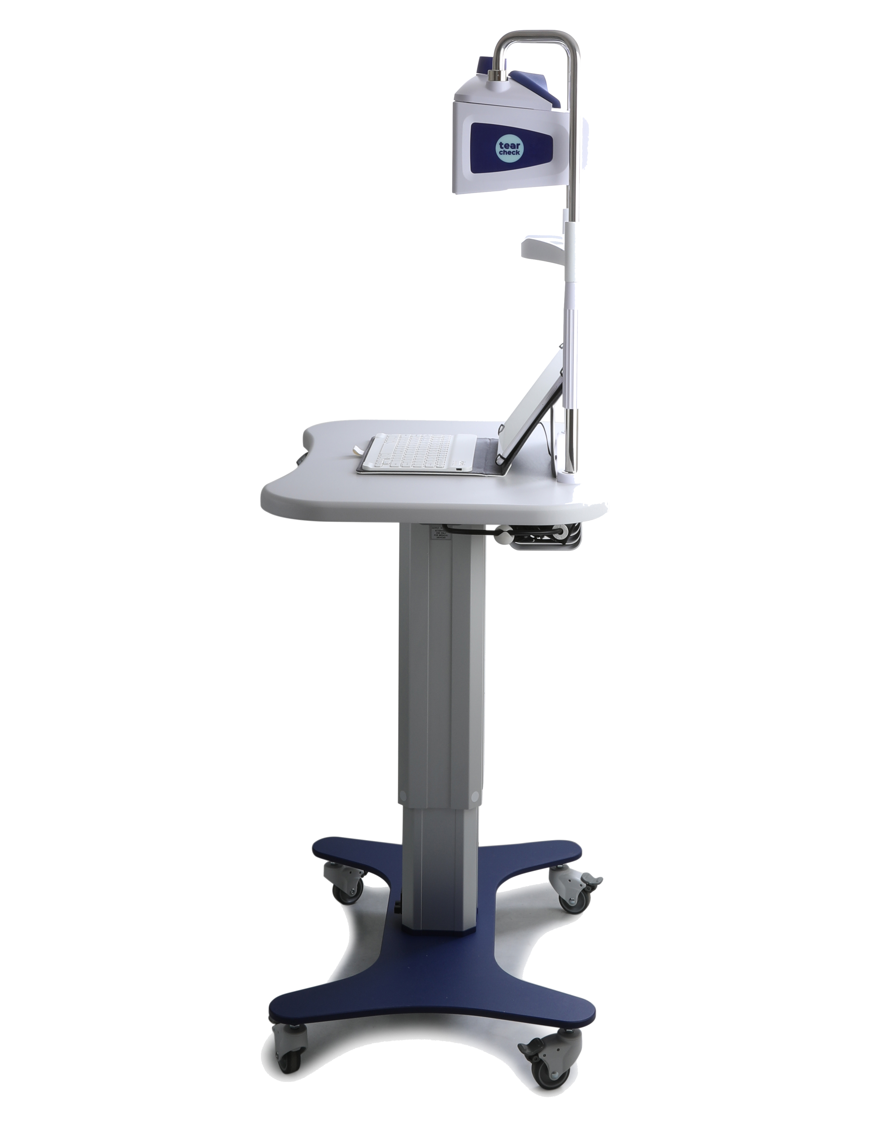
Figure 2: Courtesy of ESW Vision.
How Tearcheck operates
The ocular surface inflammatory risk assessment shows the exact dimensions and dryness score on the integrated screen; TFSE/NIBUT allows for a 10-second evaluation that gives an exact dryness score. NIBUT specifically allows for a view of where and when the surface of the tear film ruptures, while TFSE allows for visualization of micro-deformations on the surface of the tear film.
Additionally, the blink pattern is analyzed for 1 minute, which allows for incomplete blinks to be assessed for underlying causes of dry eye. Hyperemia is also assessed with images of the bulbar and palpebral conjunctiva. This specifically allows for the examination of underlying inflammatory-induced hyperemia.
The Demodex function allows for enlarged images of the base of the eyelashes to allow for visualization of the presence of Demodex mites. Further, meibography is performed by flipping the upper or lower eyelid and capturing an image.
The provider is to determine where the glands are located along with the system. They may add or subtract to the area the computer determines, and a ratio is created. The score is dependent on the provider's selection accuracy.14,15
There have been no significant studies in the literature that support Tearcheck’s use in the diagnosis and management of DED.16
Keratograph 5M (Oculus)
Created by Oculus, Keratograph 5M is a non-invasive diagnostic device that uses imagery technology to evaluate the cornea. It contains a built-in color camera and a real keratometer.
This device captures external images by projecting a ring pattern Placido disc on the tear film surface using an infrared light source. Its functions include tear meniscus height measurement, non-invasive tear film break-up time, examination of the meibomian glands, and evaluation of the lipid layer.
Figure 3 shows the Keratograph 5M device.

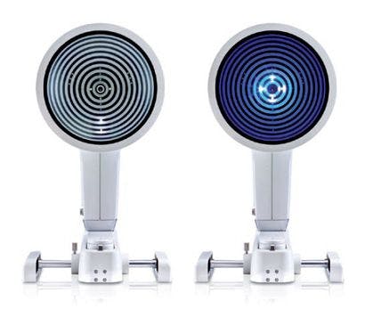
Figure 3: Courtesy of Oculus.
How was the Keratograph machine upgraded?
This version was improved from the Keratograph 4 by updating the joystick, allowing for complete control over video and image capture without the use of a mouse. Other added functions include R-scan classification for redness, lipid layer assessment, and upgraded non-invasive Keratograph tear film break-up time (NIKBUT) that prevents glare using a new infrared illumination system.
This device offers a display that is patient-friendly and enables patients to be involved in their care. They have easy-to-understand diagnostic printouts following imaging and have a database to keep videos for patient records.17
Lipiview II Interferometer (Johnson & Johnson Vision)
Made by Johnson & Johnson Vision, this new design functions to provide dynamic images of the meibomian glands and measures lipid layer thickness with nanometer accuracy. The Lipiview II Interferometer has a color-specific display for easy-to-understand lipid layer thickness results that allow patients to learn about their eye health.
It provides high-definition images and videos of the meibomian glands, as seen in the image below, due to their new surface lighting. Additionally, the lumination source changes intensity based on the difference in thickness across the lids, leading to better-quality images.
Figure 4 demonstrates the Lipiview II Interferometer device.

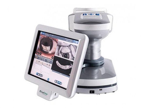
Figure 4: Courtesy of Johnson & Johnson Vision.
Recommendations for using the Lipiview II Interferometer
Their high-definition video allows physicians to evaluate the distribution of ocular lipids across the eye surface during a blink response analysis. The examination takes about 20 seconds to obtain data. This is an upgrade from the previous LipiScan, which could not detect partial blinks or have lipid layer thickness analysis.
The accuracy of the testing may be affected by oil-based drops, and it is asked that patients do not use these drops for more than 12 hours before the examination. Additionally, the use of ophthalmic ointments is not to be used for 24 hours.
Further causes of inaccuracy would be the use of oil face creams around the eyes, eye rubbing, swimming in chlorinated pools less than 12 hours ago, and contact lens use should be discontinued at least four hours prior.18
LacryDiag Ocular Surface Analyzer (Lumibird Medical)
Similar to other devices previously mentioned, the LacryDiag Ocular Surface Analyzer (Lumibird Medical) enables physicians to obtain a full diagnostic workup for dry eye disease and underlying causes. It is a sleek device with easy-to-use programming for simple applications.
Figure 5 highlights the LacryDiag Ocular Surface Analyzer device.

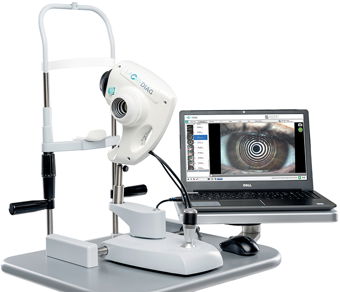
Figure 5: Courtesy of Lumibird Medical.
How the LacryDiag Ocular Surface Analyzer operates
The LacryDiag provides four non-contact exams: NIBUT, interferometry, tear meniscus height, and meibography.
With the use of its infrared LEDs, the LacryDiag Ocular Surface Analyzer can examine:
- Blepharitis and Demodex imaging
- Bulbar redness
- Staining with fluorescein with an assessment of corneal, conjunctival, and lid margins
- Pupillometry
- White-to-white measurement
- Corneal deformation.
These examinations are completed in 4 minutes, making patient results easily accessible and increasing the daily office flow.19 A study by Ward et al. showed a significant difference between observers A and B for meibography.
When compared with the Keratograph 5M, there was a significant difference in intraobserver variability for NIBUT. This study highlighted repeatability and the use of multiple outcome measures are important for the diagnosis of DED.20
Both devices are capable of very similar analysis and have the efficacy to help diagnose underlying causes of dry eye disease.
I-Pen Osmolarity System (I-MED Pharma)
The I-Pen Osmolarity System from I-MED Pharma is a portable electrical tool used to measure osmolarity. This device contacts the orbital tissues, such as the palpebral conjunctiva, measuring the electrical impedance of the tear film on tissues.
The device calculates an osmolarity after about 2 seconds on the eyelid tissue and displays the value in units of mOsms/L on its crystal display screen. The calculation uses an indirect measurement of the impedance.21
Figure 6 shows the I-Pen Osmolarity System device.

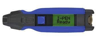
Figure 6: Courtesy of I-MED Pharma.
Clinical research on the I-Pen Osmolarity System
The accuracy and application of the I-Pen were evaluated by Park et al. in 2021. Their study found the device to be considered suitable for use in clinical settings, with good performance in diagnosing DED. They found a sensitivity of 90.9% and specificity of 90.6% for diagnosing DED.22
However, other data has been conflicting compared to other devices on the market, such as the TearLab Osmolarity System. Studies found that the TearLab was more effective in delineating DED patients than the I-Pen Osmolarity system.23
Another study’s findings suggested that utilizing either device solely led to poor reliability for diagnosing dry eye; therefore, additional testing should done for further analysis.24
IDRA (SBM SISTEMI)
IDRA ocular surface analyzer developed by SBM SISTEMI, Inc. Torino, Italy, is a diagnostic tool for DED that utilizes infrared and ultraviolet light in diagnosis. The device is mounted between the slit lamp and a biomicroscope, allowing for five non-invasive examinations.
Similar to other devices such as Tearcheck, examinations include NIBUT, lipid layer thickness, tear meniscus height, meibography, and eye blink. Additionally, IDRA provides pupillometry, white-to-white measurement, ocular redness classification, and a generic examination.
One of the key features is its ability to analyze the lipid, aqueous, and mucin layers within the tear film, which allows for a better understanding of dry eye disease causes.25
A study performed in 2023 to evaluate the accuracy of IDRA for diagnosis of DED found IDRA to have a positive concordance with routine clinical diagnostic tests and was an effective tool in diagnosing dry eye.26
Occyo Cornea Dome Lens Imaging System (Occyo, Innsbruck, Austria)
The Cornea Dome Lens Imaging System, designed by Occyo, Innsbruck, Austria, offers physicians innovative imaging unlike other devices on the market. The device contains a novel imaging lens that conforms to the curvature of the patient's eye, enabling high-resolution images of the ocular surface.
Figure 7 features the Occyo Cornea Dome Lens Imaging System device.

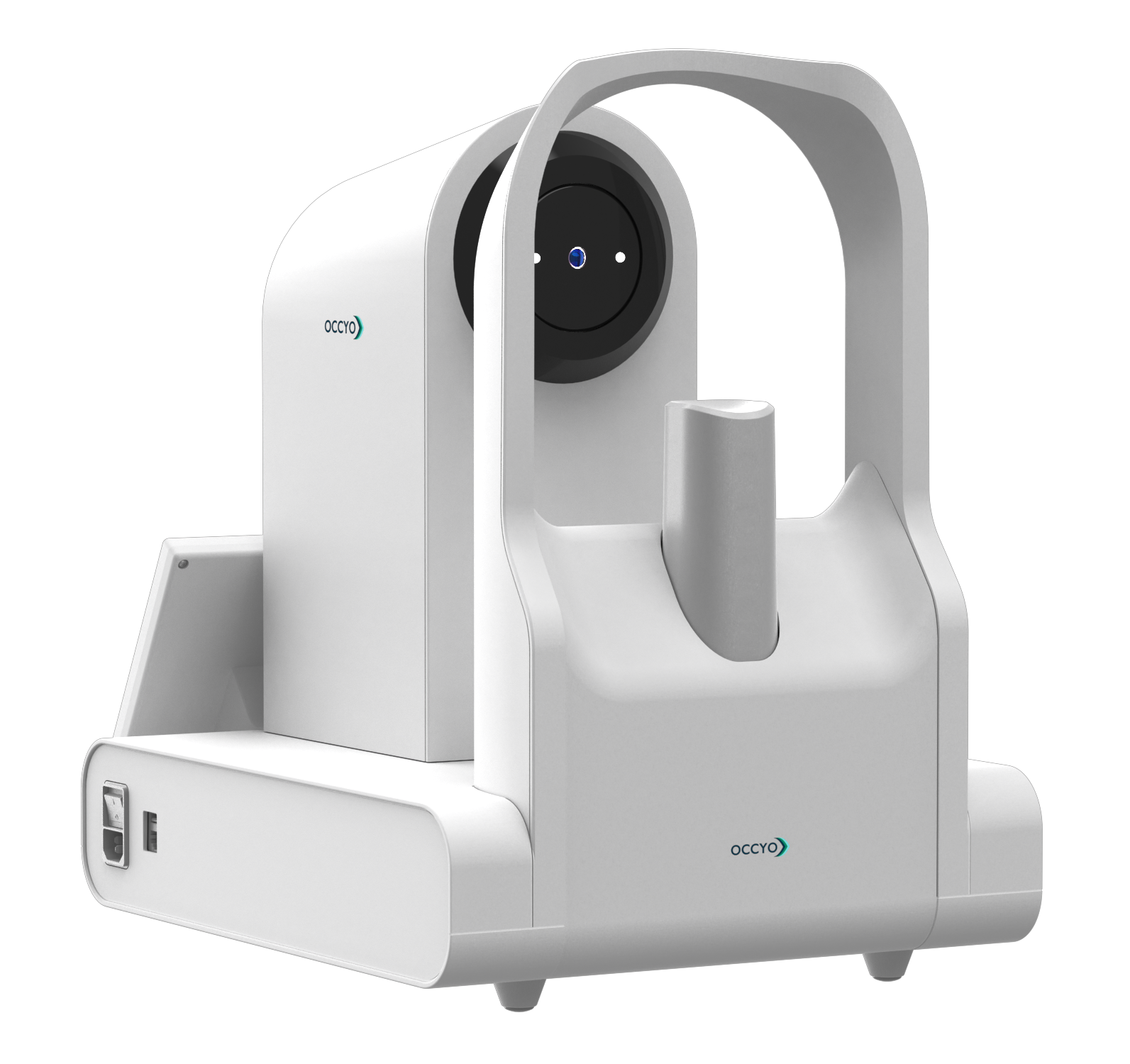
Figure 7: Courtesy of Occyo, Innsbruck, Austria.
The imaging system can fixate on a target, therefore accurately resulting in a centralized view into the lens. This results in decreased eye movement effects. The main goal of this device is to create high-definition images that are standardized and replicable without human intervention.16
In a study performed in 2021, they found the repeatability and systematic use of the Cornea Dome Lens System provided an excellent baseline for eye redness.27 However, there is little evidence for its use in the diagnosis of dry eye disease.
Cobra HD Retinal Camera (ViewLight)
The Cobra HD Retinal Camera (ViewLight) is a non-mydriatic digital fundus device that provides rapid screening for retina status. For dry eye disease diagnosis, the device contains a module for meibography.
Figure 8 shows the Cobra HD Retinal Camera device.

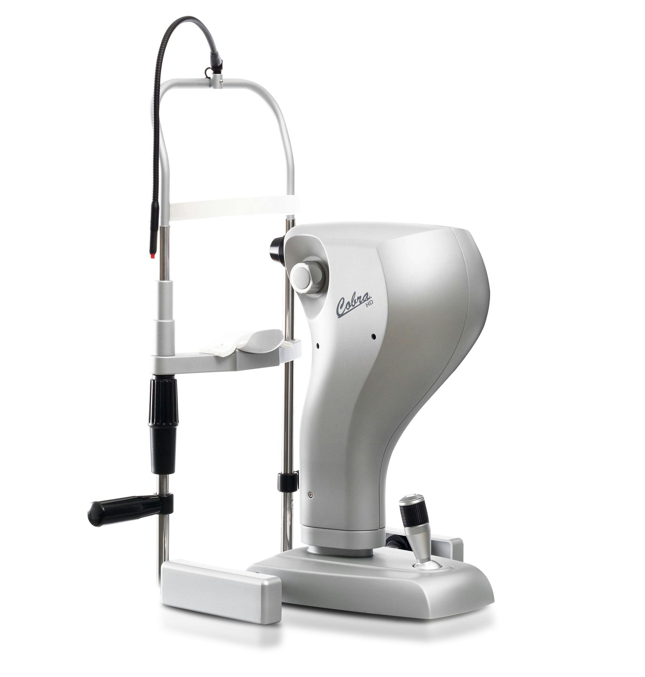
Figure 8: Courtesy of ViewLight.
Clinical research on the Cobra HD Retinal Camera
A study in 2021 comparing the Cobra HD camera and the Antares showed there was a significant difference in meibomian gland (MG) dropout in those with dry eye vs. non-dry eye with the use of either device, highlighting the accuracy of the Cobra and the interchangeability of the two devices.28
Additionally, a study looking at MG loss and the association between age, sex, and dry eye using the Cobra HD device showed substantial standard deviations in the mean MG loss between participants with and without dry eye symptoms (30 ± 17% and 45 ± 18%, respectively).29
Utility and implications of artificial intelligence in diagnostic POC
Artificial intelligence (AI) systems have begun to pop up in all areas of life. The use of AI systems in medicine is slowly being explored, and the world of ophthalmology is no exception. Within the umbrella of AI comes what is known as machine learning. This is a process in which a class of algorithms learn from data rather than being programmed with explicit rules.
Deep learning utilizing deep artificial neural networks to promote image and text recognition is a further subdivision of machine learning. Deep learning, in particular, has been optimized within the field of ophthalmology for the analysis of images, such as data from retina segments.
Of note, deep learning algorithms and over-the-counter (OTC) have already been shown to allow for the reliable detection of retinal diseases.30,31
Clinical studies on AI in DED diagnosis
With DED being one of the most underdiagnosed conditions, many have begun to explore the use of AI for image analysis when diagnosing dry eye disease. Ciężar et al. utilized a 2D Fourier transformation analysis to analyze MGD in patients presenting with dry eye disease symptoms. They showed the algorithm could correctly classify patients into healthy, intermediate, and unhealthy for gland dysfunction.32
Another study performed in 2023 utilized AI to estimate the tear film break-up time for the diagnosis of DED.33 They compared their AI results with the Asia Dry Eye Society (ADES) DED diagnostic criteria. Their results showed the accuracy of tear film breakup time estimation was 0.789 (95% CI 0.769 to 0.809), and the area under the receiver operating characteristic curve of this AI model was 0.877 (95% CI 0.861 to 0.893).33
The sensitivity and specificity of this AI model for the diagnosis of DED were 0.778 (95% CI 0.572 to 0.912) and 0.857 (95% CI 0.564 to 0.866). Therefore, they successfully developed an AI model to diagnose DED.33
Future protocols for AI in DED diagnosis
These examples provide a positive use moving forward; however, there are still considerations that need to be worked out before the full implementation of such systems.
Common guidelines should be developed for model evaluations using AI algorithms. Additionally, code and data sharing need to be implemented to allow for reproducibility of results from these systems.
Key takeaways
Dry eye disease affects a majority of individuals in the United States and throughout the world. The utilization of technological advances to help diagnose and uncover the underlying causes of DED is so important.
With the technologies mentioned above, ophthalmologists can continue to help understand their patient's symptoms and provide the best treatment possible. The growth of technology will continue through the years, and the increased awareness of artificial intelligence will continue to play a part in the medical community.
It is important to understand the technologies available and continue utilizing what works best for your practice and patients.


