


After multiple years of training in medical school and ophthalmology residency, pursuing fellowship is one of the last steps before entering clinical practice. When ultimately deciding which subspecialty fellowship to pursue, ensure that the area you choose is one that intellectually stimulates you, offers the medical and surgical treatment opportunities you can foresee yourself providing to patients, and has the opportunity for future research and innovation.
During ophthalmology residency, residents should strive to experience every subspecialty they can potentially pursue. By immersing yourself in every subspecialty rotation, you will gain insights into the patient population, pathology, and lifestyle associated with various subspecialties. Ultimately if you decide that cornea and external disease is that subspeciality for you, then this article will outline the ways in which to apply for, prepare for, and ultimately succeed in cornea fellowship!
Cornea fellowship facts and FAQs
Is cornea fellowship competitive?
Cornea fellowships are competitive, but as with any fellowship it varies from program to program. In 2020, according to the SF Match statistics, there were 101 positions offered in cornea and 89 positions filled. Vacancies have slowly decreased over the past three years.
How long is cornea fellowship?
The standard cornea fellowship is one year long, but some programs offer an additional year for research.
How do I apply to cornea fellowship?
Applying for cornea fellowship is done through the SF Match Central Application Service (CAS) and by contacting programs directly to learn about additional materials and requirements.
How to prepare for cornea fellowship during ophthalmology residency
There are various opportunities that you can embrace during residency to learn and do as much you can to prepare for cornea fellowship.
Spend time in cornea clinics
Residents should aim to spend dedicated time in clinics with their cornea attendings. Shadow a variety of attendings to gain perspectives in various pathologies, patient personalities, and treatment approaches. For instance, spend one day with a cornea specialist whose clinic is focused on cataract and refractive surgery, another day with an attending who focuses on ocular surface reconstruction and another day with an attending who specializes in endothelial dysfunction and keratoplasty.
It is important to experience the breadth of cornea and external disease ,and realize that each cornea specialist’s practice can have a very different focus and tempo. Some residents may not have cornea departments with such diverse faculty. In those scenarios, see if externships are possible through your program to other institutions (either nationally or internationally) that may have experts in areas that you have not seen through your own department.
Spend time in cornea operating rooms

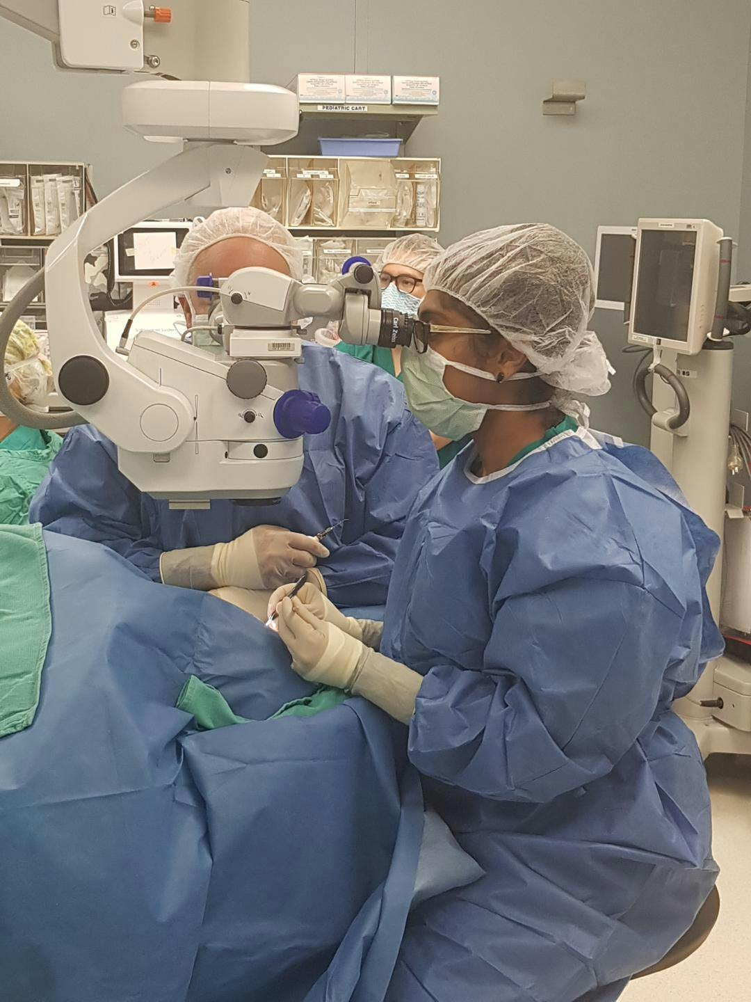
Dr. Venkateswaran performing in the operating room as a cornea fellow.
Time in the operating room is invaluable. One of the thrills and draws of cornea and external disease as a subspeciality is the intricacy and precision of corneal and anterior segment surgery.
Watch your attendings suture a penetrating keratoplasty (PKP) or deep anterior lamellar keratoplasty (DALK) or perform a Descemet’s stripping automated endothelial keratoplasty (DSAEK) or Descemet’s membrane endothelial keratoplasty (DMEK). Observe your attendings carefully insert and rotate a toric intraocular lens (IOL) into the correct position, perform laser-in situ keratomileusis (LASIK), photorefractive keratectomy (PRK) or small incision lenticular extraction (SMILE), or excise an ocular surface tumor or pterygium.
If you dedicate time throughout your residency to spend with these attendings in the OR, they will perhaps allow you to partake in steps of the surgery that they deem appropriate for your skill level. The opportunity to throw in a few sutures, perform irrigation and aspiration at the end of a cataract surgery, or even position an endothelial graft can be incredibly rewarding and educational as a resident.
Practice surgical techniques in the wet-lab
As a resident, it is crucial to practice surgical skills in the wet-lab setting. Your first time performing a procedure should not be on a real patient. Leverage the support of senior residents, fellows or even attendings to spend time with you in the wet-lab to practice skills such as corneal suturing, wound creation, or phacoemulsification.
Attend multi-day educational courses at various academic institutions (ie the Harvard Cataract Course) or industry-sponsored cataract surgery courses (i.e., Alcon, Johnson & Johnson, or Bausch and Lomb 2nd and 3rd year cataract surgery courses) where various specialists lecture on select topics and precept you in organized wet-lab sessions.
Courses are also available at the resident level at national meetings such as the annual American Academy of Ophthalmology (AAO) or American Society of Cataract and Refractive Society (ASCRS) meetings. Hard work in the wet-lab will result in more opportunities in the OR and attendings will take note of your practice and developing skills.
Conduct research
Conducting research as a resident can serve many purposes. First off, performing in-depth research in any area will expand your knowledge base exponentially. Choose an attending or mentor who focuses on an area of cornea or external disease that you enjoy and dedicate several months to a research project. Time spent in the lab or conducting clinical research can help you develop a strong relationship with an attending as well as author manuscripts and present your work at national meetings.
Such opportunities will help you stand-out as a cornea fellowship applicant and also provide you with topics to discuss with attendings during fellowship interviews or networking sessions. Start off with small projects such as case reports and expand to larger retrospective, prospective or bench research studies as your familiarity and confidence in performing research grows.
Cornea fellowship: what to expect
AUPO Certification
When applying for a one-year cornea and external disease fellowship, it is helpful to obtain a comprehensive list of accredited fellowship programs on The Association of University Professors of Ophthalmology (AUPO) website. Cornea fellowships are not governed or regulated by the Accreditation Council for Graduate Medical Education (ACGME). As such, compliance with the AUPO is a voluntary process initiated by fellowship programs and aims to create a uniform educational standard across all fellowship programs.
Metrics are set to ensure that trainees obtain adequate clinical, surgical and academic experiences at the conclusion of their fellowships and fellows complete an exit survey at the end of the year to provide feedback about their fellowship experiences. Fellowships can be applied for through the San Francisco Match System.
There are also several fellowships that are in private practice settings or academic settings that are not AUPO certified but excellent as well. In general, AUPO-certified fellowships will adhere to guidelines and be required to meet all predefined criteria while non-AUPO certified fellowships may deviate from these guidelines.
Surgical Education
When crafting your surgical experience during cornea fellowship, it is important to ensure that you are receiving training in a broad range of corneal and anterior segment pathology and surgery. Here are some major surgical categories you should aim to learn and hopefully feel confident in after a year of being a corneal fellow. Ask fellows who recently completed fellowship how much exposure they had in each of these categories to get a sense of general surgical numbers.
Refractive cataract surgery
Cataract surgery is the bread and butter of anterior segment surgery. As part of your cornea fellowship, it is important to continue to refine and hone your cataract surgery skills. Although cataract surgery will not be a focus of many cornea fellowships, you should ensure you are able to perform several cataract surgeries over the course of your fellowship year.
Exposure to femtosecond-laser-assisted cataract surgery, use of intraoperative aberrometry as well as implantation of premium IOL technologies (i.e., toric, trifocal, extended-depth of focus, or light-adjustable IOLs) will expand the armamentarium of tools you can offer for your patients at the end of fellowship. Ask your attendings to work on diversifying nuclear disassembly techniques (i.e., incorporating horizontal and vertical chop techniques, using the MiLoop (Zeiss) as well as performing manual small incision cataract surgery).

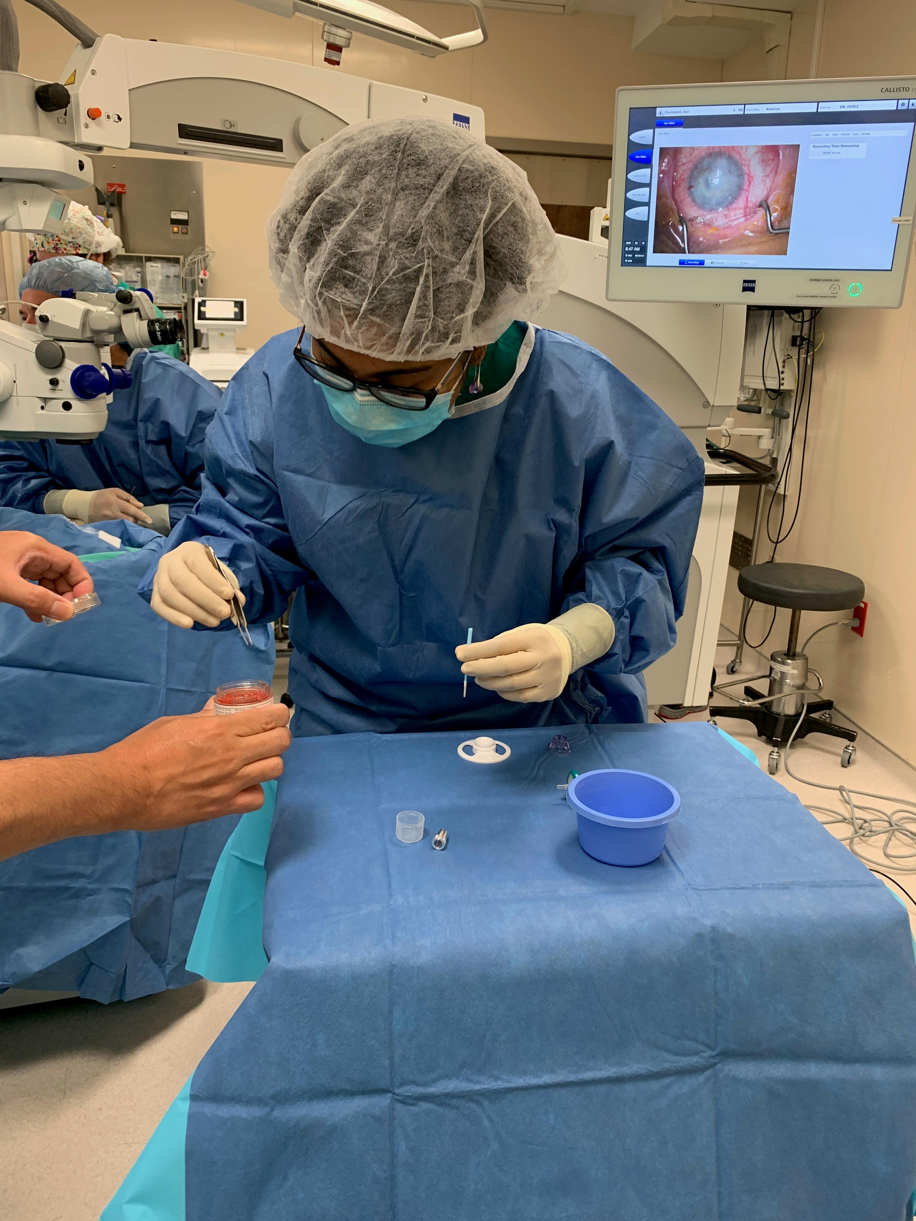
Dr. Venkateswaran performing femtosecond laser-assisted cataract surgery as a cornea fellow.
Complex cataract surgery
In addition to refractive cataract surgery, gaining exposure to complex cataract surgery cases is critical. As an anterior segment surgeon, you will often receive referrals for surgeries that are not run-of-the-mill. That means that you should become proficient with performing cataract surgery with hypermature lenses, underlying zonulopathy (i.e., trauma, pseudoexfoliation, or colobomatous eyes), intraoperative floppy iris syndrome, or corneal pathology (i.e., prior corneal scars, corneal neovascularization, ectatic corneas or even post-keratoplasty corneas) at the conclusion of your fellowship year.
Discuss intra-operative planning with your attendings, know what special instrumentation to use for these cases, when to use a pupillary expansion device or capsular tension ring or segment as well as how to decide what IOL to implant.
Keratoplasty
Keratoplasty should form a strong basis for your cornea fellowship year. As a fellow, you should be performing several PKP, DALK, DSAEK, DMEK as well as keratoprosthesis cases as the primary surgeon. Time should be also spent at your local eye bank understanding the complex processes of donor tissue harvesting and preparation. You should also practice preparing your own donor tissue (i.e. for DSAEK and DMEK cases) even though most surgeons today are using pre-stripped, pre-stained and pre-loaded tissue. Use resources through your local eye bank to practice corneal suturing and keratoplasty techniques prior to your first live cases.
As a fellow, you should also feel extremely comfortable identifying optimal surgical candidates in clinic for each of the keratoplasty procedures as well as knowing how to manage various post-operative complications (i.e. wound leaks, interface or suture-related keratitis, graft rejection, graft failure etc.).
Collagen cross-linking and intrastromal ring segments insertion
Keratoconus is a prevalent condition in cornea practice. Learning about the natural course of keratoconus, progression of disease and identifying patients for cross-linking is a critical skill to learn along with knowing how to actually perform the cross-linking procedure. Growing comfortable with safely removing the epithelium as well as administering riboflavin drops and ultraviolet light therapy during crosslinking procedures should be emphasized in fellowship.
Observing patients post-operatively, monitoring for corneal stability and/or progression after cross-linking as well as seeing rare complications such as corneal haze, refractive shifts or infectious keratitis are important learning points for cornea fellows.
Intrastromal ring segment insertion for corneal flattening, improved contact lens tolerance and reduction of myopia is another useful skill to learn when managing keratoconic patients and exposure to the planning process and insertion of these segments will benefit all cornea fellows
Anterior segment reconstruction – iris repair, secondary intraocular lenses
Oftentimes, cornea surgeons will be asked to perform iris repair techniques as well as secondary IOL insertion given the inherent complexity of these surgical cases. Time should be spent practicing iris suturing techniques (i.e., the McCannel and Seipser knot techniques) as well as performing iridodialysis repairs. Fellows should also gain exposure to various methods for secondary IOL fixation, including scleral-fixated IOL techniques (i.e., Yamane technique, glued IOL technique as well as two- and four-point goretex or prolene suture IOL fixation techniques), and iris-sutured IOLs.
While the frequency of these cases can be variable amongst fellowships, performing key steps of these procedures when they do arise can make a graduating fellow feel prepared to tackle these cases alone in practice. Similarly, wet-lab practice during fellowship using eye models is crucial to master challenging intra-operative maneuvers that are often needed to perform these surgeries successfully.
Ocular surface reconstruction/ocular surface lesions
Ocular surface reconstruction can be a large area to master during fellowship but exposure to various surgical procedures should be achieved during cornea fellowship. Limbal stem cell transplantation techniques require specialized skill sets. Learning to perform simple limbal epithelial transplantation (SLET), conjunctival-limbal autografts as well as keratolimbal autografts can prove beneficial when encountering patients in practice with significant limbal stem cell deficiency. Similarly, understanding how to utilize fresh frozen or cryopreserved amniotic membrane for ocular surface reconstruction both in the clinic and OR settings is requisite for corneal surgeons who will undoubtedly encounter various ocular surface disorders in practice.
Along with ocular surface reconstruction, excision of ocular surface lesions should be taught in corneal fellowship. Lesion removal can include removal of irregular epithelium for corneal dystrophies with concurrent diamond burr procedures or phototherapeutic keratectomy (PTK) and Salzmann’s nodules as well as pterygia and/or ocular surface tumors.
Refractive surgery
Exposure to refractive surgery is also a critical aspect of cornea fellowship. Evaluating patients preoperatively and identifying appropriate surgical candidates is the hardest aspect of refractive surgery. In addition, intra-operative planning of surgical treatments requires repeated practice; verbalizing your thought processes with your attendings will help you identify nuances in surgical planning that they employ. Having the opportunity to perform LASIK, PRK, SMILE, refractive lens exchange, or implantable collamer lens (ICL) cases will be challenging in fellowship as these patients are paying cash to have refractive surgery be performed by the refractive surgeon they have chosen.
However, several fellowship programs will offer patient discounts for those patients who elect to have their surgery performed by a fellow; recruiting these patients in attending clinics can help fellows increase their refractive surgery numbers. While cornea fellowship programs emphasis does not lie in refractive surgery, unlike refractive surgery/anterior segment fellowship programs, dedicated time should certainly be given to learning and refining refractive surgery techniques.
Tips and tricks for succeeding in cornea fellowship
Cornea fellowship is a marathon, not a race. The fellowship is 12 months of dedication, perspiration, diligence and passion for the subspeciality that you love.
Set goals
Setting goals throughout the year will help you remain on track to gain comprehensive exposure and refine your skills. If there are areas that you feel deficient in, voice concerns with your program director, organize more didactic sessions, and aim to spend more time with attendings who specialize in that area.
Learn to actively learn during clinics
Patients you encounter in cornea clinics will often have protracted clinical courses and multiple co-morbid pathologies. With time, breaking down their complex clinical history will become easier as well as identifying acute versus chronic issues that need to be addressed at a clinic visit. At the end of a clinic day, see if the plans that you devised for various patients matched up with what your attendings ultimately decided; ask your attendings why their plans differed to understand their thought processes and identify errors in your analyses.
Always aim to see pre-operative surgical evaluations and spend time listening to how your attendings counsel these patients on their surgical options as well as discuss the risks, benefits and alternatives of a given procedure. Observe a host of attendings having these important conversations and take away aspects of their delivery that you like and would want to incorporate into your own conversations with patients. Learn how they navigate challenging and/or demanding patients and discuss unexpected complications.
Anticipate your attendings’ needs
In the clinics as well as operating rooms, learn to anticipate the needs of your attendings and always be thinking one step ahead. Understand how each attendings' personality and style differs. Ensure correct instrumentation has been made available for surgical cases if you notice that your attendings often ask for them and ensure all IOLs and supplies are ordered for cases in order to avoid delays and frustrations. If you can help an OR day flow smoothly, you will naturally get to do more.
Practice surgical skills and keep a log of cases
Practice surgical skills and show that you have prepared when operating; attendings will appreciate your effort and reward you with more surgical opportunities. Solicit feedback from your attendings to ensure you are improving your weaknesses. Keep an ongoing surgical log of all your cases to ensure you are meeting (and hopefully exceeding) all the requirements for surgical education.
Engage in research
Engage in clinical and/or basic research to augment your knowledge base on areas of corneal and external disease that you enjoy in order to start to become an expert in that area.
Lean on mentors
Lastly, lean on your fellowship mentors for job advice. Attend national meetings to network for job opportunities and ask your mentors to make meaningful introductions. Cornea is a small world and who you know matters.
Preparing for your future in cornea
As you go through your cornea fellowship, begin to envision the type of career you see for yourself. Do you see yourself pursuing a career in private practice or academics? What types of patients do you want to see? Do you want to see a variety of pathologies or narrow down your area of focus? Will you be able to conduct research and if so in what area(s)?
How do you decide?
The answers to these questions require introspection. Pick the brains of your mentors who have traversed these decision trees before. Ask them for advice on how they reached the points in their career they are at today. Figure out what steps they recommend you emulate and what mistakes they recommend you avoid.
The more patients you can see and care for in fellowship, the more prepared you will be to encounter anything that comes your way in practice. However, learning and training doesn’t end in fellowship. Your first several years of practice will be the most eye-opening and intellectually challenging years that you have encountered. Turning to your colleagues for advice and assistance on tough cases will help you morph into a better clinician and surgeon.
Your career path can change at many points, and it is important to be open to the many possibilities of pursuing a career in cornea!


