


The incidence of cataracts among patients is increasing as we continue to live with an aging population. Currently, over 24 million people in the United States have cataracts and, by 2050, expected projections estimate that there will be over 50 million people with cataracts.1 Optometrists play an important pre-operative role for cataract surgery patients since cataracts are often first diagnosed during routine primary care eye exams.
An optometrist’s role is to identify patients that are in need of cataract surgery, to educate patients about their condition, to guide them into making informed treatment decisions, and to facilitate access to surgical treatment. In order to provide patients with appropriate care, it is important for optometrists to establish relationships with cataract surgeons and to make appropriate referrals to them.
Identifying patients who need surgery
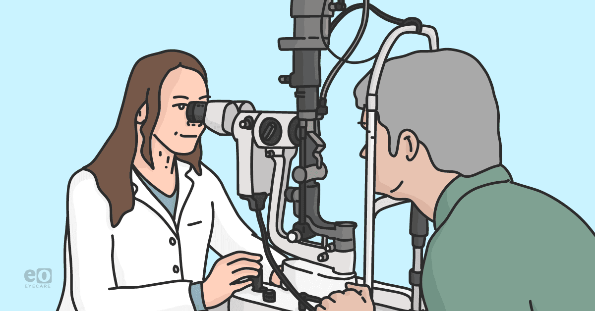
Both objective measurements and subjective concerns should be taken into consideration when determining if a patient is ready for surgery. A careful medical history should be taken from a patient to assess their lifestyle and visual symptoms, and a dilated exam should be performed to view lenticular changes and to rule out any secondary causes of vision loss.
Patients who have significant health problems and comorbidities may need medical clearance prior to surgery. Cataract surgery should be considered for patients who are no longer able to perform their daily lifestyle tasks such as reading, driving and seeing at night. When a patient’s vision has degraded to 20/30 or worse and their activities of daily living (ADLs) are affected, cataract surgery should be considered. Once it has been determined that a patient has visual impairment from cataracts, insurance companies will consider cataract removal to be a medically necessary, though an elective procedure.
Patients who desire surgery but do not meet the insurance requirements may be considered for a refractive lens exchange. Ultimately, it will be the patient’s decision whether or not to proceed with surgery and optometrists can provide them with the education they need when weighing their treatment options.
Cataract risk factors
Although there are multiple etiologies for the formation of cataracts, the most common factor is aging. Other factors that can cause cataracts to progress are smoking, an abundance of sunlight with limited UV protection, steroid medication usage, previous ocular surgeries, previous ocular trauma, radiation, and diabetes.
Overall ocular health assessment
A complete anterior and dilated posterior segment exam should be performed to determine cataract etiology and to identify any additional present pathology. Some exam considerations for these patients are:
- A patient’s best corrected visual acuity (BCVA) should be measured in both dim lighting and under glare conditions.
- Externals and lids should be evaluated for any ocular surface disease or infection that needs to be treated and under control prior to surgery.
- IOP should be measured. It is imperative to identify any glaucoma patients prior to cataract surgery since certain MIG procedures are only allowed to be done if they are performed at the same time as cataract surgery.
- Pachymetry will need to be performed to identify patients with thick corneas that may be at risk for post-operative corneal edema.
- Specular microscopy should also be considered in patients who have Fuchs’ dystrophy or a suspected low endothelial count. The cornea should be evaluated for any opacities and dry eye disease.
- Patients with traumatic cataracts should be carefully examined with gonioscopy for any signs of angle recession or secondary glaucoma.
- Additionally, patients who have narrow angles should be identified since some angle opening may occur from having cataract surgery.
- The lens should be viewed under dilated conditions to determine cataract classification using grading systems.
- A complete fundus exam needs to be performed to rule out any retinal and macular disease. Ancillary testing can be used as well.
- Macular OCT may be needed to to identify any underlying macular disease and topography can be used to identify any cornea irregularities.
- Pterygiums and Saltzman nodules can induce astigmatism and corneal irregularities and may need to be surgically removed prior to taking IOL measurements.
- A B-scan is indicated if there is no posterior view to rule out retinal detachments, vitreous hemorrhages or other vision limiting pathologies.3
Possible cataract surgery complications
Cataract surgery is a very common and low risk procedure. Worldwide, over 10 million cataract surgeries are performed each year but as with all surgeries, complications can still occur.2 Patients should be aware of the risks involved with having surgery.
Potential complications
- Retinal detachment
- Endophthalmitis
- Elevated IOP
- Glaucoma
- Iris prolapse
- TASS
- Macular edema
- Rebound iritis
- Inflammation
- Photophobia
- Corneal edema
- Dysphotopsia
- Ptosis
- Hyphema
- Eye pain
- Capsule tear
Surgical complication risk factors
It is crucial for optometrists to identify patients that have any preexisting conditions that can increase risks for surgical complications and contribute to decreased visual outcomes. Patients should be carefully evaluated for and educated on the following conditions before being referred for surgery.
| Condition | Risk | Recommendation |
|---|---|---|
| High myopia | Retinal detachment | Consider obtaining clearance from a retina surgeon prior to cataract surgery |
| History of LASIK/PRK/RK | Post-operative refractive surprise | Educate that a refractive enhancement may be needed after cataract surgery |
| Fuchs’ dystrophy | Corneal edema | Surgeon may consider low-energy phaco during surgery |
| Blepharitis/Anterior infection | Endophthalmitis | Treat any external infection prior to surgery |
| DES | Fluctuating visual outcomes | Optimize ocular surface prior to taking IOL measurements |
| Wet AMD | Macular edema | Surgeon may consider an Anti-VEGF injection prior to surgery |
| Tamsulosin usage/small pupils | Intraoperative floppy iris syndrome | Surgeon may use a Malyugin ring during surgery |
| Pseudoexfoliation | Capsule tear, lens dislocation | Surgeon may use trypan blue, iris expanders, or other instruments during surgery |
IOL discussion and patient candidacy
Depending on their candidacy, patients may have multiple IOL options to choose from and optometrists can help guide them into making appropriate decisions based on their visual desires, needs, and goals.
Patients will still need glasses after standard cataract surgery to have their best possible vision, but there are premium lens options available that may help to decrease a patient’s dependency on glasses. Having this discussion can help prepare a patient prior to their visit with the surgeon and set their expectations for their visual prognosis. Lens options that patients may elect to have are:
Monofocal IOLs: These correct a patient’s vision at either distance or near. This is the most commonly elected lens option and is the only lens option that is covered by insurance. Patients will often still need glasses to have their best vision after cataract surgery.
Toric IOLs: These lenses decrease the amount of astigmatism a patient may have following surgery. Consider these for any patient who has over 0.75D of astigmatism. Patients who have Against The Rule (ATR) astigmatism will be more symptomatic for lower amounts of astigmatism than patients who have With The Rule (WTR) astigmatism. Alternatively, Limbal Relaxing Incisions (LRIs) may be used to correct small amounts of astigmatism.
Monovision targets: This option can be used to correct distance vision in a patient's dominant eye and near vision in a patient's non dominant eye using monofocal or toric IOLs. This is recommended only for patients who have trialed and had previous success in monovision contact lenses.
Multifocal and Extended Depth of Focus IOLs: These lenses correct a patient's vision across multiple distances and may help to eliminate the need for glasses in most, but not all, visual situations. Glasses may still be needed for certain visual tasks such as reading small print or seeing in dim lighting. These lenses may cause glare and halos at nighttime. These are not recommended for patients who have any retinal or macular disease. They can cost patients several thousand dollars per eye.
Managing patient expectations
Having frank discussions about expected and realistic visual outcomes prior to surgery is crucial in setting up patients for success. Patients can expect to have visual improvement as soon as one day after cataract surgery provided that no additional complications have occurred. Any patients with pre-existing visual limiting conditions will continue to have decreased vision after cataract surgery.
In preparation of obtaining the best post operative results, it is important to treat any conditions that can reduce visual outcomes or contribute to patient discomfort prior to surgery. Patients should be aware that neural adaptation to their new intraocular lenses can often take weeks to months.
Timeline of recovery
- Patients will need to return for post-operative evaluations one day, one week, and one month after surgery for each eye. Restrictions will be in place following surgery that include refraining from physical activity, heavy lifting, bending over, wearing makeup, swimming, and using hot tubs. In order to protect the wound incision sites, patients will need to sleep with a hard shell eye patch for one week after surgery.
- Patients will be prescribed medicated eye drops to help prevent infection and to reduce inflammation after surgery. They will need to use an antibiotic, an NSAID, and a steroid that will be tapered over several weeks.
- Patients who had an antibiotic steroid injection at the time of surgery may not need any additional drops post-operatively.
- Patients with any surgical complications such as CME, high IOP, and rebound iritis may need an additional or extended drop schedule.
- Patients will need to wait 4-6 weeks for their eyes to heal and for their refraction to be stable before glasses can be prescribed.
Establishing relationships with ophthalmologists
It is important to have habitual communication between the optometrist and the cataract surgeon that is ongoing throughout the patient's pre and post-op period. Co-managing optometrists should send a detailed referral letter prior to surgery and exam notes after any co-managed post-operative visit.
Optometrists should be familiar with the surgeon to whom they are referring, as well as their previous surgical results. It may be beneficial to shadow the ophthalmologist in surgery to become more familiar with their practices and procedures. When making referrals, it is helpful to be aware of the lens options that a surgeon offers, if the surgeon performs MIGs or other concomitant surgeries alongside cataract surgery, and if the surgeon offers dropless surgery as an option to patients.
What to include in a referral letter
A referral letter to a cataract surgeon should include the patient's BCVA, anterior, and posterior segment findings, any lens recommendations that may have been discussed, the patient’s visual goals, and any current treatments the patient may be on. If co-managing surgery post-operatively, it is important to communicate the patients post-operative refraction to the surgeon. If there is a refractive surprise in a patient’s eye after their first eye surgery, the surgeon may consider adjustments to the IOL calculations for the patient’s second eye.
In conclusion
Optometrists play an important consultative and diagnostic role for patients who are in need of cataract surgery. Patients who have been educated about surgery and the expected outcomes are more likely to have end result visual satisfaction and success.
References
- “Vision 2020: The Cataract Challenge.” Edited by Allen Foster, Community Eye Health International Centre for Eye Health, 2000, https://www.ncbi.nlm.nih.gov/pmc/articles/PMC1705965/.
- “Cataract Complications.” Edited by David Yorston, Community Eye Health, International Centre for Eye Health, Mar. 2008, https://www.ncbi.nlm.nih.gov/pmc/articles/PMC2377378/.
- Kenneth M. Daniels, O.D. “Back to the Basics, Part 6: Preoperative Comanagement of Cataract Surgery Patients.” Review of Optometry, 19 Nov. 2008, https://www.reviewofoptometry.com/article/back-to-the-basics-part-6-preoperative-comanagement-of-cataract-surgery-patients.


