


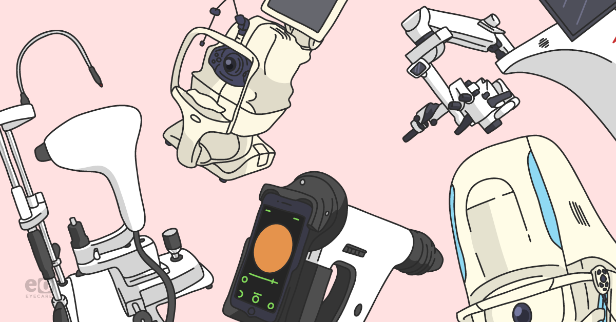
Technology and ophthalmology go hand-in-hand. Whether it's the latest wide-field fundus photography system, OCT technology, or ultrasound, imaging devices have enhanced diagnosing and managing nearly all ophthalmological conditions. While some imaging devices have become commonplace, we aim to highlight devices that may not be as common but could benefit your practice and patient outcomes. In some cases, we argue for a unique application of an imaging modality that you may already have.
Technology and diversity in imaging options in ophthalmology, and in particular retina, only continues to expand. It is estimated that by 2025, the global retinal imaging devices market will exceed six billion dollars.
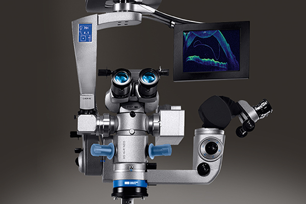
1) Intraoperative Optical Coherence Tomography (iOCT)
While OCT has revolutionized eyecare in a clinic setting, the utilization of OCT during surgery (i.e., intraoperative OCT) is still grossly underutilized. Survey data revealed that only approximately 25% of surgeons currently utilize iOCT.2-6 This is despite multiple studies showing the ability of iOCT to facilitate superior precision in surgery, superior endpoint visualization and reduction of unnecessary steps.2-6
Additionally, research shows that iOCT informs and changes surgical decision-making in anywhere from 10 to 40% of surgical cases.2-6 Whereas before handheld or portable iOCT devices were problematic and impractical, new microscope-integrated iOCT devices represent a new horizon allowing for seamless, live integration.

2) Remidio FOP NM10 Portable Fundus Camera
Smartphones cannot only benefit your practice but can help alleviate preventable eye disease burden globally. Smartphone-based handheld fundus photo systems such as Remidio’s FOP NM10 have become so sophisticated that they can capture retina images through a non-mydriatic eye with a 45-degree field of view. These devices can be used at the slit lamp, providing imaging equivalent to retina desktops in validated studies.
Additionally, the FOP NM10 can screen for diabetic retinopathy using an algorithm that takes just 10 seconds, also validated in multiple peer-reviewed journals as an evidence-based and high-quality screening tool.7-10 Having such non-mydriatic cameras in an internal medicine or family medicine practice may revolutionize the future of diabetic retinopathy screening.

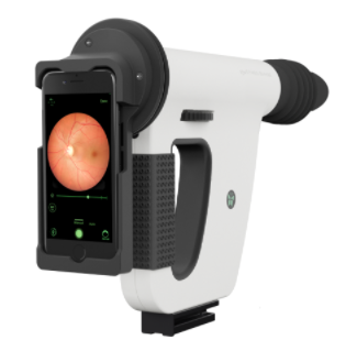
Remidio FOP NM10 Portable Fundus Camera
3) Widefield Fundus Photography, Remidio Vistaro
While to some, it may be straightforward, not all have adopted widefield fundus photography imaging. The benefits of wider-field images of the fundus are multiple. Further assessment of the periphery allows for more accurate staging of diseases like diabetic retinopathy. Additionally, it allows for superior evaluation of hereditary retinal disorders, choroidal tumors, choroidal dystrophies, and peripheral degenerative patterns, all of which directly lead to more comprehensive and accurate diagnosis, prognosis, and management.
Remidio’s Vistaro represents one particular widefield fundus camera that is unique in that it is a smartphone-based, handheld imaging device that can provide the equivalent of the traditional seven-field ETDRS image.11 Additionally, it gives a field of view that extends to/beyond 60 degrees.

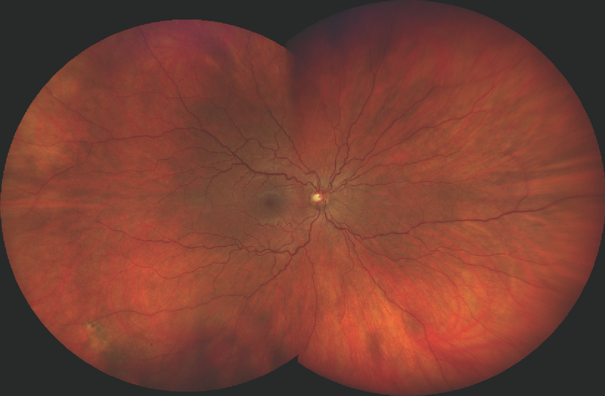
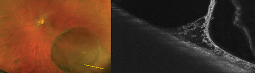
This image, captured on a ZEISS Clarus 500, demonstrates the widefield view into the outer peripheral areas of the retina. Contributed by Dan Epshtein, OD, FAAO.
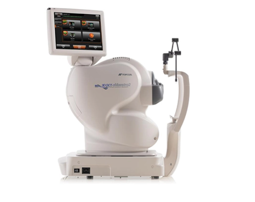
4) Maestro2 Automated OCT/Fundus Camera with OCTA
The Maestro2 from Topcon is a fully automatic OCT system that can capture a color fundus image, OCTA, and OCT with a single button, in an undilated eye. Additionally, a follow-up feature scans the exact location with high precision during subsequent visits. This integration allows for superior visualization and tracking of conditions like choroidal neovascularization, diabetic retinopathy, and microaneurysms.
The value of this type of device comes in the potential it has to optimize clinic efficiency and decrease dependency on highly skilled technicians due to ease of use.12 Additionally, the follow-up feature mitigates inter- and intra-photographer variability between follow-up visits.

Maestro2 Automated OCT/Fundus Camera with OCTA
5) Optos Silverstone
Optos Silverstone is the first and only imaging device to offer optomap®-guided swept-source OCT. Single-capture ultra-widefield (optomap) imaging has been associated with both improved clinic flow and improved patient satisfaction.13,14 Given the immense, growing burden of diabetes, hypertension, and related morbidities, clinical volumes will continue to rise, giving tremendous value to tools like Silverstone that streamline clinic flow and improve efficiency and safely.15,16

Conclusions
Major trends emerging in the latest imaging modalities and tools are integrating multiple images into a single device that is more compact, higher quality imaging, more portable and handheld devices, increased ability to capture images through the non-mydriatic eye, and increased utilization of artificial intelligence and algorithmic imaging. While some of the latest imaging has peer-reviewed data to back its use, other tools are yet to be validated. However, even in cases where validation remains to be strengthened, combination image modalities that are more portable, faster, or algorithm-based represent opportunities for increased efficiency.
References:
- Retinal Imaging Devices Market Size USD 6.3 Bn by 2025 (kbvresearch.com)
- Chavala SH, Farsiu S, Maldonado R, Wallace DK, Freedman SF, Toth CA. Insights into advanced retinopathy of prematurity using handheld spectral domain optical coherence tomography imaging. Ophthalmology. 2009 Dec;116(12):2448-56.
- Pfau M, Michels S, Binder S, Becker MD. Clinical experience with the first commercially available intraoperative optical coherence tomography system. Ophthalmic Surg Lasers Imaging Retina. 2015;46(10):1001-1008.
- Falkner-Radner CI, Glittenberg C, Gabriel M, Binder S. Intrasurgical microscope-integrated spectral domain optical coherence tomography-assisted membrane peeling. Ophthalmol Retina 2015;35(10):2100-2106.
- Ehlers JP, Dupps WJ, Kaiser PK, Goshe J, Singh RP, Petkovsek D, Srivastava SK. The Prospective Intraoperative and Perioperative Ophthalmic ImagiNg with Optical CoherEncE TomogRaphy (PIONEER) Study: 2-year results. Am J Ophthalmol. 2014 Nov;158(5):999-1007. doi: 10.1016/j.ajo.2014.07.034. Epub 2014 Jul 29. PMID: 25077834; PMCID: PMC4250395.
- Ehlers JP, Modi YS, Pecen PE, Goshe J, Dupps WJ, Rachitskaya A, Sharma S, Yuan A, Singh R, Kaiser PK, Reese JL, Calabrise C, Watts A, Srivastava SK. The DISCOVER Study 3-Year Results: Feasibility and Usefulness of Microscope-Integrated Intraoperative OCT during Ophthalmic Surgery. Ophthalmology. 2018 Jul;125(7):1014-1027. doi: 10.1016/j.ophtha.2017.12.037. Epub 2018 Mar 2. PMID: 29409662; PMCID: PMC6015779.
- Chhablani J, Kaja S, Shah VA. Smartphones in ophthalmology. Indian J Ophthalmol. 2012;60(2):127–31.
- Sosale B, Aravind SR, Murthy H, Narayana S, Sharma U, Gowda SGV, et al. Simple, Mobile-based Artificial Intelligence Algo r ithm in the detection of Diabetic Retinopathy (SMART) study. BMJ Open Diabetes Res Care. 2020 Jan;8(1):e000892.
- Natarajan S, Jain A, Krishnan R, Rogye A, Sivaprasad S. Diagnostic Accuracy of Community-Based Diabetic Retinopathy Screening With an Offline Artificial Intelligence System on a Smartphone. JAMA Ophthalmol. 2019 Oct 1;137(10):1182–8.
- Sengupta S, Sindal MD, Baskaran P, Pan U, Venkatesh R. Sensitivity and Specificity of Smartphone-Based Retinal Imaging for Diabetic Retinopathy. Ophthalmol Retina. 2019 Feb;3(2):146–53.
- Nagiel A, Lalane RA, Sadda SR, Schwartz SD. Ultra-widefield fundus imaging: A review of clinical applications and future trends. Retina 2016;36:4:660-78.
- Tornambe, The Impact of Ultra-widefield Retinal Imaging on Practice Efficiency, US Ophthalmic Review 2017
- Successful interventions to improve efficiency and reduce patient visit duration in a retina practice. Retina, 2021.
- The Impact of Ultra-widefield Retinal Imaging on Practice Efficiency. US Ophthalmic Review, 2017.
- Feasibility of peripheral OCT imaging using a novel integrated SLO ultra-widefield imaging swept-source OCT device. International Ophthalmology, 2021.
- .Feasibility and Clinical Utility of Ultra-Widefield–Navigated Swept-Source Optical Coherence Tomography Imaging. Journal of VitreoRetinal Diseases, 2021.


